I. INTRODUCTION
Titanium dioxide (TiO2) has been the subject of intense research for its broad potential applications, including photocatalytic water splitting and degradation of environmental toxic organic pollutants.Reference Fujishima and Honda1, Reference Linsebigler, Lu and Yates2 Its abundance, stability under irradiation, and strong photocatalytic activity make TiO2 a well-known candidate for energy and environmental applications. However, due to the large band gap (3.2 eV for anatase), TiO2 can only utilize the ultraviolet (UV) region of sunlight, which is about 5% of the total energy of the solar spectrum.Reference Chen and Mao3 Various doping or modification methods have been proposed to shift the absorption of TiO2 to include visible light and enhance the photocatalytic activity under visible light.Reference Asahi, Morikawa, Ohwaki, Aoki and Taga4, Reference Zhu, Qiu, Iancu, Chen, Pan, Wang, Dimitrijevic, Rajh, Meyer, Paranthaman, Stocks, Weitering, Gu, Eres and Zhang5 The photocatalytic activity of TiO2 is also highly dependent on the crystal phase, specific surface area, and the concentration of body/surface defects.Reference Carneiro, Savenije, Moulijn and Mul6 These factors are critical to prevent the recombination of photogenerated electrons and holes, which have a short diffusion length in TiO2.Reference Salvador7 Many efforts have been made to overcome this limitation, such as deposition of noble metals and transition metal cocatalysts, etc.Reference Wang, You, Shi, Li and Guo8–Reference Meekins and Kamat10 A series of recent studies on graphene have revealed that composites of two-dimensional (2D) graphene nanostructures and TiO2show enhanced photochemical activity under visible light.Reference Jang and Zhamu11–Reference Zhang, Lv, Li, Wang and Li13
Besides graphene, layered oxides are another family of materials with characteristic 2D structures, and several layered niobates and titanates are known as photoactive wide band gap semiconducting oxides.Reference Miseki, Kato and Kudo14 Recently, substantial efforts have been devoted to the soft chemical modification of oxides to achieve exfoliation of layered structures to form oxide nanosheets. The nanosheets are obtained through reactions of the layered oxides with exfoliating reagents in liquid suspension. Featured with well-defined structures and extremely high anisotropy as well as large specific surface areas, these nanosheets can exhibit unusual photocatalytic behaviors in their reassembled forms (typically restacked from suspension) and in the as-prepared colloidal suspension.Reference Ma and Sasaki15 By utilizing the huge surface area and short charge transport pathways in the colloidal nanosheet system, Osterloh et al. discovered that individually dispersed niobate and titanate nanosheets are catalysts for photochemical H2evolution in photocatalytic water splitting. The nanoscale dimensions of nanosheets cause low ohmic resistance for photoexcited charge diffusion and reduce the impact of short lifetimes of the excited state.Reference Townsend, Sabio, Browning and Osterloh16–Reference Allen, Thibert, Sabio, Browning, Larsen and Osterloh18
Composite catalysts have been studied previously, e.g., incorporation of semiconducting nanoparticles into the interlayer of layered niobates and titanates shows promising improvement of their photocatalytic activities.Reference Centi and Perathoner19 This method of fabrication of a nanocomposite usually involves utilizing large organic exfoliating agents to pillar the structure to allow incorporation of nanoparticles in the interlayer galleries. A recent study has demonstrated that the exfoliation reagent surrounding the nanosheets works as a counter cation and principle hole scavenger, which impairs the effect of sacrificial electron donors in photocatalytic systems.Reference Nakato, Yamada, Nakamura and Takahashi20 In this paper, we introduce a new method for synthesizing a composite with protonated niobate nanosheets H1.8Bi0.2CaNaNb3O10 (BCNN) supported by commercially available anatase TiO2using a unique organic-free exfoliation process. The resulting nanocomposites display remarkably enhanced photocatalytic activity for methylene blue (MB) degradation under UV and visible light.
II. EXPERIMENT
A. Materials
Parent phase Bi2CaNaNb3O12 powders were prepared using conventional solid-state synthesis. The reactants included analytical grade Bi2O3, CaCO3, Na2CO3, and Nb2O5, which were mixed in stoichiometric proportions and milled under isopropyl alcohol using a McCrone micronizing mill (McCrone Microscopes & Accessories, Westmont, IL) with alumina media. The powders were air dried and calcined at 815 °C in air for 5 h. After heat treatment, the sample batches were reground by hand, and 10 g of the material was pressed into pellets using a 1.9-cm die at ∼60 MPa. The remaining sample batch was then used to form a sacrificial powder bed for use in further heat treatments to limit bismuth volatilization at elevated temperatures. Sample pellets were then reacted under a total of three high temperature cycles. For each cycle, the pellets were heated at 1050 °C for a period of 12 h in covered MgO crucibles, followed by sequential grinding by hand, x-ray analysis and pressing into pellets.
B. Synthesis of nanosheets and composite assemblies
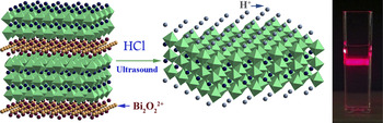
Colloidal suspensions of exfoliated BCNN nanosheets were synthesized by simultaneous ion exchange and exfoliation of the Bi2CaNaNb3O12 Aurivillius phase, as shown schematically in Fig. 1. One gram of Bi2CaNaNb3O12was mixed with 100 mL of 5 N HCl at room temperature and ultrasonicated for 6 h. The acid-sonicated products were collected and washed with deionized water followed by high speed centrifugation at 12,000 rpm. Without drying, the final product was redispersed in 50 mL HCl solution with pH = 4, and the suspension was then centrifuged at 3800 rpm to remove nonexfoliated particles and the suspension containing the nanosheets was collected. The concentration of the colloid suspension was determined by precipitating 10 mL of the liquid by heating and boiling, centrifuging with water and drying in air at 70 °C. The concentration of BCNN nanosheets was about 1 mg/mL.
Schematic of the ion exchange and exfoliation process and a photo of the resulting nanosheet suspension illuminated with a red laser.

To make the nanosheet–TiO2 composite, 0.1 g of TiO2 anatase particles (Alfa Aesar, Ward Hill, MA, 99.9%) was mixed with 1 mL BCNN nanosheet suspension in 20 mL water and ultrasonicated for 1 min. The concentration of BCNN nanosheets was about 1 mg/mL, as determined by drying the suspension and weighing the solids. The composition of the composite is therefore estimated as ∼1 wt% BCNN nanosheets with ∼99 wt% TiO2. This estimate was also confirmed by energy dispersive x-ray spectroscopy analysis. The mixtures were then calcined in air at 343, 500, 600, and 700 K, yielding four specimens referred to as TB343, TB500, TB600, and TB700.
C. Photocatalytic experiments
The photocatalytic activity of the photocatalyst was evaluated by decomposition of MB dye. The tests were carried out using a 1000 W xenon lamp (Oriel Corporation, Stratford, CT) set to 500 W output (Filter ORIEL 6628 and 6629). A 100 mL solution of 200 μM MB and 100 mg of BCNN/TiO2 composite powder was constantly stirred before and during the reaction. Before irradiation, the suspensions were magnetically stirred in the dark for 45 min to establish equilibrium between the MB molecules and the surface of the particles. The solution was then irradiated and the concentration of remaining MB was measured using a UV–visible–near-infrared (UV/Vis/NIR) spectrophotometer (Perkin Elmer Lambda 950 UV/Vis/NIR spectrometer, Waltham, MA). The UV–Vis spectra of MB in the visible light region show a main band at λmax≈ 664 nm, which shifts to slightly higher wave length as the concentration decreases. For the best accuracy, the spectra were measured over the range of 400–800 nm, and the concentration was calculated from the maximum of the spectra according to the Beer–Lambert law and normalized to the concentration of the MB solution at the beginning of each experiment.
D. Characterization
The atomic-scale structures of the nanosheets and TiO2 composite were studied using x-ray diffraction (XRD) (Bruker D8 Advance, Madison, WI; Cu Kα = 1.54 Å). The XRD patterns were recorded within the 2θ range of 5–55° with steps of 0.02° at a scan rate of 5 s per step. A Micromeritics TriStar 3000 (Norcross, GA) surface area and porosity analyzer was used to determine the Brunauer–Emmett–Teller (BET) surface area. High-resolution transmission electron microscopy (HRTEM) was used to study the microstructure of the nanosheets and BCNN/TiO2 composite using a Hitachi HF-3300 (Tokyo, Japan) cold field-emission transmission electron microscopy and scanning transmission electron microscopy at 300 kV. UV–Vis diffuse reflectance spectra of the samples were obtained for the dry samples using a UV–Vis spectrometer (Perkin Elmer Lambda 900 UV/Vis/NIR spectrometer), and the band gap energy (E g) was calculated by fitting the reflectance data using the Kubelka–Munk function.Reference Gao and Wachs21, Reference Barton, Shtein, Wilson, Soled and Iglesia22 The infrared (IR) absorption spectra of the solid samples were recorded using a Thermo Nicolet Nexus spectrometer (Waltham, MA) on single-crystal KBr sample holders.
III. RESULTS AND DISCUSSION
The common method for synthesizing metal oxide nanosheets is a multistep soft chemical process including ion exchange to produce the protonated phase, followed by inserting bulky organic ions to expand the interlayer gallery.Reference Ma and Sasaki15 The organic exfoliating agent typically used is tetrabutylammonium (TBA+). In contrast, our new ultrasonic exfoliation approach yields organic-free nanosheet suspensions that may be reassembled by drying onto planar substrates or to create nanosheet–TiO2composites. Further, our new method shortens the reaction time from days to hours and successfully produces contaminant-free mono- and multilayer niobate nanosheets.
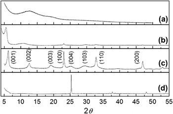
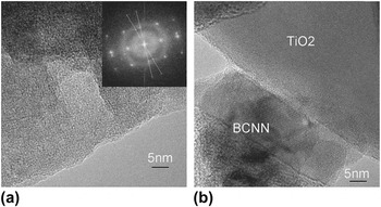
XRD patterns of the BCNN nanosheet colloidal suspension, reassembled nanosheet film, and BCNN/TiO2 composite are shown in Fig. 2. For comparison, the ion-exchanged BCNN was prepared as described in literatureReference Sugimoto, Shirata, Kuroda and Sugahara23 and the XRD data is also included [Fig. 2(c)]. As shown in Fig. 2(a), there appears to be only a very weak scattering signal from the dilute nanosheet suspension, which indicates that BCNN nanosheets were well dispersed. After drying on a sapphire single crystal surface [Fig. 2(c)], the XRD pattern for the reassembled nanosheets is similar to that of the ion-exchanged layered BCNN shown in Fig. 2(b).Reference Sugimoto, Shirata, Kuroda and Sugahara23 The strong (001) peaks and diminished (010) and (110) peaks imply the layer-by-layer reassembling of the nanosheets. Comparing to the ion-exchanged BCNN crystal, the (001) peaks of the layered nanosheets are shifted to lower angles, suggesting lattice expansion by ∼1.8 Å due to intercalation of water molecules in the interlayer space of the protonic nanosheets. The XRD pattern of the BCNN/TiO2 composite [Fig. 2(d)] is similar to that of pure TiO2 powder. No XRD peaks from the nanosheet are observed due to the extremely low concentration of the nanosheets (∼1%) and the highly disordered condition as the composite forms. HRTEM studies [Fig. 3(a)] show that the restacked nanosheets take on random orientations in-plane. The inset in Fig. 3(a) shows the diffractogram after the Fourier transformation of the main figure, with the strong reflections related to the perovskite subunit cell. Figure 2(b) shows the microstructure of the nanosheet/TiO2 composite after heat treatment at 700 K. It is clear that the nanosheets are deposited on the surface of TiO2as highly disordered clusters.
XRD patterns of (a) the BCNN nanosheet colloidal suspension; (b) reassembled nanosheets on sapphire; (c) ion-exchanged BCNN; (d) BCNN/TiO2composite.

HRTEM images of the (a) BCNN nanosheets on a holey carbon grid and (b) TiO2/BCNN nanosheets composite.

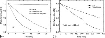
The photocatalytic activity of the BCNN/TiO2 composite under UV and visible light (λ ≥ 420 nm) was examined, with the results shown in Fig. 4 for the samples calcined at 700 K. Figure 4 compares the degradation behavior of the composite catalyst with that of the same mass of TiO2 P25 (Degussa, Evonik Industries, Essen, Germany) under identical conditions. A large increase of reaction rate is noted. Under UV irradiation, the BCNN/TiO2 composite degraded about 70% of the MB within 15 min; in contrast, it took nearly 25 min for the P25 to reach the same concentration. The BCNN/TiO2 composite shows superior activity under visible light also, with the MB degradation rate of the BCNN/TiO2 composite nearly twice that of P25. Considering the BET surface area of the composite is only 9.4 m2/g, compared to 57 m2/g for P25, the improvement of photocatalytic activity of the BCNN/TiO2composite is significant.
MB decomposition upon (a) UV and (b) visible light irradiation of the reference sample of P25 TiO2 and BCNN/TiO2composite.

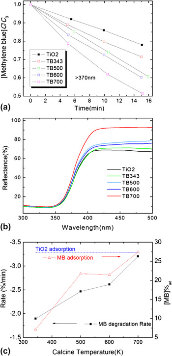
To reveal the origin of the enhanced photocatalytic properties, we investigated the influences of calcination temperature on BCNN/TiO2 composites. Figure 5(a) shows the UV light (>370 nm) photodegradation of MB over the BCNN/TiO2 composite calcined under different temperatures. This result clearly shows that the photocatalytic activity of the composites is improved by nearly 100%, as the calcination temperature increases from 343 to 700 K. UV–Vis spectroscopy was used to study the band structure of the calcined BCNN/TiO2 composite and the host TiO2. The diffuse reflectance spectra of the catalyst composites are shown in Fig. 5(b). The band gap of the catalysts was calculated by fitting the Kubelka–Munk equation:
 $${\rm{\alpha }}hv = {\rm{const}}(hv - E_{\rm{g}} )^2 \quad,$$
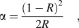 $${\rm{\alpha }} = {{(1 - R)^2 } \over {2R}}\quad,$$
Characteristics and performance of BCNN/TiO2composite prepared using different calcination temperatures. (a) MB decomposition under UV light (>370 nm); (b) UV–Vis diffuse reflectance spectra; (c) MB degradation rate and the surface adsorption.

where R is the reflectance. The band gap energy of the composite TB700 is blueshifted by only about 0.04 eV from that of the host TiO2. Clearly, the shift of the band gap is not responsible for the improved photocatalytic activity.
We also examined the MB adsorption behavior of the BCNN/TiO2 composites by measuring the concentration of MB in solution after establishing equilibrium with the added catalyst. In Fig. 5(c), the MB degradation rate and the MB adsorption rate are plotted as a function of calcination temperature and clearly show the same trend. We note from Fig. 5(c) that the MB adsorption on TB700 is the same as for pure TiO2, but the photodegradation rate for TB700 is substantially higher. These results highlight the role of the BCNN nanosheets for increased surface adsorption and enhanced composite performance.
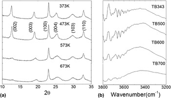
To further investigate the mechanism of the enhanced photocatalytic activity, we studied changes that occur with temperature. In situ XRD was used to study the BCNN/TiO2 composites but without success because of the very small loading of nanosheets. Therefore, restacked BCNN nanosheets were prepared to study the structural behavior at high temperature. The progression of XRD patterns up to 673 K is summarized in Fig. 6(a). The result shows signs of topochemical dehydration reactions, which have been demonstrated in the protonated Ruddlesden–Popper and Dion–Jacobson phases such as H2La2Ti3O10, HCa2Nb3O10, and H2SrTa2−xNbxO7.Reference Gopalakrishnan and Bhat24–Reference Poterala, Chang, Clark, Meyer and Messing27
(a) High temperature in situ XRD data for BCNN nanosheets deposited on a sapphire crystal and (b) IR spectra of the BCNN/TiO2composites.

In this reaction, the in-plane tetragonal lattice parameter remains unchanged with temperature (a ≈ 3.81 Å, ignoring thermal expansion), and the reflections from the perovskite structure such as (100), (110), and (103) show no changes. However, as the temperature increases from 473 to 573 K, the (00l) peaks are significantly weakened, broadened, and shifted to smaller d spacings. The XRD data clearly indicate that the structure disorders along the c-axis during heating, from the condensation of the layered 2D perovskite structure during the topochemical dehydration.
For the BCNN nanosheets, the protons are uniformly distributed on the surface to compensate the negative charge of the perovskite slab. Therefore, –OH groups presumably occupy all the surface octahedral vertices, similar to the case of acid-exchanged Ruddlesden–Popper phases. During heating, dehydration converts the interlayer M–OH groups to shared oxygen linkages M–O–M.Reference Fang, Kim and Mallouk26 The hydrated surface of TiO2 is known to be rich in Ti–OH groups due to the dissociation of water.Reference Vittadini, Selloni, Rotzinger and Grätzel28, Reference Arrouvel, Digne, Breysse, Toulhoat and Raybaud29 In the BCNN composite, the nanosheets bond to the TiO2 particle surfaces and subsequent dehydration of the Nb–OH and Ti–OH groups can therefore result in the formation of hetero Nb–O–Ti bridges, which may then improve the charge transfer between the particles and nanosheets. As shown in the in situ XRD patterns, the topochemical dehydration occurs above 473 K (and below 573 K), which coincides with in the large jump in phototcatalytic activity and surface absorption of MB [Fig. 5(c)]. Thus, the enhanced photocatalytic activity of the BCNN/TiO2 composite and its dependence on the calcination temperature are indications of the formation of a heterojunction, which enables photoinduced charge separation between the nanosheets and TiO2 host.Reference Uchida, Yamamoto, Fujishiro, Watanabe, Ito and Sato30 Further studies are in progress to confirm this hypothesis.
Besides the chemical transformation at the BCNN/TiO2 interface, the surface properties of the composite are also critical for the photocatalytic redox reactions. Surface hydroxyl groups have been proposed to act as traps for both photoexcited electrons and holes and improve the photocatalytic performance of TiO2 on the photooxidation of phenol and MB.Reference Du, Bueno-Lopez, Verbaas, Almeida, Makkee, Moulijn and Mul31 At the surface of the BCNN/TiO2 composites, the protonated BCNN nanosheets decorate the TiO2 surface with a tremendous number of hydroxyl groups. IR spectroscopy was used for the investigation of surface hydroxyl groups, with results in the high frequency region of 3800–3200 cm−1 shown in Fig. 6(b). The broad and complex absorptions lower than 3600 cm−1 are due to the –OH oscillation of undissociated water molecules coordinatively adsorbed on surface cations.Reference Minella, Faga, Maurino, Minero, Pelizzetti, Coluccia and Martra32 In the 3600–3800 cm−1 region, the series of narrow and partially-resolved peaks are related to the weak intermolecular interactions of –OH groups.Reference Minella, Faga, Maurino, Minero, Pelizzetti, Coluccia and Martra32 As the calcination temperature increases from 373 to 700 K, we note a substantial decrease of the absorption bands for the v(OH) modes in the region of 3800–3600 cm−1. This result indicates the condensation of hydroxyl groups, consistent with the results from the MB photodegradation and surface adsorption study and the in situ XRD results. We conclude therefore that the surface population of hydroxyl groups dominates the performance of the BCNN/TiO2 composite, with smaller concentrations of –OH groups yielding superior performance. This conclusion is somewhat counter to the notion that surface-hydroxylated centers act as trapping sites to improve photocatalytic reactions.Reference Linsebigler, Lu and Yates2 In the MB degradation process, the hydroxyl group absorbs a hole in the valence band and produces OH· radicals, which are the reactants for the oxidation of MB molecules.Reference Houas, Lachheb, Ksibi, Elaloui, Guillard and Herrmann33 One possible explanation is that the dehydration of the BCNN surface exposes Nb5+ and O2− acid–base pairs, similar to the Ti4+ and O2− acid–base pairs observed on the surface of the dehydroxylated TiO2.Reference Martra34 Upon mixing with a MB solution, we expect some of the sites to remain acid–base pairs, while others dissociate water molecules to rehydroxylate some surface sites. Since MB has a cationic configuration, its adsorption is favored on a negatively charged surface site. Therefore, the exposed O2−sites could serve as both hole traps and adsorption centers for MB molecules and result in enhanced photodegradation rates.
An additional driving force for enhanced photoactivity may be local ferroelectric fields within the nanosheets. Our recent studies of the structures of Aurivillius phases show that the parent BCNN phase has a calculated ferroelectric polarization of 14.97 °C/cm3 along the a-axis in space group B2cb, in other words, along the long axes of the nanosheets.Reference Nichols, Shi, Huq, Voge and Misture35 Previous work has shown that the static dipolar fields of the ferroelectric domains can effectively separate photogenerated electrons and holes and drive the charges toward the surface to participate in the catalytic reaction.Reference Burbure, Salvador and Rohrer36 If the nanosheets maintain the structural distortions that yield the ferroelectric polarization, or if the topochemcial dehydration enhances the distortions, the photoactivity might be improved.
Finally, we note that the weak photocatalytic activity under visible irradiation cannot be adequately explained by considering only the surface hydroxyl groups because visible light is well below the band gap of the BCNN/TiO2 composite. Optical absorptions in semiconductors in the below band gap region have been attributed to impurity center interactions with the lattice, which may be operating here.Reference Pankove37 In fact, we have established that some 20% of the perovskite A-site cation positions are occupied Bi3+on the perovskite A-sites, providing a large number of defect sites.Reference Houas, Lachheb, Ksibi, Elaloui, Guillard and Herrmann33
IV. CONCLUSIONS
An organic-free method was developed for the synthesis of protonic niobate nanosheets BCNN. Composite catalysts prepared from BCNN nanosheets and TiO2 (anatase) show markedly increased photodegradation rates for MB solutions. The calcination temperature of the composite is the key factor that determines the nanosheet–TiO2 chemical linkage and surface hydroxyl concentration, which together define the MB adsorption behavior and photoreaction rate. The results suggest that charge transfer at the BCNN–TiO2 interface and the presence of Nb5+ and O2− acid–base pairs are responsible for the markedly improved performance, though there may be an additional contribution from ferroelectric domains within the nanosheets. Niobate nanosheets act as low-cost cocatalysts for high performance TiO2/nanosheet composites.
ACKNOWLEDGMENTS
This material is based upon work supported by the National Science Foundation under Grant No. DMR 0606246. J.L. was supported separately by Alfred University. The TEM study performed at the Oak Ridge National Laboratory’s High Temperature Materials Laboratory was sponsored by the U.S. Department of Energy, Office of Energy Efficiency and Renewable Energy, Vehicle Technologies Program.