Coarctation of the aorta accounts for 5–8% of all CHT.Reference Hoffman and Kaplan 1
Coarctation in neonates is usually a catastrophic illness and is associated with congestive heart failure, which demands immediate and aggressive treatment because of its poor natural history. The optimal management strategy for native aortic coarctation in neonates and young infants is controversial. Surgical repair has been the standard treatment for aortic coarctation since 1944. After Lababidi performed the first balloon angioplasty in 1983, balloon angioplasty is gradually gaining acceptance.Reference Lababidi 2 In recent years, percutaneous balloon angioplasty has been found to be a less invasive treatment alternative to surgical repair for coarctation of the aorta. However, this strategy remains controversial in neonates and young infants. Balloon angiography is beneficial in neonates, but even after balloon dilatation early recoarctation is still an issue. In our study, we examined the effectiveness of the balloon angiography by histopathology.
Methods
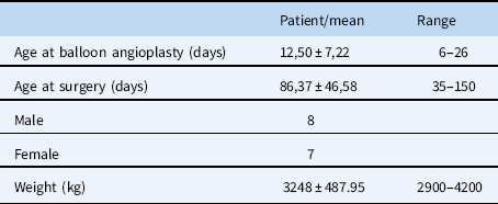
Between 01 October, 2014 and 01 August, 2017, we encountered 15 patients <30 days of age on whom surgical aortoplasty after successful balloon angioplasty for native aortic coarctation was performed. These patients had complications regarding recoarctation, following balloon angioplasty intervention at our institute and other centres. The diagnosis of coarctation was made by echocardiography or CT in all patients. Patients with complex cardiac anomalies, deep preoperative acidosis, and neonatal sepsis were excluded from the study. Overall, median time for balloon angioplasty after birth was 12 days (range, 6–26 days). Median time for balloon angioplasty to surgery was 86 days (range, 35–150 days), with a mean weight of 3.2 kg.
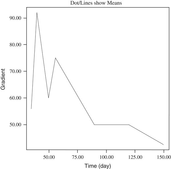
Statistical analyses were performed with SPSS for Windows (release 15), and for the continuous values the Spearman’s correlation test was used (Fig 1).
The negative relationship between echocardiographic gradient and operation time (r=−0.723**, p=0.002)

Balloon angioplasty technique
Balloon dilatation of the coarctation was performed using the standard retrograde femoral arterial approach in all the patients. The balloon diameter was selected to be not two times greater than the diameter of the stenotic area at the coarctation site and not to exceed the diameter of the aorta at the level of the diaphragm. The length of the balloon was 20–30 mm. The balloon was inflated one to three times to the pressure level recommended by the manufacturer until relief of waist was observed. Balloon angioplasty was considered successful when the waist disappeared and the post-procedure pressure gradient reached ≤ 20 mmHg.
The indications for surgery included persistent large aortic gradient, weak femoral pulse, and hypertension. Re-intervention for recurrent coarctation was performed where the arm-to-leg gradient was >20 mm Hg, the peak systolic echocardiographic gradient was >25 mm Hg, or the upper-extremity systolic blood pressure exceeded the 95th percentile for age.
Operative technique
The patient is positioned right lateral recumbent. A lateral thoracotomy incision is made through the third or fourth intercostal space. The left lung is retracted anteriorly and inferiorly with a moist swab. A longitudinal incision is made in the mediastinal pleura over the upper descending aorta posterior to the vagus nerve and is extended superiorly over the left subclavian artery, ligating and dividing the superior intercostal vein in the process.
After appropriate proximal and distal aortic clamps are chosen, the ductus arteriosus is doubly suture-ligated with a 5–0 or 6–0 polypropylene suture, which is left on a haemostat at this point. Heparin (100 u/kg) is administered intravenously, and the proximal clamp is carefully applied after 3 minutes to allow adequate heparin circulation. The location of this clamp is determined by the planned operation and by the anatomic type of coarctation. The focus must be on allowing construction of the most possible largest and extended anastomosis, and careful attention must be paid to positioning this clamp perfectly. The distal clamp is placed at or below the level of the second pair of intercostal vessels, and the snares around the intercostal vessels are placed on traction. The coarcted segment with the ductal tissue is then resected. After completion of the extended anastomosis, the distal aortic clamp is removed to allow the excluded segment to fill with blood and expel air. The proximal aortic clamp is then slowly removed, and the systemic pressure is carefully watched. The mediastinal pleura is routinely closed, and a single pleural drain is inserted before closure of the thoracotomy.
Histopathologic evaluation
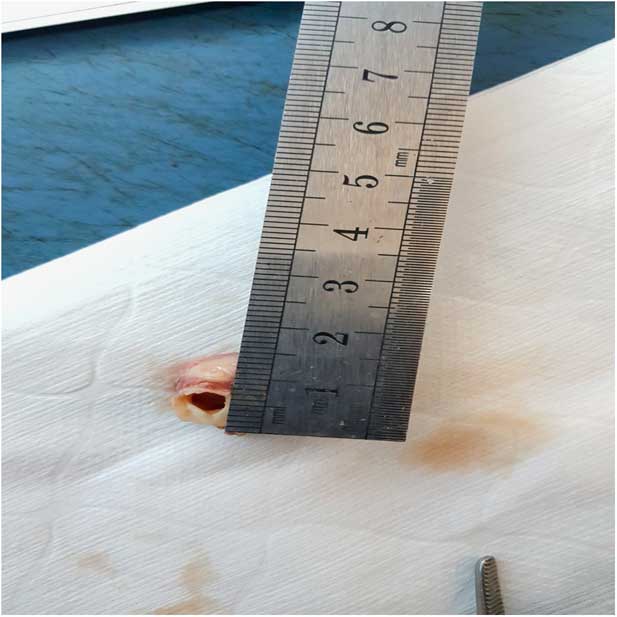
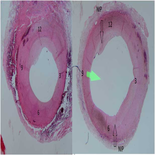
Histologic sections were prepared from resected portions of the thoracic aorta from all neonates. Such specimens had been preserved in 10% buffered formalin (Fig 2). Pathologic evaluation of the resected coarctation segments from patients having angioplasty followed by surgery was performed by the same pathologist. The specimen oriented according to ductal insertion, which could be recognised from the outer aspect of adventitia (Fig 3). If the vascular size was convenient to cut in a longitudinal direction, it was separated into two to eight pieces clockwise beginning with ductal site coded as 12 o’clock. Otherwise, transverse sections were made. At least one of the samples was passed through the insertion of ductus (ligamentum) arteriosus. After routine tissue processes, microscopic sections were prepared with haematoxylin–eosin, Verhoeff’s elastic van Gieson, and Masson’s trichrome. Slides were examined by light microscopy.
Microscopic view of coarctation.

Clockwise schema and neointimal proliferation (NIP) of the coarctation segment.

Periodic clinical evolutions; echocardiographic derivated peak pressure gradients were obtained for all neonates, initially before balloon angioplasty, after intervention, the post-procedure day, and thereafter 3th, 6th, 12th post-procedural months routinely.
Results
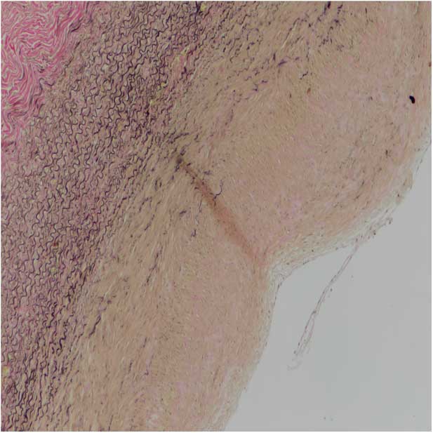
A total of 15 surgically excised specimens were available for histological examination. We evaluated ductal insertion sites and the possible tears with the findings of recovery. Histopathologic examination revealed that all healed tears at different segments reached media from intima at various grades. In all patients’ coarctation zone, there was elastic fibre loss and smooth muscle degeneration in the media layer with neointimal thickening. Loss of internal elastic lamina at different segments accompanying neointimal proliferation was the cause of increased thickness of the wall which resulted in diamater irregularities in the luminal aspect of the aortic wall (Fig 4). Disruption of elastic fibres for the whole arterial wall was mostly seen at the region of ductal insertion tissue and opposite of the aortic wall.
Section of resected aortic coarctation from the patient. Aorta has elastic fibre loss and smooth muscle degeneration in the media layer with neointimal thickening.

Cystic medial necrosis was revealed in all cases. Cystic medial necrosis was observed in each of the surgical specimens within different intensities and spread. Grade 2–3 cystic medial necrosis was found in 14 patients. Of 15 patients, only one patient had grade 1 cystic medial necrosis. The appearance of cystic medial necrosis is a consequence of balloon angioplasty. This is also one of the factors that increase the development of aneurysm in the future.
Histopathological analysis of the aorta showed a widened subendothelial region with separation of endothelial cells from the internal elastic lamina in all cases in which balloon angioplasty was used.
Patients’ demographic analyses were similar (Table 1). Median time for balloon angioplasty after birth was 12 days (range, 6–26 days). Median time for balloon angioplasty to surgery was 86 days (range, 35–150 days). No repeat intervention was required in the 15 patients who underwent surgery followed by angioplasty. Early mortality was seen in 1 patient after surgery. Chylothorax was observed in only one patient as a postoperative complication.
Patient characteristics.

As a result, the reasons for recurrence of recoarctation after balloon angioplasty are as follows: patients with higher preoperative echocardiographic gradients had recoarctation earlier, neointimal proliferation, aortic intimal fibrosis at the region of ductal insertion, and ductal residual tissue debris.
Discussion
Coarctation of the aorta accounts for 5 to 8% of neonatal CHD and represents a spectrum of aortic narrowing that varies from a discrete entity to tubular hypoplasia. Generally, native coarctation involves the isthmus and part of the transverse arch.
Coarctation of the aorta is at times missed on clinical examination, and a high index of suspicion is required to make this diagnosis. Patients with coarctation of the aorta have a poor prognosis if they do not receive surgical or catheter interventions. Campbell et al examined the natural history of coarctation of the aorta and demonstrated that the median age of death for unrepaired coarctation of the aorta is 31 years, with 76% of deaths attributable to complications of aortic coarctation (25.5% cardiac failure, 21% aortic rupture, 18% bacterial endocarditic, 11.5% intracranial haemorrhage).Reference Campbell 3 As a result, accumulating evidence shows that coarctation is a systemic vascular disease rather than a simple mechanical obstruction that can be resolved by interventions.
When coarctation is severe, immediate surgical or balloon intervention is necessary after birth. This is especially true for neonates, where despite the performance of all types of corrective intervention of the native coarctation of the aorta, repeat intervention may be required due to restenosis. Restenosis rates at balloon angioplasty vary from 5 to 24% and are higher in neonates. Recoarctation of the aorta refers to restenosis after an initially successful surgical or catheter-based repair and is thought to be secondary to either a residual obstruction or development of restenosis.
Crafoord and NylinReference Crafoord and Nylin 4 performed the first surgical intervention in 1944. The Blalock–Park procedure was reported in that same year.Reference Blalock and Park 5 Following the introduction of prosthetic patch aortoplasty in 1961,Reference Vossschulte 6 the subclavian flap aortoplasty procedure was reported in 1966 by Waldhausen and Nahrwold as a strategy to lower the high rates of restenosis.Reference Waldhausen and Nahrwold 7 We have used extended end-to-end anastomosis technique in our patients because of the low rates of restenosis.
For neonates with/without hypoplasia of the transverse arch, an extended end-to-end anastomosis using a broader longitudinal incision across the proximal aorta improved the outcome and is currently preferred because of low mortality rates and low rates of restenosis.Reference van Heurn, Wong and Spiegelhalter 8 , Reference Torok, Campbell, Fleming and Hill 9
The technique of balloon angioplasty involves expansion of the constricted coarctation site, which results in rupture of the intima and injury of the media. Balloon angioplasty was initially used in neonates with heart failure who were at a high risk for surgery. Post-procedural complications after balloon angioplasty include aortic wall complications such as dissection, intimal disruption, tear (1–7%), cerebrovascular accidents (<1%), and death (0–2%).Reference Yetman, Nykanen and McCrindle 10 – Reference Rao 12 Other complications include femoral artery injuries and thrombosis in 21% of newborns and infants, and in 9% of children in their study.Reference Beekman, Rocchini and Dick 13
As one of the most important late complications of coarctation, Rao et al. reported that neonates are particularly at risk for restenosis.Reference Rao, Galal, Smith and Wilson 14 Additionally, it has been suggested that the increased restenosis risk may be caused by the fact that balloon angioplasty causes injury to the aortic intima and media. As a consequence, harmful scar tissue might cause restenosis. Balloon angioplasty was found to be preferable over surgery in non-neonatal coarctation patients, whereas balloon angioplasty in neonates remains controversial.Reference Wong, Benson, Van Arsdell, Karamlou and McCrindle 15 In conclusion, these findings show that balloon angioplasty is effective in relieving the aortic gradient in children beyond the neonatal period. However, there are only a limited number of studies on primary balloon angioplasty for native coarctation of aorta in neonates. For this reason, we have examined the effectiveness of the balloon and its effects on the aortic wall in this study.
In native coarctation, impaired elastic fibre formation occurs in the ductus arteriosus and extends to the proximal portion of the ascending aorta. In the ductus arteriosus, intimal thickening is characterised by an area of subendothelial deposition of extracellular matrix, the disassembly of the internal elastic lamina, and loss of elastic fibre where smooth muscle cells and elastic fibres are scattered.Reference Yokoyama, Minamisawa and Quan 16 – Reference Yokoyama 19
In the patients’ histopathological examination, the received sections were divided according to the clock dial. The ductus arteriosus was examined at the 12th position along the pathological section. In 15 patients the continuity of the internal elastic lamia is disrupted mostly at the levels of 12 and 6. We believe that balloon angioplasty causes the most damage to the vessel wall in these regions. As such, therapeutic tear or controlled tear, which is a tear through the intima and at least partially into the media and vessel wall’s response to the tear, plays a role in recoarctation. All neonates’ histologic studies have shown intimal hyperplasia caused by fracture of the internal elastic lamina and the migration of smooth muscle cells and fibroblasts from tunica media to intima and their proliferation. As a result of balloon angioplasty, restenosis may occur owing to residual tissue debris. Resection of ductal tissue after surgery may be a cause of lesser restenosis.
The ductus arteriosus has greater contractile ability because of smooth muscle cells.Reference Mataıchı, Ken, Masahıko, Katsumı and Yuıchıro 20 Histopathologic sections revealed that smooth muscle cell proliferation was seen mostly at level 12. When we resected the ductal tissue surgically, recoarctation may not appear owing to muscle cell proliferation like in the balloon angioplasty group. As a result of our findings, removal of smooth muscle cells in the coarctation zone by surgery prevents early recoarctation.
In our histopathologic examination, the existence of cystic medial necrosis was found on the vessels walls on which balloon angioplasty was performed. The region where balloon angioplasty was applied, according to the aforementioned grading scheme, a grade 2–3 cystic medial necrosis was found and according to the clock dial at the mostly sections of the 12th to 3rd and 6th position was observed. Therefore, severe and widespread occurrence of cystic medial necrosis at the histopathologic segments was present, owing to balloon dilation. It was predicted that these areas could likely lead to aneurysm formation in the future.
In the course of our analysis, we observed neointimal proliferation mostly at the 12th, 4th, and 6th positions causing narrowing of the lumen, and it is thought that these findings may be the cause of recoarctation. Recoarctation occurs especially in patients who had high echocardiographic gradient before intervention. The neointimal proliferation might cause early recoarctation.
In our clinic, recoarctation is a common complication following balloon angioplasty and tends to occur in most neonates. After angioplasty procedure, surgery was required within 4–25 weeks. Recoarctation is common in neonates owing to the narrowness of the vessel lumen and excessive response to the injured vessel wall. In neonates with high gradient on echocardiography before balloon angioplasty, recoarctation occurs more rapidly (p=0,002). There was a negative relationship between the echocardiographic gradient and operation time (r=−0.723**, p=0.002) (Fig 1).
Palliative balloon angioplasty may be considered to stabilise the critically ill and urgent patient. Balloon angioplasty, when performed on patients at their earliest possible age, delays actual corrective operation to a later date, which in turn provides less risky surgical outcomes in infants who are gaining weight, growing, and do not have any haemodynamic complaints. Therefore, successful surgeries performed on these infants result in a lower rate of restenosis.
Conclusion
Even with the technical developments of endovascular techniques, recoarctation is still a problem for neonates. Histopathological findings reveal that healed intimal and medial tears, internal elastic lamia disruption in different segments, and ductal tissue left over caused neointimal fibroelastic proliferation after balloon angioplasty. These histopathological findings were evaluated as evidence of a successful dilatation when the balloon angioplasty was performed. However, after the balloon angiography, the response of the aortic wall can be excessive in newborns and early recoarctation may develop. In centres in which the neonatal ICU is inexperienced, balloon angioplasty is particularly recommended. Balloon angioplasty, when performed on patients at their earliest possible age in developing neonatal clinics, delays actual corrective operation to a later date, which in turn provides less risky surgical outcomes in infants who are gaining weight, growing, and do not have haemodynamic complaints.
Financial Support
This research received no specific grant from any funding agency or commercial or not-for-profit sectors.
Conflicts of Interest
None.
Ethical Standards
The trial was approved by the institutional ethics committee of the centre and was conducted in accordance with the Declaration of Helsinki.
Acknowledgements
None.