Telomeres are structures that cap the ends of linear chromosomes. In human cells they are composed of the hexameric DNA sequence TTAGGG tandemly repeated into arrays of up to 25 kb. A key function of telomeres is to counteract the end-replication losses that occur as a consequence of semiconservative replication of linear DNA molecules (Ref. Reference Olovnikov1). This function is mediated by the reverse transcriptase telomerase (TERT), which acts together with the telomerase RNA component (TERC) in a complex of proteins to catalyse the RNA-templated addition of telomere repeats de novo at the chromosomal terminus (Refs Reference Greider and Blackburn2, Reference Bodnar3) (Box 1).
Box 1. The telomerase complex
Telomeric function is conferred by a complex of proteins that associate with telomeres via interactions with the major TTAGGG-repeat-binding proteins TRF1 (TERF1), TRF2 (TERF2) and POT1 (Ref. Reference de Lange22). The reverse trancriptase telomerase (TERT) and the telomerase RNA component TERC, together with dyskerin (DKC1), constitute the core telomerase complex. DKC1 is an RNA-binding protein that functions to support the biogenesis of a functional TERC–TERT complex and contributes to TERC stability (Refs Reference Mitchell, Wood and Collins23, Reference Mochizuki24). This complex forms part of a larger holoenzyme complex that includes the small nucleolar proteins nucleolar protein 10 (NOP10) and nonhistone protein 2 (NHP2), both of which are associated with DKC1 and support the maturation of TERC via cooperation of the hairpin–hinge–hairpin–ACA (H/ACA) motif within TERC (Refs Reference Fu and Collins25, Reference Pogacic, Dragon and Filipowicz26). Telomerase Cajal body protein 1 (TCAB1) also forms part of the telomerase holoenzyme complex, directing the TERT, TERC and DKC1 complex to telomeres via the Cajal bodies (Ref. Reference Venteicher27).
Telomerase is expressed at high levels in immortal cell lines, the stem cell compartments of actively proliferating tissues, and over 90% of human malignancies (Refs Reference Engelhardt4, Reference Kolquist5). In some cancers, the upregulation of telomerase activity could result from the amplification of the TERT locus: gain of the chromosome arm 5p, which contains the TERT gene, is one of the most frequent chromosome gains in human cancers, found in 13% of tumours (Ref. Reference Rooney6), and, more specifically, the 5p15.33 locus of TERT (1.4 Mb from the telomere of 5p) shows evidence of amplification in many cancer types (Ref. Reference Cao, Bryan and Reddel7). For example, amplification of 5p15.33 is the most frequent event in the early-stage lesions of non-small-cell lung cancer (Ref. Reference Kang8), is predictive of recurrence of breast cancer (Ref. Reference Hwang9) and is associated with progression of bladder cancer (Ref. Reference Yamamoto10). As discussed further later, the 5p15.33 locus also contains the cleft lip and palate transmembrane 1 like gene (CLPTM1L), which is involved in the apoptotic response to genotoxic stress induced by cisplatin treatment (Refs Reference Zienolddiny11, Reference Yamamoto12).
TERT expression levels are also modulated by stringent control at the TERT promoter. Numerous transcriptional activators have been shown to interact with the TERT promoter, including HIF-1 (hypoxia-inducible factor 1), AP-1 (activator protein 1), SP1 and the oestrogen (estrogen) receptor ER (ESR1) (Refs Reference Takakura13, Reference Yatabe14, Reference Kyo15, Reference Kyo16); indeed, with regard to the ER, treatment with oestradiols can increase telomerase activity in peripheral blood lymphocytes and breast cancer cells (Refs Reference Kyo16, Reference Calado17). Repressors of this locus include p53 (TP53), MZF2 (myeloid zinc finger protein) and WT1 (Wilms tumour protein 1) (Refs Reference Kanaya18, Reference Oh19, Reference Fujimoto20). It is also apparent that in addition to CpG methylation, chromatin structure, including histone methylation and acetylation, is important for TERT transcriptional regulation (Ref. Reference Kyo21). The complex regulation of this locus indicates the possibility that variants within the promoter sequence may modulate the expression levels of the TERT gene, and thus impact upon levels of telomerase activity or the ability of cells to upregulate telomerase during tumour progression.
Telomere dynamics in vivo
The majority of human somatic cells do not express telomerase to counteract end-replication losses, and thus telomeres erode with ongoing cell division (Ref. Reference Harley, Futcher and Greider28). As predicted by the end-replication problem (Refs Reference Olovnikov1, Reference Levy29), the majority of telomere erosion is gradual (Ref. Reference Baird30); in cells cultured in vitro, the rate of erosion is around 30–120 bp per cell division (Refs Reference Harley, Futcher and Greider28, Reference Baird30, Reference Weng31). Superimposed on gradual telomere erosion are rare stochastic telomere deletion events that can, in a single mutational event, create short dysfunctional telomeres (Refs Reference Baird30, Reference Capper32, Reference Baird33, Reference Lansdorp34); however, the mutational mechanisms and biological significance of these events are not clear. The majority of telomere erosion is thus driven by end-replication losses.
Several factors can modulate telomere dynamics to affect the rates of erosion or lengthening, but in cells that express telomerase at physiological levels one key factor may be the relative levels of telomerase activity, which is considered to be limiting (Ref. Reference Cristofari and Lingner35). The evidence for this comes from the experimental overexpression of the RNA and protein components of telomerase: separate expression does not result in significant telomere lengthening, but when both components are overexpressed together the cells exhibit telomere lengthening with no overall length control (Ref. Reference Cristofari and Lingner35). These data imply that in cells where telomere length is stable, telomerase activity levels are limiting (Refs Reference Cristofari and Lingner35, Reference Teixeira36). This must require a fine balance in the levels of telomerase activity to maintain telomere length, such that subtle changes in the relative levels of telomerase expression may have a disproportionate effect on telomere length settings. However, whether telomere length would become limiting in cells in vivo, with lower levels of telomerase activity, is unclear; telomerase preferentially extends shorter telomeres (Refs Reference Teixeira36, Reference Britt-Compton37) and thus telomeres could erode to a new shorter but stable and functional state. This has been observed in many cancer-derived cell lines (Ref. Reference Cristofari and Lingner35) and also in mice that are haploinsufficient for telomerase, where although telomeres are maintained at a shorter length, they do not exhibit obvious telomere defects (Refs Reference Hathcock38, Reference Erdmann, Liu and Harrington39).
By contrast, however, it is clear from numerous cross-sectional studies of telomere length in humans that the majority of tissues exhibit gradual telomere erosion as a function of age, despite telomerase activity in the stem cell populations (Refs Reference Kolquist5, Reference Nakamura40, Reference Aida41, Reference Takubo42). As examples, haematopoietic stem cells (HSCs) express telomerase (Refs Reference Yui, Chiu and Lansdorp43, Reference Morrison44, Reference Broccoli, Young and de Lange45, Reference Chiu46), yet they undergo telomere loss as a function of age (Ref. Reference Vaziri47). In B and T cells, telomerase is upregulated following immune stimulation (Refs Reference Norrback48, Reference Norrback49, Reference Hiyama50, Reference Maini51, Reference Hodes, Hathcock and Weng52, Reference Rufer53), which is sufficient to slow the rate of telomere erosion, and can even cause telomere lengthening (Refs Reference Weng, Granger and Hodes54, Reference Hathcock55, Reference Plunkett56); this confers a proliferative lifespan extension, which allows for repeated clonal expansions in response to antigen (Ref. Reference Hathcock55). However this is not unlimited and, ultimately, telomere erosion and the loss of telomere function occur as a function of age in these cells (Refs Reference Rufer53, Reference Plunkett56, Reference Akbar, Beverley and Salmon57). Thus, in addition to the relative levels of telomerase activity, the rate of telomere erosion in human cells is also dependent upon levels of cell turnover. Neuronal cells, for example, have lower rates of cell turnover and thus exhibit longer telomeres when compared with other tissues with higher rates of turnover such as the liver (Refs Reference Takubo42, Reference Nakamura58). The loss of telomere function as a consequence of telomere shortening in proliferating cell populations is exemplified by patients with mutations within TERT such as in dyskeratosis congenita, who show extensive telomere loss and associated clinical phenotypes (discussed later).
Telomere length and disease
It has become increasingly apparent that disease states and lifestyles can modulate the rates of telomere erosion in human tissues as a function of age. The majority of data on this subject comes from correlations of telomere length in peripheral blood leukocytes (PBLs) in various disease states, including atherosclerosis (Refs Reference Benetos59, Reference Samani60), osteoporosis (Ref. Reference Valdes61), psychological stress (Ref. Reference Epel62), obesity and cigarette smoking (Ref. Reference Valdes63). Several studies have also shown that PBL telomere length is indicative of cancer risk, with associations between either short or long telomeres and increased cancer risk. Short telomeres have been observed in cases of head and neck, bladder, lung and renal carcinoma (Refs Reference Svenson, Ljungberg and Roos64, Reference McGrath65, Reference Wu66), with an increasing risk of these cancers in those with short telomeres, such that individuals within the lower quartile of telomere length showed an odds ratio (OR) of 5.11 (Ref. Reference Wu66) (Box 2). Telomeres in nonmalignant PBLs were significantly shorter in individuals with aggressive non-Hodgkin lymphomas compared with age-matched controls (Ref. Reference Widmann67). Short PBL telomere length was also associated with increased risk of oesophageal adenocarcinoma in cases of Barrett oesophagus (Ref. Reference Risques68). In contrast, however, one study of breast cancer showed a significant association between long PBL telomere length and breast cancer risk, with an OR of 5.17 in the quartile with the longest telomeres (Ref. Reference Svenson69), although another study of breast cancer showed no significant differences in PBL telomere length between affected and control individuals, or with respect to risk, even among those in the lower length quartile (the OR was 1.6 but this was not statistically significant) (Ref. Reference Shen70). PBL telomere length is positively correlated with nevi size and count (Ref. Reference Bataille71), and those with shorter telomeres exhibit a decreased risk of melanoma (Ref. Reference Han72); this may be associated with shorter telomere length conferring a shorter replicative lifespan in melanocytes, which undergo a p16 (CDKN2A)-dependent proliferative lifespan barrier, thus providing a more stringent barrier to unlimited cell division (Refs Reference Bataille71, Reference Sviderskaya73). Hence, several cross-sectional surveys show possible links between PBL telomere length and cancer (Refs Reference Svenson, Ljungberg and Roos64, Reference McGrath65, Reference Wu66, Reference Widmann67, Reference Risques68, Reference Svenson69, Reference Han72); however, it should be noted that two longitudinal studies showed no significant link between either telomere length or rates of erosion with cancer risk (Refs Reference Martin-Ruiz74, Reference Nordfjall75). This discrepancy may be a function of the lack of tumour specificity in these latter studies and thus a lack of power to detect subtle length differences in specific tumour types; alternatively, it may be due to differences in telomere measurement between laboratories.
Box 2. Terminology
Genome-wide association studies
Genome-wide association studies analyse the entire genome for association with a particular disease, without making any assumption about the loci that may be implicated. They use unrelated individuals and thus have large sample sizes to detect association, but can do this with a higher level of resolution than can be achieved by using genetic linkage studies. The genome-wide association approach has been facilitated by the International HapMap project and high-throughput genotyping technology.
Linkage disequilibrium
This is the nonrandom association of alleles from independent loci, which results in certain combinations of alleles (haplotypes) occurring more or less frequently than expected by chance.
Logarithm of the odds (LOD) score
The LOD score is a statistical estimate of the probability of genetic linkage, expressed with a logarithmic scale; a score of greater than 3 indicates strong linkage with 1000 to 1 odds in favour of linkage.
Odds ratio (OR)
In this article, odds ratio indicates the likelihood that an individual with a particular SNP allele will develop a particular type of cancer.
Quantitative trait loci (QTL)
QTL are linked to genes that contribute to nonmendelian, polygenic inheritance patterns, which result from two or more genes that produce continuously variable phenotypes.
Single-nucleotide polymorphism (SNP)
A SNP refers to a single DNA base that is variable: SNPs have been used for large genome-wide association studies.
The link between telomere length in PBLs and these various conditions including cancer is likely to be related, at least in part, to levels of inflammation modulating the degree of cell turnover in the HSC populations, which may in turn exacerbate the rates of telomere erosion (Ref. Reference Aviv76). Also, levels of oxidative stress may directly, or indirectly, modulate rates of telomere erosion (Refs Reference Baird77, Reference Britt-Compton78, Reference von Zglinicki79, Reference von Zglinicki80). Thus, in these conditions the shortening of telomeres in PBLs may not always be causal but more likely a biomarker of disease and rates of ageing in specific organ systems.
However, telomere dynamics in vivo are also determined by telomere length inheritance, and there is considerable variation in telomere length at the start of life (Refs Reference Okuda81, Reference Youngren82, Reference Baird83). In the male germline, individual-specific mean telomere lengths can vary from around 9 kb to over 16 kb in the population (Ref. Reference Baird83). If this variation is recapitulated in the zygote then this could represent a substantial individual-specific difference in replicative capacity. In this situation, different individuals can have distinct telomere-driven proliferative lifespan barriers, which may impact on cancer predisposition. There is a significant genetic component to telomere length, with heritability estimates of between 80–90% (Refs Reference Andrew84, Reference Slagboom, Droog and Boomsma85), or 36% if shared environmental influences are taken into account (Ref. Reference Andrew84). Quantitative trait loci (QTL) analysis has identified various loci that contain putative modulators of germline telomere length, including loci with significant LOD scores at 14q23.2 (Ref. Reference Andrew84) and 12q12.22 (Ref. Reference Vasa-Nicotera86), and a genome-wide association study identified single-nucleotide polymorphism (SNP) variants at 18q12.2 (Ref. Reference Mangino87) (Box 2). PBL telomere length is indicative of systemic telomere length (Refs Reference Nakamura40, Reference Takubo42, Reference Okuda81, Reference Youngren82), and thus individuals who start life with short telomeres in their PBLs are also likely to display short telomeres in their other tissue systems. The mechanism by which these QTLs affect telomere length has not been established, but they could include loci that may influence levels of telomerase activity. Such loci could determine individual-specific differences in the telomere length at which individuals start life, but may also impact on the rates of telomere erosion in tissue systems. This may in turn influence individual-specific proliferative capacity and thus the number of cell divisions that can take place before telomere dysfunction occurs, and this may have a systemic impact on disease risk, including cancer.
Consequences of telomere dysfunction
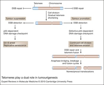
Telomeres prevent the natural ends of chromosomes from being recognised as double-stranded DNA breaks. In the presence of a functional double-stranded-DNA-break response, telomere erosion ultimately results in the loss of telomere function and the triggering of replicative senescence, a p53-dependent cell cycle arrest (Ref. Reference d'Adda di Fagagna88) (Fig. 1). Telomere-loss-dependent replicative senescence is considered to represent a stringent proliferative lifespan barrier that has to be overcome for a cell to progress to malignancy. However, the corollary of this tumour-suppressive mechanism may be an age-related accumulation of senescent cells (Refs Reference Jeyapalan89, Reference Herbig90). The presence of senescent cells will impact upon the number of mitotically active cells and thus compromise tissue homeostasis; however, by exhibiting a more catabolic phenotype, senescent cells may actively degrade the tissue microenvironment in which they reside (Ref. Reference Campisi91), which in older individuals, or those with compromised telomere maintenance, may render the tissue microenvironment more permissive for tumour progression (Ref. Reference Krtolica92).
Telomeres play a dual role in tumourigenesis. Full-length telomeres inhibit DNA-damage checkpoints, thus preventing the natural end of the chromosome from being recognised as a double-stranded break in DNA (DSB). Gradual telomere erosion will result in shorter telomeres that no longer inhibit DNA-damage checkpoints, leading to a p53-dependent DNA damage checkpoint that, via p21 (CDKN1A) and pRB (RB1) (not shown), will result in G1–S cell cycle arrest (replicative senescence). This represents a tumour-suppressive mechanism. In the absence of a functional DNA-damage checkpoint, telomeres continue to erode to a point at which they lose all the telomere end-capping function and are subject to DNA end-joining reactions, resulting in telomere fusion events that can create dicentric chromosomes. This can initiate cycles of anaphase bridging, breakage and fusion, resulting in large-scale, potentially oncogenic, genomic rearrangements such as nonreciprocal translocations.

The importance of telomere length from the standpoint of ageing and cancer is exemplified by telomerase-knockout mice. After several generations, the telomeres in these mice erode sufficiently to confer phenotypes that include defects in proliferative tissues that mirror some age-related phenotypes in humans; these include immunosenescence, alopecia, hair greying, intestinal atrophy and increased rate of tumour formation (Refs Reference Blasco93, Reference Herrera94, Reference Lee95, Reference Rudolph96). Mutations in the DNA-damage checkpoint responses rescue some of these phenotypes, but this was accompanied by an increase in the rate of tumour formation (Ref. Reference Chin97). Thus, telomere length and replicative senescence play a key role in the control of proliferative lifespan, thereby providing a tumour-suppressive mechanism.
In the absence of a functional double-stranded-DNA-break response, telomeres can continue to erode to a length at which the end-protective function is lost and widespread genomic instability ensues (Ref. Reference Counter98); at this point cell death outweighs cell growth and the culture undergoes ‘crisis’ (Ref. Reference Shay99). In these cells, widespread telomere fusion occurs between extremely short telomeres (Refs Reference Capper32, Reference Letsolo, Rowson and Baird100). Telomere fusion can create dicentric chromosomes subject to cycles of anaphase bridging, breakage and fusion, which can result in genomic rearrangements such as nonreciprocal translocations and localised amplifications that are common in a broad spectrum of tumour types (Refs Reference Murnane and Sabatier101, Reference Artandi102, Reference Mitelman103, Reference Shih104) (Fig. 1). There is circumstantial evidence to suggest that this process may occur during the progression of malignancy in humans. Many studies in humans have shown that telomere length is reduced in cancer samples compared with normal tissue (Refs Reference Engelhardt105, Reference Hastie106, Reference Meeker107), consistent with cellular proliferation in the absence of complete telomere length maintenance, but they do not demonstrate telomere dysfunction. There are in situ data showing an increase in anaphase bridging, a surrogate marker of telomere fusion, at the adenoma–carcinoma transition in breast (Ref. Reference Chin108), colorectal (Ref. Reference Rudolph109) and squamous cell (Ref. Reference Gordon110) carcinomas. Cytogenetic analysis of pancreatic carcinoma and osteosarcomas also showed that terminal breakpoints were clustered to the terminal bands, and breakpoint frequency was associated with shorter telomeres at specific chromosome arms (Ref. Reference Gisselsson111). It appears likely, but has not been formally established, that many different cancer types may be subjected to a short-telomere-driven crisis during the progression to malignancy.
Therefore, on the one hand telomere erosion can provide a tumour-suppressive function; on the other hand, telomere erosion in the absence of a functional DNA-damage response may drive tumour progression. Telomere length and its control are thus intimately linked to the process of carcinogenesis. It is therefore of interest to examine the link between telomerase variants and the risk of cancer.
TERT mutations and associated variants
The TERT gene is highly conserved (Ref. Reference Nakamura112) and in the human population there is less genetic diversity at this locus, with limited nonsynonymous SNPs and lower levels of heterozygosity, when compared with other, nontelomeric genes (Ref. Reference Savage113). These data suggest that TERT may be under an evolutionary constraint, highlighting the key role that telomerase activity and telomere length control may play in both the ageing process and tumourigenesis and indicating that subtle variation at the TERT locus may have a disproportionally large effect on telomere biology.
Bone marrow failure syndromes
Mutations in components of the telomerase complex predispose to dyskeratosis congenita, a bone marrow failure syndrome (Ref. Reference Kirwan and Dokal114). These include TERT and TERC, as well as the nucleolar proteins DKC1, NOP10 and NHP2, and TINF2, a component of the shelterin complex (Refs Reference de Lange22, Reference Kirwan and Dokal114) (Box 1). The dyskeratosis-congenita-causing K902N mutation in TERT is located within one of the reverse transcriptase motifs of telomerase, and was predicted from studies of the human immunodeficiency virus (HIV) reverse transcriptase to affect nucleotide selection in the catalytic site and to decrease enzyme activity (Ref. Reference Armanios115). In vitro reconstitution of telomerase activity showed that the K902N mutation effectively abolishes telomerase activity, but it does not act in a dominant-negative fashion; instead, telomerase is dependent on copy number of functional TERT transcripts (Ref. Reference Armanios115). The K902N mutation in TERT results in a haploinsufficiency for telomerase activity, which leads to impaired telomere length maintenance. Haploinsufficiency for telomerase activity was also apparent in dyskeratosis congenita patients with mutations in TERC (Ref. Reference Goldman116). The link between TERT gene dosage, telomerase activity and telomere length maintenance has also been exemplified in mouse models (Ref. Reference Hathcock38).
Dyskeratosis congenita patients display disease anticipation, where the severity and age of onset of the disease worsens from one generation to the next, and this is associated with progressive telomere shortening between the generations (Ref. Reference Vulliamy117). Thus, it is clear that although functional telomerase is expressed in these individuals, the lower activity levels result in shorter telomeres that shorten further between the generations. In dyskeratosis congenita, short telomeres cause a severe, heterogeneous clinical phenotype that includes reduced haematopoiesis, with reduced number of haematopoietic progenitors and colony-forming ability (Refs Reference Kirwan118, Reference Marley119), and chromosomal instability (Refs Reference Dokal and Luzzatto120, Reference Dokal and Vulliamy121). It is now apparent that dyskeratosis congenita patients suffer a predisposition to cancer and show poor survival (Refs Reference Feinberg and Coffey122, Reference Alter123). A meta-analysis of a large number of patients showed that overall there was an 11-fold increase in all cancer types, with a similar spectrum and frequency to that observed in Fanconi anaemia patients. The most common cancer site was the tongue with a 1154-fold increase in frequency, and acute myeloid leukaemia showed a 196-fold increase (Ref. Reference Alter123). The most likely cause of the defects in dyskeratosis congenita is considered to be a reduction in the proliferative potential of stem cell pools, particularly those of the haematopoietic system (Refs Reference Kirwan118, Reference Marley119, Reference Marsh124, Reference Goldman125), resulting in stem cell depletion, cell death and senescence that may manifest age-related disease and bone marrow failure (Ref. Reference Kirwan and Dokal126). Failure of telomere length maintenance has been implicated in other bone marrow failure syndromes, including Fanconi anaemia, Shwachman–Diamond syndrome, Diamond–Blackfan anaemia and aplastic anaemia (Refs Reference Dokal and Vulliamy121, Reference Ball127, Reference Thornley128, Reference Yamaguchi129). These conditions are also cancer-prone; in particular, they have higher rates of myeloid leukaemia (Refs Reference Lipton130, Reference Alter131, Reference Socie132). The mechanistic basis of cancer predisposition in dyskeratosis congenita patients and the other bone failure syndromes has not been established. Both bone marrow and fibroblasts from dyskeratosis congenita patients display large-scale genomic instability including nonreciprocal translocations, dicentric chromosomes and ring chromosomes (Refs Reference Dokal and Luzzatto120, Reference Dokal133). These types of aberrations are consistent with telomere dysfunction (Ref. Reference Letsolo, Rowson and Baird100) (Fig. 1), and are common in many types of malignancy. The inability to sufficiently counteract end-replication losses in these patients may therefore facilitate telomere dysfunction and thus lead to genomic instability and cancer.
Dyskeratosis congenita and the other bone marrow failure conditions are rare and display extreme phenotypes. However, they demonstrate that impaired telomere length maintenance in humans does confer a considerably higher risk of cancer. It is thus pertinent to consider that more subtle variation within the components involved in the maintenance of telomere length may also confer an increased risk of cancer without overt clinical phenotypes. Over recent years a combination of targeted analysis of genes associated with telomere biology, together with large-scale genome-wide association studies, has revealed that the locus that contains the TERT gene is associated with cancer risk.
Haematological malignancies
The TERC and TERT mutations found in dyskeratosis congenita are not always fully penetrant, with some patients presenting with aplastic anaemia or with myelodysplastic syndrome (Ref. Reference Yamaguchi134). The observation of an elevated incidence of myelodysplastic syndrome progressing to acute myeloid leukaemia in some families segregating aplastic anaemia with mutations in TERT led to a screen in patients with acute myeloid leukaemia for mutations within TERT (Ref. Reference Calado135). Missense mutations in TERT were observed in 11 out of 133 patients, including three homozygotes, which was significantly different from normal controls (P < 0.0001); no mutations were detected in the TERC gene. However, TERT mutations were also detected in normal control individuals, indicating that these variants may not be causal but instead confer a risk factor for acute myeloid leukaemia (Ref. Reference Calado135). From this study, the A1062T TERT variant was the most common in acute myeloid leukaemia. A separate study examining families presenting with myelodysplastic syndrome or acute myeloid leukaemia identified four out of 20 families with variants in TERT or TERC (Ref. Reference Kirwan136). Disease anticipation was also apparent in the families, indicating that progressive telomere shortening between the generations may drive disease progression. These TERT and TERC mutations were hypomorphic, resulting in haploinsufficiency for telomerase activity (Refs Reference Calado135, Reference Kirwan136), but this was very variable, with some variants showing no activity and others showing very small differences in activity (Ref. Reference Calado135). It therefore appears that telomerase activity does not predict the clinical outcome and that additional environmental and genetic factors are determining the clinical presentation (Ref. Reference Kirwan136). A screen specifically for the A1062T TERT variant, which has a minor allele frequency of 0.006, in other haematological malignancies identified diffuse large B cell lymphoma (frequency = 0.026, N = 274) and chronic lymphocytic leukaemia (frequency = 0.03, N = 195) as showing significantly higher frequencies (P = 0.0001) of the A1062T variant (Ref. Reference Hills and Lansdorp137). These studies have data to indicate that TERT variants may impact upon telomere length, suggesting the possibility of an increase in the number of short dysfunctional telomeres, which may facilitate genomic instability and thus progression in the early stage of leukaemogenesis (Refs Reference Calado135, Reference Hills and Lansdorp137). This is important as it provides a primary mechanism for disease development in acute myeloid leukaemia in addition to the known roles of RUNX1 transcription factor fusion genes and dominant-negative mutations in the transcription factor CEBPA (Refs Reference Smith138, Reference Song139).
Genome-wide association studies
Numerous genome-wide association studies have been carried out to look for associations between cancer risk and single-nucleotide variants. The evidence for 5p15.33 containing risk factors for cancer is compelling, with multiple studies all showing significant associations with many different types of cancer.
Lung cancer
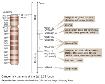
The cholinergic nicotinic receptor genes at 15q25 have been implicated in nicotine dependence (Ref. Reference Saccone140) and this locus was also independently identified in other genome-wide association studies as a susceptibility locus for lung cancer (Refs Reference Hung141, Reference Amos142, Reference Thorgeirsson143). A further genome-wide association study to identify additional susceptibility loci in the Caucasian population revealed eight SNPs; seven were within 15q25, but one, rs402710 (P = 2 × 10⁻⁷), was located at 5p15.33, the locus that includes both the TERT and CLPTM1L genes (Ref. Reference McKay144) (Fig. 2). Two other SNPs at this locus, rs31489 and rs401681, which also showed evidence of association, were in strong linkage disequilibrium (LD) with rs402710. rs401681 was also identified in an independent genome-wide association study for association with lung cancer (Ref. Reference Wang145). An additional SNP at 5p15.33, rs2736100, also showed independent association with lung cancer (risk) in Caucasian populations (Refs Reference McKay144, Reference Landi147) and the Chinese population (Ref. Reference Wang146), with risk specifically identified for adenocarcinoma (OR = 1.23, P = 3.07 × 10⁻⁷) but not with other histological subtypes in one of these studies (Ref. Reference Landi147); however, a subsequent meta-analysis showed a very weak association between rs2736100 and lung cancer risk (Ref. Reference Broderick148). The same meta-analysis revealed strong association (P = 2.99 × 10⁻⁹) of lung cancer risk and the rs4975616 SNP located between the CLPTM1L and TERT genes (Ref. Reference Broderick148). A study looking for association with a range of cancer types identified a strong association (OR = 1.20, P = 3.2 × 10⁻⁵) between lung cancer risk and the A allele at rs2736098, a synonymous SNP in the second exon of the TERT gene (Ref. Reference Rafnar149).
Cancer risk variants at the 5p15.33 locus. Exons of the TERT and CLPM1L genes at this locus are illustrated as light blue lines/blocks. Risk variants for cancer identified from genome-wide association studies are detailed together with cancer predispositions.

Despite the multiple strong associations that have been reported, there is no direct evidence that these SNPs are causative for lung cancer. rs2736100 is located within intron 2 of TERT and rs402710 is located in intron 16 of the CLPTM1L gene but is within a region of LD that includes the TERT promoter (Fig. 2). The association of rs402710 with lung cancer susceptibility has been independently confirmed; however, the underlying cause may be independent of telomere biology as this variant was associated with higher levels of the bulky DNA adducts that are formed from the binding of metabolites of the key carcinogens in tobacco smoke such as polycyclic aromatic hydrocarbons (Ref. Reference Zienolddiny11). CLPTM1L has been documented to be upregulated in cisplatin-resistant cell lines and this was linked with cisplatin-induced apoptosis (Ref. Reference Yamamoto12). Thus, the involvement of variants in the CLPTM1L gene in lung cancer may be more compelling than variants in the TERT gene; indeed, another study found an association of a SNP in intron 13 of CLPTM1L with lung cancer. However, these markers are all within intronic regions and they are within a region of strong LD that encompasses CLPTM1L and the 5′ end of TERT and thus the contribution of TERT to lung cancer predisposition cannot be ruled out.
A study of the Korean population examining genes implicated in telomere function and lung cancer risk identified rs2736098, which was in LD with rs2853669 within the TERT promoter; in addition, it identified rs2735940 also in the TERT promoter (Fig. 2) and rs6985140 in intron 3 of tankyrase (TNKS1), another gene implicated in telomere function (Ref. Reference Choi150). This study also indicated that the effects of these variants may be additive, with the TERT rs2736098/rs2735940 A/T haplotype together with TNKS1 rs6985140 G being associated with an increased risk of lung cancer (OR = 1.69, P < 0.001) (Ref. Reference Choi150). The rs2853669 variant is located in the TERT promoter within a binding site for the transcription factor ETS2; ETS2-mediated transcription is considered to regulate TERT expression and may be required for TERT upregulation during tumour progression (Refs Reference Hsu151, Reference Dwyer and Liu152). As TERT is considered to be limiting for telomerase activity, variants that may control TERT expression levels are potentially tantalising candidates for association with cancer risk. However, rs2853669 has been identified as a risk variant only in this single study.
The link between telomerase variants and lung cancer is also indicated by idiopathic pulmonary fibrosis, an inflammatory disease that creates large-scale fibrosis of the lung; individuals with this disease frequently develop lung cancer (Ref. Reference Park153). Telomerase mutations account for the familial disease (Refs Reference Armanios154, Reference Tsakiri155), and a genome-wide association study for susceptibility loci for sporadic idiopathic pulmonary fibrosis identified rs2736100 (Ref. Reference Mushiroda156).
The TERT gene contains several variable noncoding minisatellites (Ref. Reference Leem157). The MNS16A minisatellite, located downstream of the TERT gene, contains putative binding sites for GATA1 transcription factor and it has been shown to have promoter activity controlling the expression of antisense TERT transcripts (Ref. Reference Wang158). This locus is a variable number tandem repeat (VNTR) and the promoter activity was modulated by repeat length; as such, variation in MNS16A repeat length could regulate the antisense expression and thus the levels of TERT expression. When the four different-length alleles at MNS16A were classified into long (L) and short (S) groups, the L/L genotype was found to be associated with an increased risk of lung cancer (OR = 2.18) (Ref. Reference Wang158).
Basal cell carcinoma
The lung-cancer-susceptibility variant rs401681 (Ref. Reference Wang145), which was in the same LD block that contains rs402710, is also associated with risk for basal cell carcinoma, with the C allele showing an OR of 1.25 (P = 3.7 × 10⁻¹²) (Ref. Reference Rafnar149) (Fig. 2). This study examined a total of 33 800 cancer cases and 45 800 controls spread among 16 additional cancer sites, and four of these showed a significant association with the rs401681 C allele: in lung (OR = 1.15, P = 7.2 × 10⁻⁸), bladder (OR = 1.12, P = 5.7 × 10⁻⁵), prostate (OR = 1.07, P = 3.6 × 10⁻⁴) and cervix (OR = 1.31, P = 2.6 × 10⁻⁴). No significant association was found with breast and colon cancer. Also, no association was detected for endometrium, kidney, ovary, pancreas, stomach and thyroid cancer or for lymphoma, multiple myeloma and squamous cell carcinoma; however, this may be accounted for by the sampling sizes (Ref. Reference Rafnar149). Interestingly, the rs401681 C allele conferred protection from melanoma (OR = 0.86, P = 5.0 × 10⁻⁸) (Ref. Reference Rafnar149), which was confirmed in a subsequent study (Ref. Reference Stacey159). Melanoma protection may be associated with the reduction of nevi size and count in individuals with shorter PBL telomeres (Ref. Reference Bataille71), which is also associated with a reduced risk of melanoma (Ref. Reference Han72).
Further analysis of this locus in basal cell carcinoma revealed an additional variant, rs2736098 – a synonymous SNP in exon 2 of TERT that has also been associated with lung cancer risk in the Korean population (Ref. Reference Choi150). The rs2736098 A allele was, with the exception of cervical cancer, significantly associated with the same cancer types as rs401681: basal cell (OR = 1.21, P = 1.3 × 10⁻⁵), lung (OR = 1.20, P = 3.2 × 10⁻⁵), bladder (OR = 1.16, P = 1.3 × 10⁻⁴) and prostate (OR = 1.13, P = 1.3 × 10⁻⁴) (Ref. Reference Rafnar149) (Fig. 2). Interestingly, despite the very high LD between rs2736098 and rs401681 (D′ = 0.94), the association with the cancer types remained significant for each variant after pair-wise adjustments for each allele. Thus, these alleles cannot account for all of this association, which indicates that an additional variant may capture the effect of both these variants. Analysis of the known variants within the TERT coding sequences, the promoter and three of the VNTRs in this region showed no significant association with the cancer types associated with rs2736098 and rs401681. An analysis of telomere length from peripheral blood samples in females born between 1925 and 1935 revealed significantly shorter telomere lengths in those homozygous for the rs2736098 A allele (P = 0.027) or for the rs401681 C allele (P = 0.017) (Ref. Reference Rafnar149); however, this association was not apparent in younger women, born between 1940 and 1950 (Ref. Reference Rafnar149). While these data need to be independently verified, they do indicate the possibility that these variants may confer a differential rate of telomere erosion that only becomes apparent in older individuals, which may in turn increase the risk for certain cancers.
Glioma
A large genome-wide association study looking for variants that confer a risk for glioma identified the rs2736100 variant in intron 2 of TERT (OR = 1.27, P = 1.50 × 10⁻¹⁷); this variant or others in LD has also been associated with other cancer types (Refs Reference Wang146, Reference Rafnar149, Reference Shete160) (Fig. 2). A second variant in intron 2, rs2853676, was also identified (OR = 1.26, P = 4.21 × 10⁻¹⁴) but is in weak LD and risk association appeared to be independent of rs2736100 (Ref. Reference Shete160). The causal variant has not been identified but it is pertinent to note that TERT expression correlates with grade and prognosis of glioma (Ref. Reference Wager161), and thus there may be a biological basis to these associations.
The MNS16A minisatellite, implicated in lung cancer risk, also showed an association of specific-length alleles with differences in survival time of individuals with glioblastoma multiforme: those with the S/S genotype had a median survival of 25.1 months, whereas the L/L and L/S genotypes had a survival of 14.6 and 14.7 months, respectively (P = 0.039) (Ref. Reference Wang162). The molecular basis of this association is not clear and the role that TERT antisense expression plays in regulating telomerase activity has not been established. Furthermore, there are conflicting data: an independent study showed no association of the MNS16A genotypes with survival time, with all genotypes varying between 15.3–16.8 months. However, this study did show that the S allele was significantly (P = 0.012) more common in glioma patients (OR = 2.05 for the S/S genotype) (Ref. Reference Carpentier163). A larger study on both glioma and meningioma showed no significant differences in the MNS16A genotypes with the risk of developing either of these cancers (Ref. Reference Andersson164). This study also showed that survival with the L/L genotypes was significantly greater than for the L/S but not the S/S genotype. All three studies on the MNS16A genotypes and glioma show different associations, and thus the role that MNS16A may play in these diseases is unclear and this marker currently has no prognostic value. Associations of the other TERT minisatellites (Ref. Reference Leem157) and cancer risk have not been reported.
Breast cancer
Since telomere dysfunction has been hypothesised to play a role in breast cancer development (Refs Reference Chin108, Reference Fordyce165), variants within five genes involved in telomere biology were examined for association with breast cancer (Ref. Reference Savage166). This analysis included rs2736098, identified by genome-wide association with risk to lung cancer as well as basal cell, bladder and prostate cancer (Refs Reference Rafnar149, Reference Choi150), but not breast cancer. Consistent with this genome-wide association study (Ref. Reference Rafnar149), no association with breast cancer was detected (Ref. Reference Savage166). However, there was an apparent reduction in risk with three SNPs within TERT with borderline significance, in families with a history of breast cancer: rs2736098 (OR = 0.57, P = 0.01), rs2735940 (OR = 0.73, P = 0.04) and rs2853669 (OR = 0.66, P = 0.05), which were identified as lung cancer risk loci in the Korean population (Ref. Reference Choi150). However, the apparent association with reduction in inherited breast cancer risk of rs2853669 located in the TERT promoter was not reproduced by another, well-powered study (Ref. Reference Varadi167).
It is interesting to note that no association has been reported for variants at the 5p15.33 locus with risk for other notable cancers, including in particular colorectal carcinoma in which telomere biology has been implicated (Refs Reference Hastie106, Reference Gertler168, Reference Gertler169): large genome-wide association studies looking for risk alleles in colorectal carcinoma did not identify any genes implicated in telomere function (Refs Reference Tomlinson170, Reference Tenesa171).
Pancreatic cancer
One genome-wide association study showed no association with the rs401681 SNP in pancreatic cancer; however, this may have been an underpowered study (Ref. Reference Rafnar149). Subsequently, a larger genome-wide association study has shown association of the rs401681 T allele with pancreatic cancer (OR = 1.19, P = 3.66 × 10⁻⁷) (Ref. Reference Petersen172). Interestingly, it is the rs401681 C allele and not the T allele that is associated with lung, bladder and prostate cancer (Ref. Reference Rafnar149).
Clinical implications/applications
It is clear that there is a spectrum of disorders associated with the TERT locus. This includes dyskeratosis congenita and other bone marrow failure syndromes, where rare hypomorphic mutations in TERT clearly result in telomere shortening and telomere dysfunction, and where the clinical manifestations are severe and include a considerable elevation in cancer risk. The mechanistic basis of the cancer risk in these conditions has not been established.
Several independent well-powered genome-wide association studies have identified common alleles at noncoding SNPs in the vicinity of the TERT gene that confer an elevated risk for a variety of cancers. The central role that telomere length and telomerase expression play in tumour progression makes the involvement of SNPs at the TERT locus in conferring risk of cancer tantalising. However, the causal variants linked to these SNPs have not been identified and thus the role of variants within TERT or other genes in this region, such as CLPTM1L, has not been established. Therefore, the clinical implications of these findings have not been established. It is clear, however, that telomere length is an important risk factor for cancer and individual-specific differences in telomere length may thus impact upon cancer risk. Telomere length is a complex trait, with multiple protein interactions likely to control individual-specific telomere length. Telomerase is a key component of telomere length control. The noncoding variants identified in the genome-wide association studies may directly, or indirectly via additional variants in LD, modulate aspects of TERT transcription. This may in turn regulate levels of telomerase activity and thus the ability of the cell to maintain or slow the rate of telomere erosion.
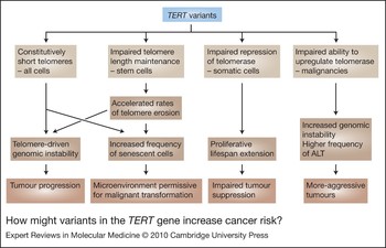
How could mutations and variants at the TERT locus predispose to cancer? The circumstantial evidence from basic observations on telomere dynamics in cells in culture and in human tumours, together with the effect of short or dysfunctional telomeres in mouse models, are consistent with the view that telomere dysfunction can drive tumour formation. The constitutively short telomeres in individuals with the bone marrow failure syndromes may therefore become prematurely dysfunctional and drive the observed chromosomal instability (Fig. 3). If the more subtle variants at this locus impact upon the regulation of telomerase expression, this could also lead to constitutively short telomeres, as well as an impairment of somatic telomere length maintenance. This in turn may lead to premature telomere dysfunction and genomic instability.
How might variants in the TERT gene increase cancer risk? The figure presents speculation as to the possible mechanisms by which variants/mutations in the TERT gene may predispose to cancer. The inheritance of constitutively short telomeres may either facilitate premature telomere dysfunction that drives genomic instability and tumour progression, or the accumulation of senescent cells that may render the tissue microenvironment more permissive for malignant transformation. If telomere length is not sufficiently maintained in stem cell populations, this may lead to accelerated rates of telomere erosion as a function of age, also leading to telomere dysfunction and/or replicative senescence. The repression of telomerase provides a stringent tumour-suppressive function. The inability to repress telomerase in somatic tissues, may impair suppression by extending proliferative lifespan. If telomerase is not efficiently upregulated during tumour progression, this may allow greater telomere dysfunction, leading to increased genomic instability, or to the upregulation of the alternative lengthening of telomeres (ALT) pathway; both these situations could result in more-aggressive tumours.

It is also apparent that short telomeres, in the context of a functional DNA-damage response, can provide a tumour-suppressive function. Individual-specific differences in telomere length and/or the ability of cells to regulate telomerase activity, may impact the efficacy of this mechanism. This tumour-suppressive mechanism manifests itself in the generation of senescent cells, which accumulate in tissues as a function of age. The accumulation of senescent cells can render the tissue more permissive for malignant transformation. Thus, an individual with compromised telomere maintenance and constitutively short telomeres may also exhibit an increase in the frequency of senescent cells as a function of age, which may facilitate tumour progression (Fig. 3). Sequence variants within the TERT promoter and control regions could impair the repression of telomerase in the majority of somatic cells, which would have the effect of slowing the rates of telomere erosion and extending proliferative lifespan. This would diminish the impact of telomere-mediated tumour suppression (Fig. 3).
An impaired ability to upregulate telomerase during tumour progression may result in additional telomere dysfunction prior to telomere stabilisation. This in turn could increase the extent of genomic instability and thus the probability that oncogenic mutation could occur and drive progression. In this situation there would be a strong selection pressure within the tumour cell milieu to stabilise telomere length, either via amplification of the TERT locus, or via the induction of the alternative lengthening of telomeres (ALT) pathway, a recombination-based mechanism for telomere maintenance in the absence of telomerase (Ref. Reference Bryan173) (Fig. 3).
Strategies to slow telomere erosion or reset individual-specific telomere lengths may therefore be a useful intervention in individuals with a deficiency in telomere length maintenance. In this respect, compounds that can transiently induce telomerase expression, by interfering with transcriptional activators/repressors of the TERT promoter, may have therapeutic applications. Indeed, the search for agents that can modulate telomerase expression is an active area of research. One such compound, TAT2 (cycloastragenol), was obtained from a screen of Chinese medicine plant extracts for compounds that upregulate telomerase activity. TAT2 has been shown to reversibly enhance telomerase activity in cultured CD8+ T cells, retarding telomere erosion and increasing replicative capacity (Ref. 174). This may be mediated via the MAPK/ERK (mitogen-activated protein kinase or extracellular-signal-regulated kinase) pathway and the ETS transcription factors that interact with the TERT promoter, or by post-translational modification of telomerase (Ref. Reference Fauce174). Importantly, there was no evidence of loss of growth control in TAT2-treated T cells. The success of these types of compounds in the clinic remains to be seen.
Research in progress and outstanding research questions
The TERT mutations identified in forms of dyskeratosis congenita, aplastic anaemia and other bone failure syndromes together with those that predisposed to myelodysplastic syndrome or acute myeloid leukaemia and to chronic lymphocytic leukaemia have been shown to result in haploinsufficiency for telomerase activity. This facilitates an understanding of the aetiology of these conditions. Data on the effect of synonymous TERT variants on telomerase activity are lacking, and thus whether these variants or others in LD with them affect telomerase activity or telomere length maintenance is not known. It therefore remains open to question whether it is the TERT locus or CLPTM1L or indeed another gene in LD with these variants that is causal for risk associations to the various different cancer types described. To progress this issue, a complete re-sequencing of the 5p15.33 locus will be required to identify the causal variants, and this will need to include all the regulatory regions. It is clear that the control of telomerase expression is paramount in its function to control proliferative lifespan of human cells. It will then need to be established how these variants affect telomerase activity, or the ability to repress or upregulate telomerase, and importantly how this may impact upon telomere length maintenance, inheritance and relative rates of telomere erosion.
Acknowledgements and funding
Research in my laboratory is supported by Cancer Research UK, Leukaemia Research UK, the Welsh Office of Research and Development and the Association for International Cancer Research, and I am a Cancer Research UK Senior Fellow. I thank the peer reviewers for their helpful comments and suggestions.