Introduction
Echinococcus granulosus sensu lato (s.l.) is the causative agent of cystic echinococcosis (CE), a neglected zoonotic disease distributed worldwide. Echinococcus granulosus s.l. is a species complex that includes Echinococcus granulosus sensu stricto (s.s.) (G1 and G3 genotypes), Echinococcus equinus (G4 genotype), Echinococcus ortleppi (G5 genotype) and Echinococcus canadensis cluster (G6/7, G8 and G10 genotypes) (Nakao et al., Reference Nakao, McManus, Schantz, Craig and Ito2007; Hüttner et al., Reference Hüttner, Nakao, Wassermann, Siefert, Boomker, Dinkel, Sako, Mackenstedt, Romig and Ito2008; Thompson, Reference Thompson2008; Romig et al., Reference Romig, Ebi and Wassermann2015). In the current nomenclature, the E. canadensis cluster can be divided into E. intermedius (G6/7), E. borealis (G8) and E. canadensis (G10) (Romig et al., Reference Romig, Deplazes, Jenkins, Giraudoux, Massolo, Craig, Wassermann, Takahashi and de la Rue2017; Casulli et al., Reference Casulli, Siles-Lucas and Tamarozzi2019).
The life cycle of E. granulosus s.l. involves canids, especially dogs, and livestock such as sheep, cattle and goats. Humans are dead-end accidental hosts and do not contribute to the continuity of the life cycle of the parasite (Eckert et al., Reference Eckert, Gemmell, Meslin and Pawlowski2001). Although the larval form or the hydatid cyst most commonly develops in the liver, it can be detected in any internal organs as well. The majority of CE cases have a single hydatid cyst that affects only an organ (Brunetti et al., Reference Brunetti, Kern and Vuitton2010).
CE is mostly endemic in rural areas where animal breeding is conducted and humans are in close contact with the life cycle of the parasite such as in western China, central Asia, South America, eastern Europe and Mediterranean region, including Turkey (Deplazes et al., Reference Deplazes, Rinaldi, Alvarez Rojas, Torgerson, Harandi, Romig, Antolova, Schurer, Lahmar, Cringoli, Magambo, Thompson and Jenkins2017). The incidence of human CE from endemic areas varies from 1 to 200 per 100 000 (Wen et al., Reference Wen, Vuitton, Tuxun, Li, Vuitton, Zhang and McManus2019). In Turkey, as in most countries, the incidence of human CE is mostly based on hospital records and does not reflect real prevalence value (Altıntaş, Reference Altintas2008). According to a population-based ultrasound screening study conducted in Turkey, Bulgaria and Romania, around 106 000 people living in rural areas of Turkey might be infected with abdominal CE (Tamarozzi et al., Reference Tamarozzi, Akhan, Cretu, Vutova, Akinci, Chipeva, Ciftci, Constantin, Fabiani, Golemanov, Janta, Mihailescu, Muhtarov, Orsten, Petrutescu, Pezzotti, Popa, Popa, Popa, Velev, Siles-Lucas, Brunetti and Casulli2018). As reported by the Ministry of Health, between the years 1955 and 1972, 12 206 human CE cases have been recorded in Turkey and, between 1975 and 1994, 40 242 surgically confirmed CE cases have been reported (Altıntaş, Reference Altıntaş2003). More recently, in a study based on the examination of different hospital and health directorship documents and the Turkish Ministry of Health records, a total of 14 789 human CE cases were recorded in Turkey between 2001 and 2005. In that study, the distribution of CE cases varied by region as follows: 2534 (13.13%) in Marmara region, 2114 (16.94%) in Aegean region, 2578 (16.09%) in Mediterranean region, 5404 (38.57%) in Central Anatolia region, 428 (5.70%) in Black Sea region, 844 (6.80%) in Eastern Anatolia region and 887 (2.75%) in South-Eastern Anatolia region (Yazar et al., Reference Yazar, Ozkan, Hökelek, Polat, Yilmaz, Özbilge, Ustun, Koltaş, Ertek, Sakru, Alver, Cetinkaya, Koç, Demirci, Aktaş, Parsak, Ozerdem, Sakman, Cengiz, Ozer, Keklik, Yemenici, Turan, Daştan, Kaya, Tamer, Girginkardeşler, Türk, Sinirtaş, Evci, Kiliçturgay, Mutlu and Artiş2008).
Several papers indicated that G1 and G3 genotypes (E. granulosus s.s.) are predominant in the human host in Turkey (Utuk et al., Reference Utuk, Simsek, Koroglu and McManus2008; Snabel et al., Reference Snabel, Altintas, D'Amelio, Nakao, Romig, Yolasigmaz, Gunes, Turk, Busi, Hüttner, Sevcová, Ito, Altintas and Dubinský2009; Ergin et al., Reference Ergin, Saribas, Yuksel, Zengin, Midilli, Adas, Arikan, Aslan, Uysal, Caliskan, Oner, Kucukbasmaci, Kaygusuz, Torun and Kocazeybek2010; Simsek et al., Reference Simsek, Kaplan and Ozercan2011; Eryıldız and Şakru, Reference Eryildiz and Şakru2012; Bakal et al., Reference Bakal, Simsek and Kazez2015; Eroğlu et al., Reference Eroglu, Genc and Koltas2016; Erdoğan et al., Reference Erdoğan, Özkan, Mutlu, Karaca and Şahin2017; Oral Babaoğlu et al., Reference Oral Babaoğlu, Soyder, Malatyalı, Ertuğ and Ertabaklar2018; Orsten et al., Reference Orsten, Boufana, Ciftci, Akinci, Karaagaoglu, Ozkuyumcu, Casulli and Akhan2018). Echinococcus canadensis (G7 genotype), also known as pig strain, was reported from Turkey for the first time by Snabel et al. in 2009. Following this record, E. canadensis cluster (G6/7) was notified from Thrace Region, Aydın Province and Elazığ Province (Simsek et al., Reference Simsek, Kaplan and Ozercan2011; Eryıldız and Şakru, Reference Eryildiz and Şakru2012; Oral Babaoğlu et al., Reference Oral Babaoğlu, Soyder, Malatyalı, Ertuğ and Ertabaklar2018).
The genetic diversity of E. granulosus s.l. might affect the life-cycle pattern, host specificity, development rate, pathogenicity, etc. (Eckert et al., Reference Eckert, Gemmell, Meslin and Pawlowski2001). However, information on the relationship between genetic diversity of E. granulosus (s.l.) in human hosts and the clinical features remain limited and unclear. This study aimed to investigate the correlation between the genetic variation of E. granulosus s.l. and cyst characteristics such as their number, location, diameter, volume and type according to WHO-IWGE classification.
Materials and methods
Sample collection, genomic DNA isolation and PCR amplification
Hydatid cyst samples were collected from 94 patients at the Faculty of Medicine, Hacettepe University. This cohort of patients received percutaneous treatment for active cysts from May 2018 to June 2019 according to WHO-IWGE cyst type classification (Brunetti et al., Reference Brunetti, Kern and Vuitton2010). Ethical approval was provided by the Ethics Committee from the Faculty of Medicine, Hacettepe University (GO 18/366-15). Patient demographics and clinical features such as cyst type, number, location, size and volume were recorded. Size of the cysts was calculated as the largest diameter measured among three dimensions from right to left in the axial plane by ultrasound machine software. Volume of the cysts was calculated according to the ellipse formula (V = (4/3) × π × R 1 × R 2 × R) by measuring three dimensions.
Hydatid cyst fluids were aseptically collected, and stored at −80°C. The cyst fluid was centrifuged to precipitate protoscolices and the gDNA extraction was performed from protoscolices using the Thermo Blood and Tissue DNA Extraction Kit (Qiagen, Hilden, Germany) according to the manufacturer's instructions. The primers forward: 5′TTGAATTTGCCACGTTTGAATGC3′ and reverse: 5′GAACCTAACGACATAACATAATGA3′ were used for amplification of partial the cytochrome c oxidase subunit 1 (mt-CO1) gene, as previously described (Nakao et al., Reference Nakao, Sako, Yokoyama, Fukunaga and Ito2000). Polymerase chain reaction (PCR) products were visualized under ultraviolet light after electrophoresis in 1.5% agarose gels. A PCR result was considered positive at gel electrophoresis when the band size of ~875 bp was obtained. All amplicons were characterized by sequencing.
Sequence data and haplotype analyses
A BLAST algorithm (http://www.ncbi.nlm.nih.gov/BLAST/) was used to compare obtained sequences in this study with that present in the NCBI database. Obtained sequences were analysed as previously described by Boufana et al. (Reference Boufana, Lahmar, Rebai, Ben Safta, Jebabli, Ammar, Kachti, Aouadi and Craig2014). Briefly, data alignment was performed in Mega version 7 (Tamura et al., Reference Tamura, Stecher, Peterson, Filipski and Kumar2013) and ClustalX (Larkin et al., Reference Larkin, Blackshields, Brown, Chenna, McGettigan, McWilliam, Valentin, Wallace, Wilm, Lopez, Thompson, Gibson and Higgins2007), then transported into DnaSP 5 to determine haplotype numbers (h n), haplotype diversity (h d) and nucleotide diversity (π d) (Librado and Rozas, Reference Librado and Rozas2009). Haplotype networks were created by Hapview (Salzburger et al., Reference Salzburger, Ewing and Haeseler2011).
Statistical analyses
Statistical analysis was carried out using IBM SPSS Statistics Ver. 20 (SPSS Inc., Chicago, IL, USA). For group comparisons categorical data were tested by the chi-square test and quantitative variables were tested by Mann–Whitney U test analysis.
Results
Genetic characteristics of Echinococcus spp. and haplotype analysis
gDNA was extracted from a total of 110 hydatid cysts belonging to 94 patients and amplified 875 bp fragment within the mt-CO1 gene. All the isolates were confirmed by the BLAST algorithm as E. granulosus s.l. The majority of isolates (104/110) belonged to E. granulosus s.s. (G1 and G3). Besides, six of isolates belonged to E. canadensis cluster (G6/7). The 828 bp sequences of E. canadensis cluster (G6/7) were submitted to GenBank (GenBank accession numbers: MT103156 and MT103155).
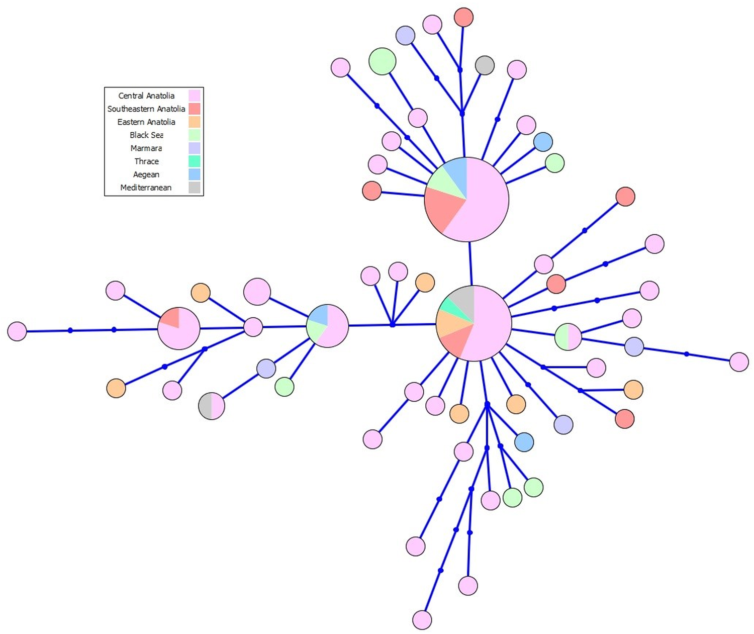
Thirty-five polymorphic sites, of which 23 were parsimony-informative, were detected in the analysed nucleotide sequences. There were no indels or gaps. The haplotype analysis exhibited the presence of 57 haplotypes with high genetic diversity (h d = 0.937 ± 0.00024) (Fig. 1). In the most dominant cluster the main haplotype was 100% identical to E. granulosus which is the most common haplotype, worldwide (accession no. AB491414) (Nakao et al., Reference Nakao, Yanagida, Okamoto, Knapp, Nkouawa, Sako and Ito2010). The second most dominant haplotype was 100% identical to E. granulosus s.s. (G1) (accession no. KU925412). The third most common haplotype was 100% identical to E. granulosus s.s. (G3) (accession no. MG682532). In addition, a phylogenetic tree with some reference sequences was constructed (Supplementary figure).
Haplotype network of the Echinococcus granulosus sensu stricto derived from mitochondrial data (n = 104).

In this study, we have also documented mixed-haplotype infection in a single patient. These haplotypes belonged to first and third haplotype clusters, respectively.
The nucleotide diversities and neutrality indices are shown in Table 1. The Tajima's D (−23 478) and Fu's Fs (−34 029) were both negative and statistically significant (P < 0.01). As previously described, these results indicate negative selection or population expansion (Nakao et al., Reference Nakao, Yanagida, Okamoto, Knapp, Nkouawa, Sako and Ito2010; Casulli et al., Reference Casulli, Interisano, Sreter, Chitimia, Kirkova, La Rosa and Pozio2012; Yanagida et al., Reference Yanagida, Mohammadzadeh, Kamhawi, Nakao, Sadjjadi, Hijjawi, Abdel-Hafez, Sako, Okamoto and Ito2012; Boufana et al., Reference Boufana, Lahmar, Rebai, Ben Safta, Jebabli, Ammar, Kachti, Aouadi and Craig2014; Hassan et al., Reference Hassan, Meerkhan, Boufana, Hama, Ahmed, Mohammed, Mero, Orsten, Interisano, Pozio and Casulli2017).
Haplotype numbers (h n), haplotype diversity (h d) and nucleotide diversity (π d) of cytochrome c oxidase subunit 1 (cox 1) mitochondrial gene of Echinococcus granulosus sensu stricto isolates from patients

P < 0.01.
Demographics of patients
A total of 94 patients with 110 hydatid cysts (46 female, 48 male, mean age ± 38 years) were included in this study. The distribution of the number of people by regions were 53 (56.3%) from Central Anatolia, 12 (12.7%) from South-eastern Anatolia, 9 (9.6%) from Eastern Anatolia, 6 (6.4%) from Black Sea, 6 (6.4%) from Marmara (including 1 patient from Thrace region), 4 (4.3%) from Aegean and 4 (4.3%) from Mediterranean.
Cyst characteristics
Cyst number and location
The majority of cysts were located in the liver (98/110), especially the right lobe (83/98). The CE cysts were also noted in spleen (5/110), kidney (3/110), pelvis (3/110) and bone (1/110). Among 94 patients, 68 had a single CE cyst, 20 had two cysts and 8 had more than two cysts.
Cyst type
Using the WHO-IWGE expert consensus classification system, 54 of the 110 CE cysts were identified as CE1, 32 as CE2 and 24 as CE3 (6 CE3a and 18 CE3b). For statistical analysis, we classified the cysts into two main groups as without daughter vesicles (CE1 and CE3a) and with daughter vesicles (CE2 and CE3b). The inactive CE patients such as CE4 and CE5 were not included in the patient group due to no need for intervention.
Cyst diameter and volume
The mean diameter of CE cysts was 7.97 cm. In detail, according to Echinococcus species, the mean diameter of cysts was 8.31 and 6.96 cm for E. granulosus s.s. (G1 and G3 genotypes) and E. canadensis cluster (G6/7), respectively.
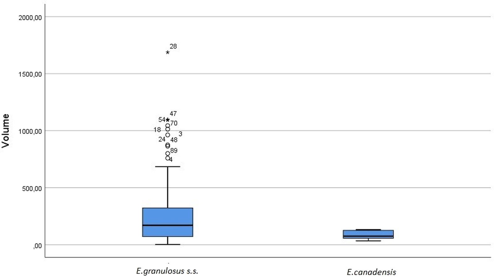
The average volume of cysts was recorded as 263.08 cm3. According to Echinococcus species, the mean volume of cysts was 273.47 cm3 and 83.03 cm3 for E. granulosus s.s. (G1 and G3 genotypes) and E. canadensis cluster (G6/7), respectively. Cyst characteristics are given in the Supplementary table.
Relationship between demographics, cyst characteristics and genetic diversity
We were able to divide E. granulosus s.l. isolates into two main groups, E. granulosus s.s. (G1 and G3) and E. canadensis cluster (G6/7). There was no relationship between genetic diversity and demographics such as age, sex and regions from where CE patients originated. Also, there was no relationship between genetic diversity and cyst features such as cyst number, cyst diameter and cyst type according to the WHO classification (P > 0.05). The relationship between haplotype diversity and cyst features was not statistically significant, as well (P > 0.05).
On the other hand, cyst volume was significantly higher for patients with E. granulosus s.s. compared to those who were infected with E. canadensis cluster (G6/7) (P < 0.05) (Fig. 2).
Comparison of volumes of cysts belonging to E. granulosus s.s. (G1 and G3 genotypes) (n = 104) and E. canadensis (G6/7 cluster) (n = 6).

Besides, the hydatid cyst location was significantly different for E. canadensis cluster (G6/7) compared to E. granulosus s.s. (P < 0.05). In fact, half of the cysts that belong to the G6/7 cluster were located other than the liver (two in the spleen and one in the bones), cysts belonging to E. granulosus s.s. were found mostly in the liver (98/104, 94.2%).
Discussion
This study was conducted to determine the relationship between genetic diversity of E. granulosus s.l. and cyst features of active CE cyst. Using mt-CO1 mitochondrial gene sequences, we demonstrated intra-specific variability in E. granulosus s.l. There is a lot of research conducted on molecular identification of E. granulosus s.l. isolates from sheep, cattle, goats and humans from different regions of Turkey (Schneider et al., Reference Schneider, Gollackner, Schindl, Tucek and Auer2010; Utuk et al., Reference Utuk, Simsek, Koroglu and McManus2008; Šnàbel et al., Reference Snabel, Altintas, D'Amelio, Nakao, Romig, Yolasigmaz, Gunes, Turk, Busi, Hüttner, Sevcová, Ito, Altintas and Dubinský2009; Ergin et al., Reference Ergin, Saribas, Yuksel, Zengin, Midilli, Adas, Arikan, Aslan, Uysal, Caliskan, Oner, Kucukbasmaci, Kaygusuz, Torun and Kocazeybek2010; Simsek et al., Reference Simsek, Kaplan and Ozercan2011; Eryildiz and Şakru, Reference Eryildiz and Şakru2012; Bakal et al., Reference Bakal, Simsek and Kazez2015; Eroğlu et al., Reference Eroglu, Genc and Koltas2016; Orsten et al., Reference Orsten, Boufana, Ciftci, Akinci, Karaagaoglu, Ozkuyumcu, Casulli and Akhan2018). According to these studies, E. granulosus s.s. (G1–G3 genotypes) was the most common species causing CE in Turkey. The results obtained in this study are consistent with the findings of the previous reports. Among 110 isolates included in this study, 104 were confirmed as E. granulosus s.s. (G1–G3 genotypes). In detail, 84 of 104 were determined as G1 genotype and 20 as G3 according to the BLAST algorithm. Even though in the present study, E. granulosus G2 genotype has not been detected, it had been demonstrated from two water buffaloes in Turkey (Beyhan and Umur Reference Yazar, Ozkan, Hökelek, Polat, Yilmaz, Özbilge, Ustun, Koltaş, Ertek, Sakru, Alver, Cetinkaya, Koç, Demirci, Aktaş, Parsak, Ozerdem, Sakman, Cengiz, Ozer, Keklik, Yemenici, Turan, Daştan, Kaya, Tamer, Girginkardeşler, Türk, Sinirtaş, Evci, Kiliçturgay, Mutlu and Artiş2011). Although some studies have already suggested that G2 is a microvariant of G1–G3 genotypes (Busi et al., Reference Busi, Snabel, Varcasia, Garippa, Perrone, De Liberato and D'Amelio2007; Vural et al., Reference Vural, Baca, Gauci, Bagci, Gicik and Lightowlers2008; Snabel et al., Reference Snabel, Altintas, D'Amelio, Nakao, Romig, Yolasigmaz, Gunes, Turk, Busi, Hüttner, Sevcová, Ito, Altintas and Dubinský2009; Casulli et al., Reference Casulli, Interisano, Sreter, Chitimia, Kirkova, La Rosa and Pozio2012; Yanagida et al., Reference Yanagida, Mohammadzadeh, Kamhawi, Nakao, Sadjjadi, Hijjawi, Abdel-Hafez, Sako, Okamoto and Ito2012; Nakao et al., Reference Nakao, Yanagida, Konyaev, Lavikainen, Odnokurtsev, Zaikov and Ito2013), a recent study demonstrated by using mitogenome analysis that G2 genotype does not exist and should be regarded as a microvariant of G3 genotype (Kinkar et al., Reference Kinkar, Laurimäe, Sharbatkhori, Mirhendi, Kia, Ponce-Gordo, Andresiuk, Simsek, Lavikainen, Irshadullah, Umhang, Oudni-M'rad, Acosta-Jamett, Rehbein and Saarma2017).
The haplotype analysis has been performed for 104 sequences of E. granulosus s.s. They were divided into 57 haplotypes with high genetic diversity (H d = 0.937 ± 0.00024). The main haplotypes detected in this study have been found previously in Turkey from different intermediate hosts. Tajima's D and Fu's Fs were significantly negative. Neutrality test results supported a possible population expansion after a recent bottleneck (Nakao et al., Reference Nakao, Yanagida, Okamoto, Knapp, Nkouawa, Sako and Ito2010; Casulli et al., Reference Casulli, Interisano, Sreter, Chitimia, Kirkova, La Rosa and Pozio2012; Yanagida et al., Reference Yanagida, Mohammadzadeh, Kamhawi, Nakao, Sadjjadi, Hijjawi, Abdel-Hafez, Sako, Okamoto and Ito2012; Boufana et al., Reference Boufana, Lahmar, Rebai, Ben Safta, Jebabli, Ammar, Kachti, Aouadi and Craig2014; Hassan et al., Reference Hassan, Meerkhan, Boufana, Hama, Ahmed, Mohammed, Mero, Orsten, Interisano, Pozio and Casulli2017).
On the other hand, E. canadensis cluster (G6/G7) was also detected in the isolates originated from Southeastern Anatolia and Central Anatolia in this study and their sequences were submitted to the GenBank. Echinococcus canadensis G7 genotype had been reported from Izmir Province in the Aegean region (Šnàbel et al., Reference Snabel, Altintas, D'Amelio, Nakao, Romig, Yolasigmaz, Gunes, Turk, Busi, Hüttner, Sevcová, Ito, Altintas and Dubinský2009) and Edirne in the Thrace region (Eryildiz and Şakru, Reference Eryildiz and Şakru2012). Besides, E. canadensis G6 genotype has been reported in two histopathologically confirmed patients from the Eastern Anatolia region from Turkey. The present study is the first report of E. canadensis cluster (G6/7) from human host originated from Southeastern Anatolia and Central Anatolia.
In human CE cases which hydatid material has been characterized for genotypes, the majority of cases (88.44%) is caused by E. granulosus s.s., followed by E. canadensis cluster (G6/G7) (11.07%), E. ortleppi (G5) (0.36%), E. canadensis (G8) (0.06%) and E. canadensis (G10) (0.06%) (Alvarez Rojas et al., Reference Alvarez Rojas, Romig and Lightowlers2014). Although there are a lot of genetic characterization studies of E. granulosus s.l. the data of the relationship between clinical features and genetic diversity remain limited and unclear. For instance, Sadjiadi et al. (Reference Sadjjadi, Mikaeili, Karamian, Maraghi, Sadjjadi, Shariat-Torbaghan and Kia2013) identified a cohort of 10 cases of cerebral CE caused by G6 genotype (E. canadensis) suggesting a cerebral localization for this genotype. In another retrospective study, clinical records of the patients were examined, and it was determined that G7 genotype (E. canadensis) causes smaller hydatid cysts compared to the G1 genotype (E. granulosus s.s.) which is the most common infectious agent in humans with an average of 5.9 cm vs 10.7 cm in diameter (Schneider et al., Reference Schneider, Gollackner, Schindl, Tucek and Auer2010). In our study, no statistically significant difference in cyst diameter between these two groups was noticed. In detail, average cyst diameters were found to be 8.31 and 6.96 cm for E. granulosus and E. canadensis cluster (G6/7), respectively. In addition, whereas giant hydatid cyst (>10 cm) was not recorded from the E. canadensis cluster (G6/7), 26.2% of hydatid cysts identified as E. granulosus s.s. were giant. In detail, most of the giant cysts belonged to the main haplotype cluster. Although no statistically significant relationship has been shown between cyst diameter and genetic diversity, cyst volume's belong to E. granulosus s.s. were significantly higher than cysts which belong to E. canadensis cluster (P = 0.042). This result showed that calculating the cyst volume will be more accurate when examining the cyst features.
On the other hand, half of (50%) G6/7-infected patients significantly harboured CE cysts other than liver localization (spleen and bone) (P < 0.05). In this study, 94.2% of the cysts were located in the liver belonged to E. granulosus s.s. Although this relationship is significant, a larger sample size of E. canadensis cluster (G6/7) is required for an accurate evaluation.
Conclusion
This study is one of the few studies investing the relationship between genotypes and cyst characteristics such as cyst number, location, diameter, volume and type according to WHO-IWGE classification. In conclusion, this study suggests that E. canadensis cluster (G6/7) can be detected in non-liver localizations more frequently than E. granulosus s.s., although this association may be related to the small sample size of E. canadensis cluster (G6/7) detected in humans. Cyst volume has been found to be an important variable which gives more accurate information than cyst diameter. Further studies are required with a larger sample set to investigate the relationship between the genetic variability of E. granulosus s.l. and cyst features. On the other hand, to the best of our knowledge, this is the first study reporting E. canadensis cluster (G6/7) in the human host from Central Anatolia and Southeastern Anatolia in Turkey.
Supplementary material
The supplementary material for this article can be found at https://doi.org/10.1017/S0031182020001535
Acknowledgements
The authors are grateful to all nurses and technicians for their kind help during sample collection.
Financial support
This study was supported by Hacettepe University Scientific Research Projects Coordination Unit (Grant No. THD-2018-16873).
Conflict of interest
The authors report no conflicts of interest in this study.
Ethical standards
Not applicable.