Self-inflicted injury (SII) in adolescence is an increasingly prevalent phenomenon that marks prospective vulnerability to a wide range of adverse outcomes, including academic underachievement, internalizing and externalizing psychopathology, suicide attempts, and eventual death by suicide (see Beauchaine, Hinshaw, & Bridge, Reference Beauchaine, Hinshaw and Bridgein press; Jacobson & Gould, Reference Jacobson and Gould2007; Klonsky, Victor, & Saffer, Reference Klonsky, Victor and Saffer2014). SII affects between 15% and 20% of adolescents in North America, with some studies reporting even higher percentages (Brent, Reference Brent2011; Heath, Baxter, Toste, & McLouth, Reference Heath, Baxter, Toste and McLouth2010; Hilt, Cha, & Nolen-Hoeksema, Reference Hilt, Cha and Nolen-Hoeksema2008; Somer et al., Reference Somer, Bildik, Kabukçu-Başay, Güngör, Başay and Farmer2015). Age of onset, which is associated with lethality of episodes and hospitalizations, is also decreasing (Ammerman, Jacobucci, Kleiman, Uyeji, & McCloskey, Reference Ammerman, Jacobucci, Kleiman, Uyeji and McCloskey2018). Moreover, lifetime prevalence rates of suicidal ideation and attempts, which are often preceded by SII, are now 12.1% and 4.1%, respectively, and increasing among teens (Nock et al., Reference Nock, Green, Hwang, McLaughlin, Sampson, Zaslavsky and Kessler2013). These findings are especially troubling given that SII is a strong predictor of eventual death by suicide (Joiner, Brown, & Wingate, Reference Joiner, Brown and Wingate2005; Swannell, Martin, Page, Hasking, & St. John, Reference Swannell, Martin, Page, Hasking and St. John2014). Yet despite the urgency of this public health concern, our understanding of etiology and associated neurobiology remains limited (e.g., Beauchaine, Crowell, & Hsiao, Reference Beauchaine, Crowell and Hsiao2015).
SII is often associated with extreme psychological distress (see Crowell, Beauchaine, & Linehan, Reference Crowell, Beauchaine and Linehan2009; Derbidge & Beauchaine, Reference Derbidge, Beauchaine, Lewis and Rudolph2014). Adolescents who self-injure are more anxious, more depressed, and more hostile than clinically referred peers who do not self-injure (Andover, Pepper, Ryabchenko, Orrico, & Gibb Reference Andover, Pepper, Ryabchenko, Orrico and Gibb2005; Klonsky, Oltmanns, & Turkheimer, Reference Klonsky, Oltmanns and Turkheimer2003). Furthermore, nearly all adolescents who self-injure are affected by at least one psychiatric disorder, and nearly half have two or more comorbid conditions (Bodfish & Lewis, Reference Bodfish, Lewis, Schroeder, Oster-Granite and Thompson2002; Haw, Hawton, Houston, & Townsend, Reference Haw, Hawton, Houston and Townsend2001). Diagnoses of depressive disorders are most common, but externalizing behaviors, often accompanied by clinically significant impairment, are also observed (e.g., Crowell et al., Reference Crowell, Beauchaine, McCauley, Smith, Stevens and Sylvers2005; Nock, Joiner, Gordon, Lloyd-Richardson, & Prinstein, Reference Nock, Joiner, Gordon, Lloyd-Richardson and Prinstein2006).
Of note, however, self-injury is not, in and of itself, a psychiatric disorder, and SII is often not associated with attempts or desire to kill oneself (Wilkinson, Reference Wilkinson2013). Nevertheless, self-injury is classified as a condition for further study in the Diagnostic and Statistical Manual of Mental Disorders (DSM-5; American Psychiatric Association, 2013), and is a symptom of both major depressive disorder (MDD) and borderline personality disorder (BPD). Given longstanding proscriptions against diagnosing personality disorders among youth, BPD remains a controversial diagnosis for children and adolescents (see Beauchaine, Klein, Crowell, Derbidge, & Gatzke-Kopp, Reference Beauchaine, Klein, Crowell, Derbidge and Gatzke-Kopp2009). These and other nosologic concerns have led some to suggest that the DSM-5 does not effectively integrate self-injury into the existing psychiatric nomenclature (see Kaufman, Crowell, & Lenzenweger, Reference Kaufman, Crowell, Lenzenweger, Beauchaine and Hinshaw2017).
Despite unresolved diagnostic issues, there is growing consensus that etiological precursors to BPD, including SII, often appear by adolescence (see Beauchaine et al., Reference Beauchaine, Klein, Crowell, Derbidge and Gatzke-Kopp2009; Derbidge & Beauchaine, Reference Derbidge, Beauchaine, Lewis and Rudolph2014; Hinshaw et al., Reference Hinshaw, Owens, Zalecki, Huggins, Montenegro-Nevado, Schrodek and Swanson2012). A plurality of adolescents who self-injure already meet full diagnostic criteria for BPD (e.g., Crowell et al., Reference Crowell, Beauchaine, McCauley, Smith, Stevens and Sylvers2005). Following from these and other findings, our research group has proposed a developmental model whereby SII and BPD emerge from a common etiology. According to this perspective, heritable trait impulsivity confers vulnerability to both SII and BPD, but only when coupled with deficiencies in emotion regulation (Beauchaine et al., Reference Beauchaine, Klein, Crowell, Derbidge and Gatzke-Kopp2009, Reference Beauchaine, Hinshaw and Bridgein press; Crowell et al., Reference Crowell, Beauchaine and Linehan2009). As described immediately below, evidence for emotion dysregulation in SII is extensive. This, coupled with (a) the role that self-injury serves in marking prospective vulnerability to BPD and other psychiatric disorders, and (b) overlapping neural correlates of emotion dysregulation and BPD, provides a basis for hypothesizing about cortical volumetric abnormalities among adolescents who self-injure, even though no such analyses have been reported in the literature to date.
Self-Inflicted Injury, Emotion Dysregulation, and Borderline Personality Development
Over the past 25 years, research on SII and borderline personality development has been influenced heavily by Linehan's (Reference Linehan1993) biosocial theory. Linehan proposed that self-injury functions to downregulate overwhelmingly negative affect, and usually precedes development of BPD (see Bentley, Nock, & Barlow, Reference Bentley, Nock and Barlow2014; Klonsky, Reference Klonsky2007, Reference Klonsky2009; Korfine & Hooley, Reference Korfine and Hooley2000). Support for an emotion regulation function of SII is substantial, and spans neural, physiological, self-report, and behavioral levels of analysis (for reviews, see Klonsky, Reference Klonsky2007, Reference Klonsky2009). Similar to adults with BPD, adolescents who engage in SII report high levels of emotion dysregulation and affective distress, which are dampened by self-injurious behaviors (Lloyd-Richardson, Perrine, Dierker, & Kelley, Reference Lloyd-Richardson, Perrine, Dierker and Kelley2007; Turner, Chapman, & Layden, Reference Turner, Chapman and Layden2012).
At the central nervous system level, an extensive literature implicates functional subdivisions of the prefrontal, anterior cingulate, and insular cortices in volitional downregulation of negative affect (e.g., Tone, Garn, & Pine, Reference Tone, Garn, Pine and Cicchetti2016; Zilverstand, Parvaz, & Goldstein, Reference Zilverstand, Parvaz and Goldstein2017). For example, in their foundational paper, Goldin, McRae, Ramel, and Gross (Reference Goldin, McRae, Ramel and Gross2008) demonstrated that effortful reappraisal of negative emotion elicits increased blood oxygen level dependent (BOLD) responding across a wide range of frontal structures, including the dorsolateral, medial, and ventrolateral prefrontal cortices (PFC), the lateral orbitofrontal cortex (OFC), the inferior frontal gyrus (IFG), and the insular cortex (see also Giuliani, Drabant, & Gross, Reference Giuliani, Drabant and Gross2011; Grecucci, Giorgetta, Bonini, & Sanfey, Reference Grecucci, Giorgetta, Bonini and Sanfey2013). Furthermore, effective PFC recruitment when regulating negative emotions dampens amygdala reactivity, a subcortical neural substrate of strong affective responses (see Ochsner et al., Reference Ochsner, Ray, Cooper, Robertson, Chopra, Gabrieli and Gross2004). Moreover, both adolescents who engage in SII and adults with BPD show bilateral amygdala reactivity abnormalities to a host of eliciting events (e.g., Hazlett et al., Reference Hazlett, Zhang, New, Zelmanova, Goldstein, Haznedar and Chu2012; Sauder, Derbidge, & Beauchaine, Reference Sauder, Derbidge and Beauchaine2016).
Although our focus here is on brain structure rather than function, it should be noted that adults with BPD exhibit functional abnormalities in many of the cortical structures listed above when regulating or attempting to regulate negative affect. These regions include the anterior cingulate cortex (ACC), the dorsolateral PFC (dlPFC), the medial PFC (mPFC), the ventrolateral PFC (vlPFC), the OFC, and the insular cortex (e.g., Domsalla et al., Reference Domsalla, Koppe, Niedtfeld, Vollstädt-Klein, Schmahl, Bohus and Lis2014; King-Cases et al., Reference King-Casas, Sharp, Lomax-Bream, Lohrenz, Fonagy and Montague2008; Krause-Utz, Winter, Niedtfeld, & Schmahl, Reference Krause-Utz, Winter, Niedtfeld and Schmahl2014; Lis, Greenfield, Henry, Guilé, & Dougherty, Reference Lis, Greenfield, Henry, Guilé and Dougherty2007; Malhi et al., Reference Malhi, Tanious, Fritz, Coulston, Bargh, Phan and Das2013; Niedtfeld et al., Reference Niedtfeld, Kirsch, Schulze, Herpertz, Bohus and Schmahl2012; Ruocco, Amirthavasagam, Choi-Kain, & McMain, Reference Ruocco, Amirthavasagam, Choi-Kain and McMain2013). Thus, BPD, a disorder characterized by emotion dysregulation, is associated with functional deficiencies in frontal structures that are ordinarily recruited to regulate emotion (see Beauchaine, Reference Beauchaine2015).
Strutural Neuroimaging Findings
Structural analyses of cortical volumes are consistent with functional data. Adults with BPD show reduced gray matter volumes across a wide range of cortical regions including the ACC, the vlPFC, the OFC, and the insular cortex (e.g., Morandotti et al., Reference Morandotti, Dima, Jogia, Frangou, Sala, De Vidovich and Brambilla2013; Rossi et al., Reference Rossi, Pievani, Lorenzi, Boccardi, Beneduce, Bignotti and Frisoni2013; Soloff, Nutche, Goradia, & Diwadkar, Reference Soloff, Nutche, Goradia and Diwadkar2008; Soloff et al., Reference Soloff, Pruitt, Sharma, Radwan, White and Diwadkar2012). Moreover, adolescents who are already diagnosed with BPD show reduced gray matter volumes in the dlPFC and OFC compared with controls (Brunner et al., Reference Brunner, Henze, Parzer, Kramer, Feigl, Lutz and Stieltjes2010). We are aware of no studies conducted with samples recruited specifically for self-injury. Important for purposes of the present study, however, where our objective is to evaluate premorbid neural vulnerability among self-injuring youth, are findings of decreased gray matter volumes in the insular cortices of adolescents at first presentation with BPD (Takahashi et al., Reference Takahashi, Chanen, Wood, Yücel, Tanino, Suzuki and McGorry2009). This suggests that at least some reductions in cortical volume may precede development of more serious psychiatric impairment among vulnerable adolescents. Accordingly, the primary objective of this study was to evaluate cortical volumes among adolescent girls, ages 13–19 years, who were recruited based solely on histories of SII, compared with age-matched peers. As reviewed above, these girls suffer from considerable impairments in emotion regulation (Crowell et al., Reference Crowell, Beauchaine, McCauley, Smith, Stevens and Sylvers2005, Reference Crowell, Beauchaine and Linehan2009), and are vulnerable to developing BPD, yet it is currently unknown whether neural deficiencies observed in adolescents and adults with BPD are observed among self-injuring samples. Consistent with the literature outlined above, we hypothesized reduced frontal gray matter volumes among participants who engage in SII. If confirmed, this would be the first such finding among girls who are at significant prospective vulnerability to BPD and suicide attempts. Given the wide range of frontal cortex regions implicated in emotion dysregulation and BPD, we conducted a whole-brain analysis, not a region of interest analysis.
Method
Participants
Self-injuring (n = 22) and control (n = 22) adolescent females, ages 13–19 years, were enrolled. One control participant was taking an SSRI and therefore excluded. Of the remaining participants, 2 self-injuring and 1 control were excluded due to excessive motion or scanner artifact. Twenty-eight participants identified as Caucasian, 4 as Hispanic Caucasian, and 8 as biracial. We did not include males given lower prevalence rates of self-injury and an inadequate sample size for evaluating sex effects. Participants were recruited from previous studies, direct mailings to families, and advertisements/letters sent to mental health providers, community centers, and pediatricians’ offices. Study procedures were approved by the local institutional review board. Informed consent was obtained from both adolescents and their parents.
To be included in the self-injury group, adolescents were required to report at least three self-injury episodes in the last year or five or more in their lifetime. At least one of these episodes had to have a lethality rating of 2 or higher on the Lifetime Suicide Attempt and Self-Injury Interview (Linehan & Comtois, Reference Linehan and Comtois1996).Footnote 1 Potential control group participants were screened out if they endorsed DSM-IV (American Psychiatric Association, 1994) criteria for a depressive disorder on the Diagnostic Interview Schedule for Children (Shaffer, Fisher, Lucas, Mina, & Schwab-Stone, Reference Shaffer, Fisher, Lucas, Mina and Schwab-Stone2000), or if they reported any lifetime self-injury event. Those who reported a history of bipolar disorder or schizophrenia, possible mental retardation, IV drug use, left-handedness, and/or magnetic resonance imaging (MRI) contraindications (e.g., metal implants or braces) were excluded from both groups.
Evaluation
Potential participants completed a preliminary phone screen, which included questions that assessed lifetime and current self-injurious behaviors, previous psychiatric diagnoses, current MDD, possible mental retardation, IV drug use, current medications, handedness, and MRI safety screening. Respondents who met preliminary phone screen criteria were evaluated more stringently during a subsequent lab visit.
The Diagnostic Interview Schedule for Children was used to evaluate current MDD and substance use disorders. Other current disorders, including attention-deficit/hyperactivity disorder (ADHD), conduct disorder, generalized anxiety disorder, panic disorder, obsessive–compulsive disorder, posttraumatic stress disorder, social phobia, schizophrenia, dysthymia, bipolar disorder, anorexia, and bulimia, were assessed using the Youth's Inventory (YI; Gadow et al., Reference Gadow, Sprafkin, Carlson, Schneider, Nolan, Mattison and Rundberg-Rivera2002), an adolescent self-report measure. The YI produces both symptom counts and diagnosis cutoffs. In addition, adolescents completed the Youth Self-Report (Achenbach, Reference Achenbach1991), which provides T scores for internalizing and externalizing problems. The Structured Clinical Interview for DSM-IV, Axis II (First, Gibbon, Spitzer, Williams, & Benjamin, Reference First, Gibbon, Spitzer, Williams and Benjamin1997) was used to assess BPD symptoms.
Participants also completed measures of self-injury, suicide attempts, and emotion dysregulation. The Lifetime Suicide Attempt and Self-Injury Interview (formerly the Lifetime Parasuicide Count; Linehan & Comtois, Reference Linehan and Comtois1996) is a structured interview that assesses lethality, suicidal intent, level of medical treatment received for, and specific circumstances surrounding adolescents’ first, most recent, and most severe SII episode. It includes ratings of both the number and the lethality of both nonsuicidal and suicidal behaviors. The Suicidal Ideation Questionnaire (Reynolds, Reference Reynolds1987, Reference Reynolds1988) was used to assess suicidal ideation at each study visit. The Difficulties in Emotion Regulation Scale (DERS; Gratz & Roemer, Reference Gratz and Roemer2004) was also administered. Although validated initially among adults, the DERS is effective in assessing individual differences in emotion dysregulation among adolescents (Neumann, van Lier, Gratz, & Koot, Reference Neumann, van Lier, Gratz and Koot2010). Finally, participants completed the Kaufman Brief Intelligence Test, second edition (Kaufman & Kaufman, Reference Kaufman and Kaufman2004).
MRI scanning
All participants completed a mock scanning session to familiarize them with MRI procedures. Structural scans were acquired as part of an hour-long session that included two functional tasks that are not presented herein (see Sauder et al., Reference Sauder, Derbidge and Beauchaine2016). Scans were performed on a 3 Telsa Philips Achieva MR System (version 2.63, Philips Medical Systems, Best, the Netherlands) with dual Quasar gradients (80 mT/m at a slew rate of 110 mT/m/s; or 40 mT/m at a slew rate of 220 mT/m/s) and an 8-channel SENSE head coil. High-resolution three-dimensional fast field echo T1-weighted magnetization prepared-rapid gradient echo fast imaging sequences generated 200 contiguous axial slices spanning the entire brain (repetition time = 7.7 ms; echo time = 3.6 ms; flip angle = 8°; field of view = 220 × 220 × 200; matrix size = 220 × 205; voxel dimension = 1 × 1.07 × 1 mm; SENSE factor = 1).
MRI analysis
Data were analyzed using statistical parametric mapping software (SPM12; http://www.fil.ion.ucl.ac.uk/spm/software/spm12/). Anatomical scans were segmented, spatially normalized, and bias corrected in the same model, using default parameters. The process corrects for image intensity nonuniformity and provides better results than serial application of these steps (Ashburner & Friston, Reference Ashburner and Friston2005). Segmented gray matter volumes were aligned to a common group space prior to normalization to Montreal Neurological Institute (MNI) space using Diffeomorphic Anatomical Registration Through Expotentiated Lie Algebra (Ashburner, Reference Ashburner2007), a high dimensional warping process. Data were smoothed using an 8-mm Gaussian kernel. To examine regional differences in gray matter volumes between self-injuring adolescents and controls, data were compared in a mixed-effects second level analysis. A between-groups t test was computed including total intracranial volumes and age as covariates, masked to remove areas of noninterest (e.g., ventricles, skull, and large white-matter bundles [corpus callosum]). Results were evaluated with a voxel threshold of p < .001, uncorrected with a minimum cluster size of 500 voxels (~1700 mm3), and subsequently corrected for multiple comparisons and nonstationarity using a family-wise error cluster extent threshold of p < .05.
Results
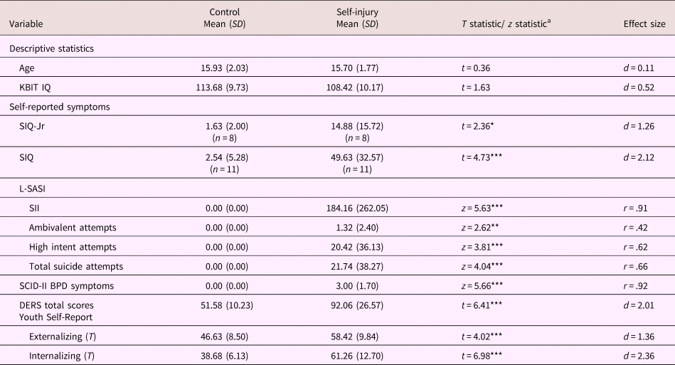
As indicated in Table 1, the SII group engaged in significant levels of self-harm, with large effect sizes separating them from the control group on all measures. The average number of lifetime nonsuicidal self-injurious behaviors among those in the SII group, as defined as self-injury in the absence of intent to die, was 180 but widely variable (SD = 256). Self-injuring adolescents also reported a substantial number of self-injurious behaviors with ambivalent intent to die (M = 19.60), and an average of 1.25 suicide attempts with high intent to die. On average, those in the SII group endorsed three symptoms of BPD on the Structured Clinical Interview for DSM-IV, Axis II (five are required for a diagnosis). The SII group also scored higher on both the Youth Self-Report internalizing and externalizing scales, and on emotion dysregulation as assessed by the DERS. These effects were significant and large, all ts (39) ≥ 4.02, all ds ≥ 1.36. As reported elsewhere, similar effect sizes were observed for DSM-IV symptoms of ADHD, conduct disorder, MDD, and dysthymia on the YI (Sauder et al., Reference Sauder, Derbidge and Beauchaine2016).
Demographic variables, levels of self-injury and suicidal ideation, and self-reported internalizing and externalizing scores, by group

Note: KBIT, Kaufman Brief Intelligence Test, second edition (KBIT-2; Kaufman & Kaufman, Reference Kaufman and Kaufman2004). SIQ, Suicide Ideation Questionnaire (Reynolds, Reference Reynolds1987, Reference Reynolds1988), standardized by grade level. SIQ-Jr (Grades 7–9) raw scores ≥31 and SIQ (Grades 10–12) raw scores ≥41 indicate significant clinical concern regarding suicide risk. L-SASI, Lifetime Suicide Attempt Self-Injury Count (Linehan & Comtois, Reference Linehan and Comtois1996). SCID-II, Structured Clinical Interview for DSM-IV Axis II (First et al., Reference First, Gibbon, Spitzer, Williams and Benjamin1997). DERS, Difficulties in Emotion Regulation Scale (Gratz & Roemer, Reference Gratz and Roemer2004). SII, self-inflicted injury. SA, suicide attempt. aT tests were conducted on normally distributed data; Mann–Whitney U tests (z) were conducted on skewed data. *p ≤ .05. **p .01. ***p ≤ .001.
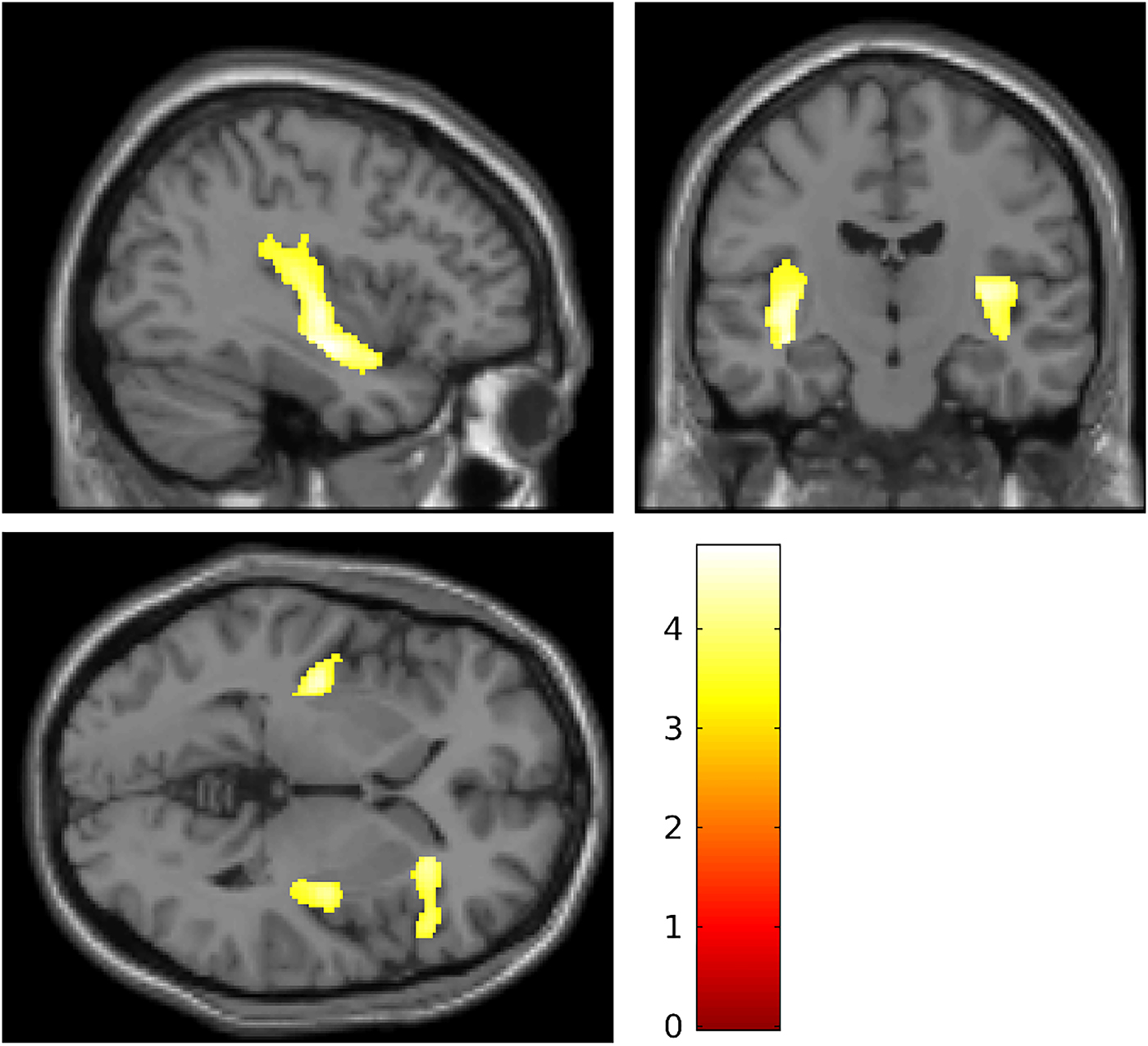
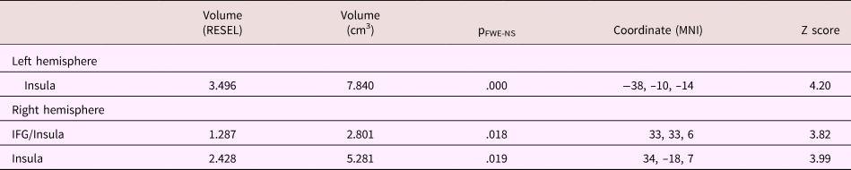
To evaluate our structural hypothesis, we first conducted a whole-brain analysis with a liberal voxel-wise correction of p < .001 uncorrected, and a strict family wise error-corrected cluster extent threshold of p < .05. This error rate was chosen to balance probabilities of Type I and Type II error given the modest sample size (n = 40). No group difference was observed in total brain volumes, t (39) = 0.33, p > .25. However, the cluster extent threshold was exceeded bilaterally for the posterior insula and the right anterior insula/inferior frontal gyrus (rIFG; see Table 2 and Figure 1). The group difference spanned multiple anatomical structures; the two larger clusters are almost entirely insula, whereas the anterior portion spans the insula and IFG. Of note, the IFG is adjacent to the insula, and is implicated in both emotion and self-regulation more broadly (e.g., Depue, Burgess, Bidwell, Willcutt, & Banich, Reference Depue, Burgess, Bidwell, Willcutt and Banich2010; Grecucci et al., Reference Grecucci, Giorgetta, Bonini and Sanfey2013). In each case, regional reductions in gray matter were observed for SII versus control participants. There were no regions of greater gray matter volume among self-injuring adolescents than controls, and no significant differences between groups in regional white matter volumes. Inspection for outliers revealed three z scores that exceeded 2.07 (98th percentile), one in each brain region (rIFG and insula bilaterally). Each outlier came from a different participant in the self-injury group. However, as all outliers were positive, these data worked against our hypotheses and were retained.
Regional reductions in gray matter volume for self-injuring adolescents relative to controls [–37, –14, 0]. Shaded regions depict bilateral reductions in posterior insula volumes, and reductions in volumes spanning and portions of the anterior insula and the right inferior frontal gyrus.

Gray matter volume differences between self-injuring and control participants for regions identified by whole brain analysis

Note: Self-injuring adolescents had reduced gray matter densities in each region, indicating reduced gray matter volumes. Volumes given in RESEL, reflecting volume corrected for smoothness (Worsley, Evans, Marrett, & Neelin, Reference Worsley, Evans, Marrett and Neelin1992), and cm3. P values reflect cluster-level corrections for family wise error (FWE) rate, corrected for nonstationarity (Hayasaka, Phan, Liberzon, Worsley, & Nichols, Reference Hayasaka, Phan, Liberzon, Worsley and Nichols2004). Coordinates are given in Montreal Neurological Institute (MNI) standard brain space, and reflect locations of maximal gray matter differences, with corresponding z scores at each location. Hemisphere labels are derived from the automated anatomical labeling (AAL) atlas (Tzourio-Mazoyer et al., Reference Tzourio-Mazoyer, Landeau, Papathanassiou, Crivello, Etard, Delcroix and Joliot2002), and reflect anatomical regions most associated with the regional difference in gray matter density. IFG, inferior frontal gyrus.
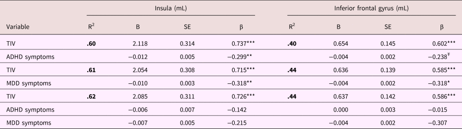
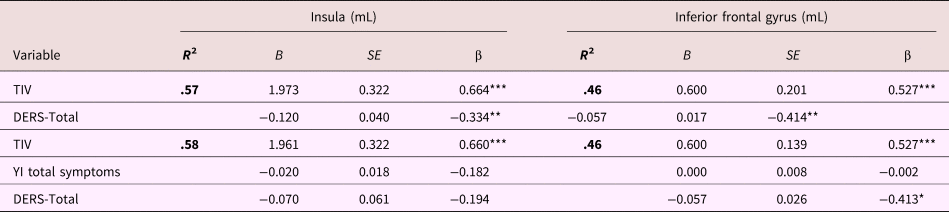
Findings of reduced insular and IFG gray matter volumes among SII participants could be attributable to overall levels of psychopathology rather than emotion dysregulation per se. Because we had no psychiatric controls without SII for comparison, we addressed this possibility by conducting regression analyses in which (a) associations between regional gray matter volumes and both ADHD and MDD were evaluated (see Table 3), and (b) regional gray matter volumes were correlated with emotion dysregulation scores from the DERS, controlling for all symptoms of psychopathology from the YI (see Table 4). As reported in Table 3, when entered alone, both ADHD and MDD symptoms accounted for significant variance in bilateral insula volumes and rIFG volumes, over-and-above effects of total intracranial volume. However, when entered into regression models together, neither ADHD nor MDD symptoms made independent contributions. They therefore contributed overlapping variance.
Linear regression of total intracranial volumes, attention-deficit/hyperactivity disorder (ADHD) symptoms, and major depressive disorder (MDD) symptoms on gray matter volumes in areas of group difference centered within the left insula and right interior frontal gyrus

Note: TIV, total volume in liters. ADHD and MDD symptom scores defined as the sum score of DSM-IV symptoms. Gray matter volumes are reported for the left insula and right interior frontal gyrus. Similar results were found for the right insula, and are not reported. *p < .05. **p < .05. ***p < .001. Ŧp = .084.
Linear regression of total intracranial volume, symptoms of psychopathology, and emotion dysregulation on gray matter volumes in areas of group difference centered within the left insula and right inferior frontal gyrus.

Note: TIV, total volume in liters. DERS, Difficulties with Emotion Regulation Scale (Gratz & Roemer, Reference Gratz and Roemer2004), YI, Youth Inventory (Gadow et al., Reference Gadow, Sprafkin, Carlson, Schneider, Nolan, Mattison and Rundberg-Rivera2002). Gray matter volumes are reported for the left insula and the right interior frontal gyrus. Similar results were found for the right insula, and are not reported. *p < .05. **p < .05. ***p < .001.
As reported in Table 4, DERS scores also accounted for significant variance in bilateral insula volumes and rIFG volumes, over-and-above effects of total intracranial volume. Furthermore, when entered into a regression with all YI symptoms, DERS scores accounted for independent variance in rIFG volumes. Thus, emotion dysregulation accounted for variance in rIFG volumes, over-and-above effects of general psychopathology.
Discussion
SII is a significant public health concern that confers prospective vulnerability to depression, borderline personality development, and eventual suicide (see, e.g., Beauchaine et al., Reference Beauchaine, Hinshaw and Bridgein press; Crowell et al., Reference Crowell, Beauchaine and Linehan2009; Jacobson & Gould, Reference Jacobson and Gould2007; Klonsky et al., Reference Klonsky, Victor and Saffer2014). To date, however, relatively little is known about neurobiological vulnerabilities to SII. Characterizing this neurobiology may improve our understanding of traits that predispose to significant distress and impairment, with direct implications for improved treatments (Beauchaine, Neuhaus, Brenner, & Gatzke-Kopp, Reference Beauchaine, Neuhaus, Brenner and Gatzke-Kopp2008). We hypothesized that self-injuring adolescents would exhibit reduced cortical volumes compared with controls, consistent with findings from adult samples with emotion regulation difficulties, including but not limited to BPD. Volumetric abnormalities in the bilateral insula and rIFG are consistent with etiological models that emphasize emotion dysregulation as a predisposing vulnerability to both SII and later BPD (Crowell et al., Reference Crowell, Beauchaine and Linehan2009; Derbidge & Beauchaine, Reference Derbidge, Beauchaine, Lewis and Rudolph2014; Klonsky, Reference Klonsky2007; Linehan, Reference Linehan1993). Insular cortex function is implicated consistently in emotion regulation (e.g., Goldin et al., Reference Goldin, McRae, Ramel and Gross2008; Grecucci et al., Reference Grecucci, Giorgetta, Bonini and Sanfey2013; Zilverstand et al., Reference Zilverstand, Parvaz and Goldstein2017). In this study, regional reductions in gray matter volume were localized to the insular cortex and rIFG, perhaps reflecting abnormalities in neural development of emotional processing. To date, there is limited evidence to suggest that reduced insular volumes are associated directly with suicidality (e.g., Hwang et al., Reference Hwang, Lee, Tsai, Chen, Yang, Lirng and Tsai2010). Any such link would almost certainly be indirect. Insular and IFG dysfunction predispose to emotion dysregulation, which likely interacts with environmental adversities to potentiate vulnerability to more debilitating psychiatric impairment (see, e.g., Beauchaine et al., Reference Beauchaine, Klein, Crowell, Derbidge and Gatzke-Kopp2009).
Volumetric abnormalities in the insula are observed in a wide range of psychiatric syndromes characterized by emotion dysregulation. For example, internalizing and externalizing boys and girls exhibit reduced insular volumes (Fairchild et al., Reference Fairchild, Passamonti, Hurford, Hagan, von dem Hagen, van Goozen and Calder2011, Reference Fairchild, Hagan, Walsh, Passamonti, Calder and Goodyer2013; Snyder, Hankin, Sandman, Head, & Davis, Reference Snyder, Hankin, Sandman, Head and Davis2017; Sterzer, Stadler, Poustka, & Kleinschmidt, Reference Sterzer, Stadler, Poustka and Kleinschmidt2007), as do adults who incur cumulative life stress and more acute forms of trauma (e.g., Herringa, Phillips, Almeida, Insana, & Germain, Reference Herringa, Phillips, Almeida, Insana and Germain2012). These latter findings are of particular interest given the increasingly well-documented role of trauma and correlated environmental adversities in development of both (a) SII and suicidal behaviors (e.g., Guendelman, Owens, Galan, Gard, & Hinshaw, Reference Guendelman, Owens, Galan, Gard and Hinshaw2016; Hinshaw et al., Reference Hinshaw, Owens, Zalecki, Huggins, Montenegro-Nevado, Schrodek and Swanson2012), and (b) cortical brain growth (e.g., Ansell, Rando, Tuit, Guarnaccia, & Sinha, Reference Ansell, Rando, Tuit, Guarnaccia and Sinha2012; Hair, Hanson, Wolfe, & Pollak, Reference Hair, Hanson, Wolfe and Pollak2015). Taken together, accumulating research suggests that the insula and other cortical structures are sensitive to neuroplastic degenerative effects of stress and trauma, with harmful consequences for emerging emotion regulation skills (see above; Kohn et al., Reference Kohn, Eickhoff, Scheller, Laird, Fox and Habel2014; Zilverstand et al., Reference Zilverstand, Parvaz and Goldstein2017). Moreover, reduced insula and other cortical volumes across various disorders are consistent with the transdiagnostic nature of emotion dysregulation in psychopathology (Beauchaine & Zisner, Reference Beauchaine and Zisner2017; Kring & Sloan, Reference Kring and Sloan2009).
Gray matter reductions extended to the rIFG, an adjacent neural structure implicated in both emotion- and self-regulation, particularly in social contexts (Grecucci et al., Reference Grecucci, Giorgetta, Bonini and Sanfey2013; Kohn et al., Reference Kohn, Eickhoff, Scheller, Laird, Fox and Habel2014; Urgesi, Mattiassi, Buiatti, & Marini, Reference Urgesi, Mattiassi, Buiatti and Marini2016). Lesions to this region result in impaired behavioral inhibition and impulsive decision making (Aron, Robbins, & Poldrack, Reference Aron, Robbins and Poldrack2004; Chamberlain & Sahakian, Reference Chamberlain and Sahakian2007). Individuals with ADHD show reduced rIFG gray matter volumes, and smaller rIFG volumes are associated with slower processing speed and reduced response inhibition (Depue et al., Reference Depue, Burgess, Bidwell, Willcutt and Banich2010). Thus, reduced rIFG gray matter volumes may help to account for high rates of impulsivity and other externalizing behaviors observed among adolescents with SII.
It is important to acknowledge that abnormalities in neural structure are not necessarily manifested in neural or behavioral function. It is relatively common to see atypical morphology in subsets of individuals who undergo MRI, despite no identifiable abnormalities in neural function or behavior. As mentioned above, however, reductions in rIFG volumes are associated with impairments in inhibition, whereas healthy emotion awareness and regulation are associated with larger insular volumes (Giuliani, Drabant, Bhatnagar, & Gross, Reference Giuliani, Drabant, Bhatnagar and Gross2011). Given the large body of evidence indicating bilateral volumetric abnormalities in the insula among emotionally dysregulated samples, it is reasonable to conclude that reduced gray matter in this brain region is a vulnerability to emotional lability and its psychiatric correlates.
In contrast to observed insular cortex volumes in several forms of psychopathology, as reviewed immediately above, insular volumes correlate positively with self-control (Rosso et al., Reference Rosso, Makris, Britton, Price, Gold, Zai and Rauch2010; Uchida et al., Reference Uchida, Del-Ben, Busatto, Duran, Guimarães, Crippa and Graeff2008). Such findings lend more support to the apparent emotion regulation/emotion suppression function of the insula, and may help to explain consistent associations between emotion dysregulation and vagally mediated autonomic dysfunction observed across these differing manifestations of psychopathology, including SII (e.g., Crowell et al., Reference Crowell, Beauchaine, McCauley, Smith, Stevens and Sylvers2005, Reference Crowell, Butner, Wiltshire, Munion, Yaptangco and Beauchaine2017), given insular involvement in regulating the peripheral nervous system (see, e.g., Beauchaine & Thayer, Reference Beauchaine and Thayer2015).
Of note, reduced volumes were not observed in many other cortical structures that are consistently compromised in BPD samples (e.g., ACC, dlPFC, mPFC, vlPFC, and OFC). There are at least three explanations for this finding. First, only a subset of self-injuring adolescences will go on to develop BPD. Although that fraction is likely to be high in the present sample given (a) the number of BPD symptoms and degree of emotion dysregulation endorsed (see Table 1) and (b) the severity of self-injury that participants were recruited for, it is nevertheless possible that heterogeneity in vulnerability to BPD diluted cortical effects.
A second possibility is that broader PFC structural deficiencies were not yet evident because neuromaturational trajectories for the SII and control groups had not yet diverged enough. It is possible that girls in the SII group will be “left behind” their typically developing peers in frontal neuromaturation. Effects of emerging cortical structural deficiencies across early adolescence have been observed for other disorders (e.g., de Brito et al., Reference de Brito, Mechelli, Wilke, Laurens, Jones, Barker and Viding2009; Shaw et al., Reference Shaw, Malek, Watson, Greenstein, de Rossi and Sharp2013). Addressing this possibility will require future longitudinal studies with vulnerable samples.
Third and finally, although many studies conducted with adults show volumetric abnormalities in BPD using whole-brain analyses of similarly sized samples, statistical power in a sample of n = 40 is limited. It is therefore possible that structural abnormalities in cortical regions other than the insular cortex and IFG were missed. Addressing this question will also require additional research.
There are a number of caveats to consider in interpreting our findings. As is usually the case with cross-sectional data, we cannot know whether insular and IFG gray matter reductions observed preceded or followed SII onset, or whether they mark prospective vulnerability to BPD. Our article represents one small step toward disentangling such questions by demonstrating, for the first time, that reduced insular cortex volumes are present among emotionally dysregulated adolescents who self-injure. Nevertheless, more longitudinal studies of gray matter development are needed to identify prospective vulnerabilities, especially regarding development of self-injury. As alluded to above, gray matter densities follow typical neurodevelopmental time courses across adolescence and young adulthood (see Gogtay & Thompson, Reference Gogtay and Thompson2010). Insular volumes increase throughout early and middle adolescence, with later reductions in late adolescence and early adulthood (Shaw et al., Reference Shaw, Kabani, Lerch, Eckstrand, Lenroot, Gogtay and Giedd2008).
It is important to restate that this is not a study of existing BPD. Rather, it is a study of likely prospective vulnerability. Such vulnerability is almost certainly more general than specific. Girls who participated are vulnerable to a range of adverse multifinal outcomes, including depression, externalizing behaviors, and substance use disorders, in addition to BPD. They are also vulnerable to developing other personality disorders. Nevertheless, given our recruitment strategy, the severity of SII, and the fact that many girls recruited using very similar inclusion criteria have been diagnosed with BPD in our previous work (e.g., Beauchaine et al., Reference Beauchaine, Crowell and Hsiao2015; Crowell et al., Reference Crowell, Butner, Wiltshire, Munion, Yaptangco and Beauchaine2017), we are confident our findings are relevant to evaluating premorbid vulnerability to BPD. That said, very few neural vulnerabilities are specific to single disorders, and the expectation that a specific neural signature could predict single disorders may be misplaced. Rather, most psychiatric disorders share neural vulnerabilities that transcend traditional diagnostic categories. Accordingly, specifying neural substrates of transdiagnostic vulnerability traits, such as emotion dysregulation, may be more fruitful in the upcoming years than persistently searching for pathognomonic signs (see Beauchaine & Constantino, Reference Beauchaine and Constantino2017).
Finally, it is important to note that cortical thickness measures and the voxel-based morphometry methods used here are not necessarily interchangeable. Although the measures yield similar results, cortical thickness may be more sensitive to normative developmental changes (Hutton, Draganski, Ashburner, & Weiskopf, Reference Hutton, Draganski, Ashburner and Weiskopf2009). Future studies are therefore needed to determine how our findings fit within contexts of typical and atypical neural development.
Caveats aside, this is the first study to demonstrate volumetric abnormalities in the insula and IFG among adolescent girls who are vulnerable to depression, suicidal behavior, and BPD. Our findings indicate that brain abnormalities that are characteristic of adults with BPD are already present among adolescents who self-injure, which should motivate the field to intervene earlier (see Beauchaine et al., Reference Beauchaine, Neuhaus, Brenner and Gatzke-Kopp2008, Reference Beauchaine, Hinshaw and Bridgein press). Cortical structures in particular follow protracted neuromaturational time courses (Brain Development Cooperative Group, 2012), and are responsive to environmental input through mechanisms of neural plasticity (e.g., Hair et al., Reference Hair, Hanson, Wolfe and Pollak2015). Prevention and early intervention programs may hold great promise in redressing abnormal patterns of neural structure and function during adolescence (a critical period for cortical neuromaturation; e.g., Casey & Caudle, Reference Casey and Caudle2013).
Financial support
Research reported in this article was supported by Grants MH86198 and DE025980 from the National Institutes of Health, and by the National Institutes of Health Science of Behavior Change (SoBC) Common Fund.