Case report
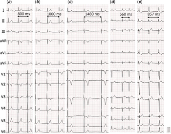
The patient was born after 38 weeks and 1 day of gestation with the induction of delivery because of fetal bradycardia. His 12-lead electrocardiogram (ECG) showed a sinus rhythm at the age of 3 months (Fig 1a) and junctional escape rhythm at the age of 11 months (Fig 1b). The 24-hour Holter monitoring at the age of 4 years showed remarkable bradycardia (average heart rate: 45 bpm), and the chest X-ray showed cardiomegaly. The 12-lead ECG showed no P wave and small ultra-high-frequency fibrillated wavelets at V1 and V2 and negative T wave in V1–4 with a QTc interval of 341 ms (Fig 1c).
12-lead electrocardiograms of the patient (a, b, c, e) and his mother (d). (a): Sinus rhythm at the age of 3 months. (b ): Junctional escape rhythm at the age of 11 months. (c): Atrial fibrillation with small ultra-high-frequency fibrillated wavelets at V1 and V2 and negative T wave in V1–4. (d): The 12-lead electrogram of his mother with the same mutations, presenting atrial fibrillation with similar ultra-high-frequency fibrillated wavelets at V1 and V2. (e): Ventricular pacemaker rhythm by the right-sided conduction system pacing.

His genetic analysis revealed double heterozygous missense mutations in SCN5A and in KCNQ1. The SCN5A mutation was p. Arg1193Gln (G to A base substitution at nucleotide 3578 in exon 20) and the KCNQ1 mutation was p. Ser140Gly (A to G base substitution at nucleotide 418 in exon 2).
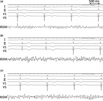
His mother had the same mutations and similar 12-lead ECG findings with small ultra-high-frequency fibrillated wavelets at V1 and V2 (Fig 1d). With the parents’ consent, we decided to implant a pacemaker for the management of marked bradycardia and cardiomegaly. During surgery, we checked intracardiac electrograms using a permanent pacemaker lead (Select Secure, Medtronic, MN, USA). We found ultra-high-frequency fibrillated wavelets in the whole right atrium (Fig 2a–c), and subsequently, we implanted a VVIR (rate-response ventricular demand type) pacemaker to the right-sided conduction system to obtain a narrow QRS complex during pacing (Fig 1e).
Intracardiac electrograms in the right atrium. Rapidly fibrillated wavelets at frequencies of approximately 12 to 30 Hz were obtained at the lower interatrial septum (a), the higher interatrial septum (b), and the right atrial appendage (c) in the right atrium.

Discussion
Atrial fibrillation is the most common form of cardiac arrhythmia and occurs predominantly in the adult population. However, familial atrial fibrillation is inherited and sporadically occurs in the pediatric population. Previous studies have described more than 15 gene mutations associated with atrial fibrillation. And having a first-degree relative with atrial fibrillation is associated with a 40% increased risk for the arrhythmia. Reference Lubitz, Yin and Fontes1 Chen et al. investigated Chinese large kindred with autosomal dominant atrial fibrillation and identified a gain-of-function mutation in the potassium channel gene of KCNQ1 (p.S140G). Reference Chen, Xu and Bendahhou2,Reference Hong, Piper and Diaz-Valdecantos3 It is reasonable to expect that the KCNQ1 mutation would shorten the atrial action potential and cause atrial fibrillation. In other investigations, some familial atrial fibrillation could be caused by loss- or gain-of-function in the sodium current due to SCN5A mutations. Reference McNair, Ku and Taylor4–Reference Wilde and Amin6 The SCN5A (p.R1193Q) mutation which was identified in our case was known to be associated with the Brugada syndrome by a loss-of-function of cardiac sodium channels. Reference Vatta, Dumaine and Varghese7 There is a general consensus that in the Brugada syndrome, contrary to LQTS3, loss-of-function due to SCN5A mutations causes a decrease in the peak cardiac sodium current and slowed upstroke of the action potential during phase 0. Reference Wilde and Amin6 The double missense mutations in SCN5A (p.R1193Q) and KCNQ1 (p.S140G) could decrease the sodium current and increase the potassium current, resulting in the remarkable shortening of the atrial action potential and in unique ultra-high-frequency atrial fibrillated wavelets on the electrocardiograms.
Conclusion
A rare case of familial atrial fibrillation presented an unusual 12-lead ECG finding of ultra-high-frequency fibrillated wavelets associated with double missense mutations in SCN5A (p.R1193Q) and KCNQ1 (p.S140G).
Acknowledgements
The authors thank John Martin for his contributions to the manuscript.
Financial support
This research received no specific grant from any funding agency, commercial, or not-for-profit sectors.
Conflicts of interest
None.
Ethical standards
Consent for publication of the case report obtained from the family.