Introduction
Sub-clinical necrotic enteritis (NE) is a form of dysbacteriosis. Dysbacteriosis is a disease condition caused by an imbalance of the normal microbial flora in the proximal parts of the small intestine and the pathogenesis can be initiated by a mixture of opportunistic pathogens. C. perfringens organisms are the main aetiological factor in sub-clinical necrotic enteritis. Although C. perfringens may cause the initial pathogenicity, as the condition progresses, necrosed tissues detach from the mucosal surface together with C. perfringens organisms. This may predispose the growth of the other opportunistic pathogens in the damaged intestinal mucosa and the condition may then be only identified as a general dysbacteriosis.
The sub-clinical form of NE is a cause of major economic loss to the broiler production industry throughout the world (Kaldhusdal, 2000). It is not usually detected or treated due to the absence of clinical signs or change in mortality rates, so it prevails to cause poor growth and feed conversion efficiency (Lovland and Kaldhusdal, Reference LOVLAND and KALDHUSDAL2001), condemnation of the affected livers and the risk of contamination of the poultry products during the slaughter process (Craven et al., Reference CRAVEN, STERN, BAILEY and COX2001). There is a need for the broiler production industry to understand how to effectively reduce its incidence in commercial broiler flocks. The objective of this review is to explain current knowledge on the aetiology of the disease and to discuss the predisposing factors that cause sub-clinical NE.
Diagnosis of sub-clinical NE
Sub-clinical NE is difficult to diagnose. The disease may be suspected from a reduced weight gain and feed conversion efficiency in a broiler flock (Lovland and Kaldhusdal, Reference LOVLAND and KALDHUSDAL2001) and high moisture content of the droppings or wet litter conditions (Hermans and Morgan, Reference HERMANS and MORGAN2007). Sub-clinical NE is more likely to cause these effects at 2-5 weeks of age but often without an increase in mortality. Disease diagnosis requires sacrificing randomly sampled birds from a suspected flock and identification of intestinal and liver lesions in post mortem examination (Kaldhusdal, 2000).
Macroscopically visible necroses in the small intestines can be used as the main diagnostic tool of sub-clinical NE. Small circular, 1-2 mm diameter; white, focal, necrotic lesions in the small intestinal mucosa initially occur (Kaldhusdal and Hofshagen, Reference KALDHUSDAL and HOFSHAGEN1992). Such lesions may enlarge and become ulcers when the damaged superficial layers and the debris are sloughed off to denude the underlying tissue. These large necroses often coalesce and sometimes will cover the whole mucosal surface. The affected small intestine is flaccid, friable and often distended with gas. However, unlike in clinical NE, the lesions in sub-clinical NE are not deep in the muscular layers of the intestinal wall. Debris is adhered to larger areas and this appears as a pseudo-membrane. The lesions are self limiting and recover at around five weeks post hatch.
The counts of C. perfringens in the small intestinal digesta or the mucosal scrapings are not closely correlated with disease severity (McReynolds et al., Reference MCREYNOLDS, BYRD, ANDERSON, MOORE, EDRINGTON, GENOVESE, POOLE, KUBENA and NISBET2004; Olkowski et al., Reference OLKOWSKI, WOJNAROWICZ, CHIRINO-TREJO and DREW2006; Palliyeguru et al., Reference PALLIYEGURU, ROSE and MACKENZIE2010), whereas median counts above 106 colony forming units (cfu) of C. perfringens per g caecal contents indicate a high probability of concurrent NE specific gut lesions (Kaldhusdal et al., Reference KALDHUSDAL, HOFSHAGEN, LOVLAND, LANGSTRAND and REDHEAD1999; Palliyeguru and Rose, Reference PALLIYEGURU and ROSE2009).
Disease confirmation needs the presence of the necrotic lesions on the small intestinal mucosa with identifiable aggregates of pathogenic C. perfringens. Gross and histopathological lesion identification and immunofluorescence or immunohistochemical confirmation of aggregated C. perfringens or toxins within the small intestinal epithelium (Das et al., Reference DAS, MAZUMDER, DUTTA, SHOME, BUJARBARUAH and KUMAR2008; Schrader et al., Reference SCHRADER, BROUSSARD, HANSEN, DIERKS, OETTING and PETERSEN2008) can confirm the disease. Necrotic debris of the intestinal lesions or haemorrhagic lymph nodes (14-30 days), suspected livers (20-30 days) or caecal contents can also be cultured. Suspected specimens must be collected anaerobically and cultured promptly after collection (Quinn et al., Reference QUINN, MARKEY, CARTER, DONNELLY and LEONARD2002) or immediately frozen and stored at -70°C. C. perfringens counts exceeding 106 colony forming units per gram (cfu/g) in the caecal contents above 14 days of age are further taken as confirmatory evidence. The presence of comparatively high plasma antibody titres against C. perfringens toxins in 25-35 day old broilers is also an indicator (Kulkarni et al., Reference KULKARNI, PARREIRA, SHARIF and PRESCOTT2007).
Aetiological factors in sub-clinical NE
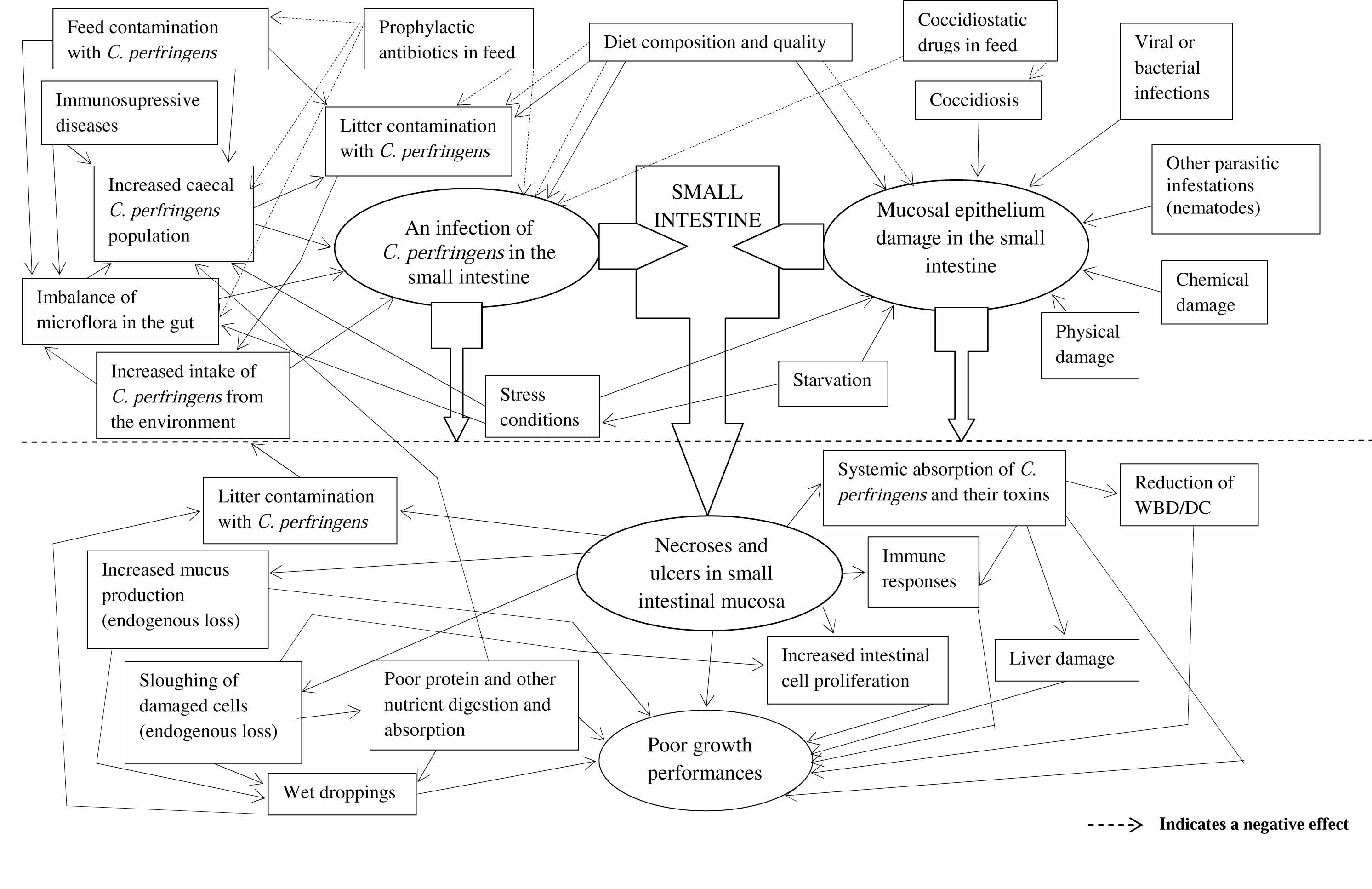
Not only the presence of C. perfringens strains capable of producing related toxins but also a number of predisposing factors are important in the aetiology of NE (Figure 1). Predisposing factors play a major role in the aetiology by disturbing the integrity of the intestinal mucosal barrier by disrupting the protective mucus layers or damaging epithelial cells. The opportunistic pathogen, C. perfringens, may then adhere to extracellular matrix molecules (Martin and Smyth, Reference MARTIN and SMYTH2010) or the necrotic cells and proliferate to produce toxin in the dying tissues and debris.
When the gut epithelium damage progresses the other intestinal commensals that also can be opportunistic pathogens such as the various serovars of E. coli and Salmonella enterica may invade the damaged epithelium and also proliferate in the intestinal contents. Such multiplication of the pathogens further deviate the intestinal microbial balance towards an infectious stage of various species of pathogens or disbacteriosis. This may then lead to further increase the moisture content in the droppings or ultimately into diarrhoeic conditions that is clinically identified as dysbacteriosis. Although the terminology dysbacteriosis is used in poultry to describe diarrhoea or extremely wet droppings with the gross lesions that are similar to the sub-clinical necrotic enteritis, such as ballooned intestine with thin walls and watery gut contents, dysbacteriosis, is not clearly defined.
Clostridium perfringens
C. perfringens is an opportunistic pathogen that is ubiquitous throughout the environment (Wages and Opengart, Reference WAGES and OPENGART2003). The main habitats of the organism are soils and the intestines of humans and animals (Stevens and Rood, Reference STEVENS and ROOD2006). The quantity of C. perfringens in soil is approximately proportional to the intensity and duration of animal husbandry in the area (Stevens and Rood, Reference STEVENS and ROOD2006) thus indicating that the gastrointestinal tract is a favoured habitat of C. perfringens. The organism is typically found up to 5x10⁵ cfu/g in soil and 102-109 cfu/g in human faeces and decaying organic matter (Stevens and Rood, Reference STEVENS and ROOD2006).
C. perfringens are thick, gram-positive, rod shaped organisms with slightly rounded (blunt) ends and remain as a single cell or in groups of two or three loosely attached chains or palisade forms. They can form large, oval, sub-terminal spores. C. perfringens can vary in size (ranging from 0.6-2.4 x 1.3-9.0 µm), and are larger than others in the genera. C. perfringens is the only non-motile species among the Clostridium genera that produce a capsule in animal tissue (Quinn et al., Reference QUINN, CARTER, MARKEY and CARTER1994).
C. perfringens can be readily isolated on anaerobic blood agar plates that are incubated anaerobically at 37°C (Wages and Opengart, Reference WAGES and OPENGART2003). They produce round, slightly opaque, shiny, double haemolytic colonies with a clear inner zone of theta (θ) toxin haemolysis and a misty outer zone of alpha (α) toxin haemolysis (Granum and Peck, Reference GRANUM and PECK2006). Thioglycollate broth, tryptose-sulfite-cycloserine (TSE) agar, egg-yolk agar and heart brain infusion agar with sheep blood (5%) also can be used for isolation of C. perfringens in an atmosphere containing 10% H2, 10% CO2 and 80% N2. C. perfringens grow vigorously at temperatures between 12-50°C with an optimum growth at 43-47°C for most strains but their growth is very slow at less than 20°C (Johansson, Reference JOHANSSON2006) and stops at 6°C (McClane, Reference MCCLANE1997). Under optimum conditions, the bacteria grow extremely rapidly with a generation time of 8-10 minutes, accompanied by heavy gas production (Stevens and Bryant, Reference STEVENS and BRYANT2002). The optimum growth occurs between pH 6 and 7 and growth is inhibited at pH less than 5 or more than 8.3 (McClane, Reference MCCLANE1997). C. perfringens prefers to grow in environments with high water activity (aw). The lowest aw allowing vegetative growth of the bacteria is reported to be 0.93 (McClane, Reference MCCLANE1997).
There have been a number of studies of specific C. perfringens genotypes and their toxin production but conclusive results have not been obtained, probably due to gene mutation of the quickly multiplying bacteria. There is a large variation in the genetic epidemiology of C. perfringens that have been identified from healthy chickens, but there is only small genetic variation in the bacterial strains that cause NE (Engstrom et al., Reference ENGSTROM, FERMER, LINDBERG, SAARINEN, BAVERUD and GUNNARSSON2003). After birds recover from sub-clinical NE, their gut flora again has a wide variation in C. perfringens genotypes (Nauerby et al., Reference NAUERBY, PEDERSEN and MADSEN2003). Until recently, all the isolates from the intestine and liver of naturally diseased or pathogen-dosed birds have produced type A toxin, with the gene coding for alpha toxin (cpa) (Gholamiandekhordi et al., Reference GHOLAMIANDEKHORDI, DUCATELLE, HYNDRICKX, HAESEBROUCK and VAN IMMERSEEL2006; Yitbarek et al., Reference YITBAREK, ECHEVERRY, BRADY, ERNANDEZ-DORIA, CAMELO-JAIMES, SHARIF, GUENTER, HOUSE and RODRIGUEZ-LECOMPTE2012). Chalmers et al. (Reference CHALMERS, BRUCE, HUNTER, PARREIRA, KULKARNI, JIANG, PRESCOTT and BOERLIN2008) and Das et al. (Reference DAS, MAZUMDER, DUTTA, SHOME, BUJARBARUAH and KUMAR2008) found that clinically ill and apparently healthy birds (probably carrying sub-clinical NE) were both positive for the cpβ2 gene. The novel toxin NetB (Keyburn et al., Reference KEYBURN, BOYCE, VAZ, BANNAM, FORD and PARKER2008) and a large cytotoxin (tpeL) gene (Chalmers et al., Reference CHALMERS, BRUCE, HUNTER, PARREIRA, KULKARNI, JIANG, PRESCOTT and BOERLIN2008) were identified in type A C. perfringens in clinically affected birds. Some of these large plasmid genes (NetB, tpeL) are related to antibiotic resistant genes (Chalmers et al., Reference CHALMERS, BRUCE, HUNTER, PARREIRA, KULKARNI, JIANG, PRESCOTT and BOERLIN2008; Bannam et al., Reference BANNAM, YAN, HARRISON, SEEMANN, KEYBURN, STUBENRAUCH, WEERAMANTRI, CHEUNG, MCCLANE, BOYCE, MOORE and ROOD2011). Pathogen dosing experimental models may replace the normal flora with previously mutated antibiotic resistant C. perfringens strains (Barbara et al., Reference BARBARA, TRINH, GLOCK and SONGER2008). There is no published information on the genetic epidemiology of C. perfringens that cause spontaneously-occurring sub-clinical NE.
The alpha toxin and NetB toxin produced by type A C. perfringens are most probably the responsible elements for the pathological lesions. An intra-duodenal infusion of crude toxins or broth cultures of type A C. perfringens reproduced the disease (Al-Sheikhly and Truscott, Reference AL-SHEIKHLY and TRUSCOTT1977a; Reference AL-SHEIKHLY and TRUSCOTT1977b). Pathological lesions develop quicker with the toxin and the lesion severity is correlated to the alpha toxin concentration (Al-Sheikhly and Truscott, Reference AL-SHEIKHLY and TRUSCOTT1977b; Schrader et al., Reference SCHRADER, BROUSSARD, HANSEN, DIERKS, OETTING and PETERSEN2008). Keyburn et al. (Reference KEYBURN, BOYCE, VAZ, BANNAM, FORD and PARKER2008) indicated that NetB was the main virulence factor but the findings of Cooper and Songer (Reference COOPER and SONGER2010) were inconclusive. Crespo et al. (Reference CRESPO, FISHER, SHIVAPRASAD, FERNANDEZ-MIYAKAWA and UZAL2007) observed that cpβ2 did not have a role in the pathogenesis of NE as cpβ2-negative strains produced lesions but cpβ2-positive strains did not. Abildgaard et al. (Reference ABILDGAARD, SONDERGAARD, ENGBERG, SCHRAMM and HØJBERG2010) concluded that C. perfringens strains that carried the NetB toxin gene did not all produce the toxin. The role of toxins and various genotypes in the disease is yet to be fully elucidated.
Predisposing factors that facilitate colonisation of C. perfringens
There are predisposing factors that allow C. perfringens to colonise the intestinal mucosa. The following section identifies and evaluates the major factors that predispose growing chickens to sub-clinical NE.
diseases
Coccidiosis has been identified in a survey of commercial broiler farms to have a strong association with the prevalence of NE (Hermans and Morgan, Reference HERMANS and MORGAN2007). Mucosal damage of the intestine as a consequence of merogonous and gametogonous multiplication stages of the coccidia lifecycle is a predominant factor that predisposes birds to NE (Williams et al., Reference WILLIAMS, MARSHALL, LA RAGIONE and CATCHPOLE2003; Collett, Reference COLLETT2004). Necrotic lesions were extensive and mortality was significantly higher in birds infected with both coccidia and C. perfringens than birds infected with C. perfringens alone (Al-Sheikhly and Al-Saieg, Reference AL-SHEIKHLY and AL-SAIEG1980). Shane et al. (Reference SHANE, GYIMAH, HARRINGTON and SNIDER1985) observed a high mortality in birds fed rations containing C. perfringens, along with an oral dose of E. acervulina compared to birds fed the two pathogens at different time intervals or C. perfringens alone. Park et al. (Reference PARK, LILLEHOJ, ALLEN, PARK, FITZCOY, BAUTISTA and LILLEHOJ2008) also identified a synergistic relationship of coccidiosis with C. perfringens.
Ionophore coccidiostatic drugs have a similar control over the caecal C. perfringens as antibiotic growth promoters (Elwinger et al., Reference ELWINGER, BERNDTSON, ENGSTOM, FOSSUM and WALDENSTEDT1998). These compounds might either prevent coccidial damage or have a direct effect on the proliferation of C. perfringens. However, Williams et al. (Reference WILLIAMS, MARSHALL, LA RAGIONE and CATCHPOLE2003) found that vaccination against coccidia with live attenuated coccidia oocysts also prevented NE. Attenuated live coccidia oocysts at very high doses have been used to induce sub-clinical NE (Pedersen et al., Reference PEDERSEN, BJERRUM, O.E. WONG and NAUERBY2008). Some amount of coccidiosis is unavoidable in commercial broiler chicken production so it is probably a major predisposing factor for sub-clinical NE.
Various other diseases predispose the birds to sub-clinical NE. Ascaridia dissimilis larval stages and C. perfringens have been isolated from intestinal scrapings of turkeys on commercial farms with NE outbreaks (Norton et al., Reference NORTON, HOPKINS, SKEELES, BEASLEY and KREEGER1992). Ascarid larvae migrance, leading to intestinal damage, was suggested to be a predisposing factor. Other diseases that commonly occur among poultry, for example salmonellosis (Shivaramaiah et al., Reference SHIVARAMAIAH, WOLFENDEN, BARTA, MORGAN, WOLFENDEN, HARGIS and TÉLLEZ2011), has been associated with NE and immune-suppressive syndromes (Kahn and Line, Reference KAHN and LINE2005) may contribute to NE development.
environmental factors
Management-related factors such as feeding, water provision, temperature control and ventilation systems have been suggested to be precursors of NE (Collett, Reference COLLETT2004). Delayed initial feeding severely impairs the development of gut associated lymphoid tissue in the hind gut and slightly impedes the fore-gut during the first 35 days of life (Bar-Shira et al., Reference BAR-SHIRA, SKLAN and FRIEDMAN2003). Food deprivation of mature birds severely depresses their immune system (Ben-Nathan et al., Reference BEN-NATHAN, DRABKIN and HELLER1981). In addition, the physical form of the feed is a factor, for example Branton et al. (Reference BRANTON, REECE and HAGLER-JR1987) found that crushed wheat reduced the mortality attributed to NE when compared with ground wheat.
A strong relationship has been identified between litter quality and the prevalence of NE on UK broiler farms (Hermans and Morgan, Reference HERMANS and MORGAN2007). Although wet litter can be a consequence of NE, this condition may predispose birds to the disease, as high water activity in litter can increase the proliferation of C. perfringens. Although diet composition is a major cause of variation in litter quality, poor ventilation systems and leakages from drinkers and roofs increase litter moisture.
Poultry often consume some of their floor-litter material and this may cause physical intestinal damage that predisposes birds to the disease (Al-Sheikhly and Truscott, Reference AL-SHEIKHLY and TRUSCOTT1977c). Rough litter materials, such as paddy husk, which is commonly used in the Asian broiler industry, may predispose birds to the disease, and the low moisture carrying capacity of this type of litter material could contribute to the disease incidence (Williams, Reference WILLIAMS2005). Turkey breeders reared on previously used litter material have an increased mortality associated with NE (Droual et al., Reference DROUAL, SHIVAPRASAD and CHIN1994). Treating litter with acid reduced the caecal and ileal C. perfringens counts significantly (Garrido et al., Reference GARRIDO, SKJERVHEIM, OPPEGAARD and SØRUM2004), probably due to the growth inhibition of C. perfringens below pH 5. Overstocking, inappropriate heating systems and poor ventilation systems have been suggested as contributing factors (Collett, Reference COLLETT2004; Williams, Reference WILLIAMS2005).
diet
The chemical composition of the diet modifies the populations of microorganisms in the gastrointestinal tract and the integrity of intestinal epithelium (Smith and Macfarlane, Reference SMITH and MACFARLANE1998). Apajalahti et al. (Reference APAJALAHTI, KETTUNEN, BEDFORD and HOLBEN2001) found that diet is the strongest determinant of the mix of caecal bacterial populations. Nairan and Bamford (Reference NAIRAN and BAMFORD1967) found that when birds were orally dosed with a suspension of C. perfringens, only those fed a commercial diet developed NE whereas those fed an experimental diet did not. This indicated that there was a major effect of diet composition on the precipitation of the disease. It is frequently difficult to decide what factors in diets exactly influence the disease, as the effects are often contradictory. The major diet components that may predispose to sub-clinical NE are discussed below.
Polysaccharides
Cereal grain provides the majority of starch and polysaccharides in chicken feed, and different cereals affect NE development. Branton et al. (Reference BRANTON, REECE and HAGLER-JR1987) found an increased mortality from NE in broilers fed wheat when compared with those fed maize. Kaldhusdal and Hofshagen (Reference KALDHUSDAL and HOFSHAGEN1992) detected a higher number of macroscopically visible focal necrotic lesions and higher C. perfringens counts in the small intestinal mucosa of birds fed high barley diets than in those fed on high maize diets. Hence, barley and wheat-based diets also have been identified as risk factors for NE in broiler chickens compared to maize in a survey of Norwegian broiler producers (Kaldhusdal and Skjerve, Reference KALDHUSDAL and SKJERVE1996). Craven (Reference CRAVEN2000) studied the colonisation of an antibiotic resistant strain of C. perfringens in the gut of broilers when the birds were fed with a maize-based diet and a diet with 50% rye, the latter increased the number of C. perfringens in the ileum, caeca and the faeces. Annett et al. (Reference ANNETT, VISTE, CHIRINO-TREJO, CLASSEN, MIDDLETON and SIMKO2002) compared the effect of wheat, barley and maize on the proliferation of bacteria in an in vitro study and found that the bacterial proliferation in the barley and wheat were higher than with maize.
Although it is evident that different cereal grains predispose birds to NE, the exact underlying mechanism is not clear. Higher levels of non-starch polysaccharides (NSP) in wheat, barley and rye are considered to increase the viscosity of the gut contents (Bedford and Classen, Reference BEDFORD and CLASSEN1992; Choct and Annison, Reference CHOCT and ANNISON1992) and the feed retention time in the intestine (Gohl and Gohl, Reference GOHL and GOHL1977). High dietary NSP thickens the unstirred water layer of the mucosa by interacting with mucopolysaccharides (Johnson and Gee, Reference JOHNSON and GEE1981) which may reduce the absorption and increase the nutrient supply to the microbes in the hind gut. Also, the thickening of the intestinal mucous layer may decrease its oxygen tension thereby promoting the adhesion of C. perfringens onto the mucosal surface. Complex carbohydrates, such as pectin and guar gum, when added to wheat diets together with C. perfringens, thickened the gut wall but reduced the necrotic lesion scores in broiler chickens (Branton et al., Reference BRANTON, LOTT, DEATON, MASLIN, AUSTIN, POTE, KEIRS, LATOUR and DAY1997).
Langhout et al. (Reference LANGHOUT, SCHUTTE, DE JONG, SLOETJES, VERSTEGEN and TAMMINGA2000) fed chickens a diet with citrus pectin, a complex, branched-polysaccharide fibre that is rich in galactoside residues. This resulted in deleterious effects on the intestinal epithelial cells and reduced nutrient digestibility but no effects were observed when the same diet was fed to germ-free chickens. This suggests a possible interaction of microbes with NSP within the intestinal environment. Certain NSPs have the ability to bind bile salts and some lipids in the intestine (Vahouny and Cassidy, Reference VAHOUNY and CASSIDY1986).
Lipids
The amount of dietary lipid and its digestibility as well as bile acid secretion, oxidative status of fats and the presence of volatile essential oils may have some effect on the incidence of NE. Soya oil supplementation in diets reduced C. perfringens counts when compared with a mixture of lard and tallow (Knarreborg et al., Reference KNARREBORG, SIMON, ENGBERG, JENSEN and TANNOCK2002a), hence oxidised fat in diets may predispose birds to NE (Dahiya et al., Reference DAHIYA, WILKIE, VAN KESSEL and DREW2006). Some essential oils have antimicrobial effects in vitro (Rao and Nigam, Reference RAO and NIGAM1970). Two different blends of essential oils reduced C. perfringens in the intestinal contents of broiler chickens (Mitsch et al., Reference MITSCH, ZITTERL-EGLSEER, KOHLER, GABLER, LOSA and ZIMPERNIK2004). However, Gutierrez et al. (Reference GUTIERREZ, BARRY-RYAN and BOURKE2008) concluded that the effects of essential oils mainly depend on interactions with other dietary ingredients. Bile acids have antibacterial effects (Inagaki et al., Reference INAGAKI, MOSCHETTA, LEE, PENG, ZHAO, DOWNES, YU, SHELTON, RICHARDSON, REPA, MANGELSDORF and KLIEWER2006), and C. perfringens is able to hydrolyse them within the intestinal environment (Knarreborg et al., Reference KNARREBORG, ENGBERG, JENSEN and JENSEN2002b).
Proteins
There is growing experimental evidence that dietary protein supply is a major causative factor for sub-clinical NE in commercial broiler production. There may be an interaction between the concentration of the dietary protein, the biological availability of the protein and antinutritional factors that have protease inhibitory activity. Palliyeguru et al. (Reference PALLIYEGURU, ROSE and MACKENZIE2010) fed practical, nutritionally-balanced diets formulated with different protein sources to broiler chickens, and found an increased incidence of sub-clinical NE with diets that included a proportion of potato protein compared to diets that contained soybean meal as the only protein concentrate. The incidence of sub-clinical NE was not correlated to the contents of amino acids or non-starch polysaccharides in the experimental diets. Similar results were also found by Fernando et al. (Reference FERNANDO, ROSE, MACKENZIE and SILVA2011). Palliyeguru et al. (Reference PALLIYEGURU, ROSE and MACKENZIE2010) suggested that these differences may have been primarily due to the high trypsin inhibitor contents in the potato protein diets. The same authors (Palliyeguru et al., Reference PALLIYEGURU, ROSE and MACKENZIE2011) demonstrated that trypsin inhibitor activities in different soya bean samples were correlated to the incidences of sub-clinical NE in broiler chickens fed practical feeds. Inhibition of trypsin enzyme leads to poor protein digestion, especially in the jejunum and the upper ileum where digestion and absorption should be maximised. The higher levels of undigested protein flowing to the hind gut in the birds fed diets with high trypsin inhibitor activity may allow greater proliferation of C. perfringens in the digestive tract, as Clostridium spp. are well known to need high protein availability to form large colonies.
There is little experimental evidence that examines the effect of dietary protein on the number of C. perfringens in chickens. An increased number of C. perfringens type A, from 102 up to 106 cfu/g, was observed in the intestines of pigs fed a high protein diet (300 g/kg) compared to a lower protein (200 g/kg) diet (Mansson and Olhagen, Reference MANSSON and OLHAGEN1967). A high protein diet (630 g/kg) was compared to a 510 g/kg protein diet and increased the C. Perfringens numbers from 106 up to 109cfu/g in the digesta of dogs even though the Lactobacilli and Streptococci counts were not affected (Zentek et al., Reference ZENTEK, ROHDE and AMTSBERG1998). This indicated that the intestinal C. perfringens populations may be more sensitive to dietary protein sources than the other intestinal flora.
Diets that provide an excess of dietary protein may increase protein levels in the ileal digesta and so predispose to high proteolytic C. perfringens populations. Broiler chickens fed diets high in fish meal (400-570 g/kg) proliferated C. perfringens bacteria within the small intestine (Drew et al., Reference DREW, SYED, GOLDADE, LAARVELD and VAN KESSEL2004). However, at lower inclusion rates of 230-315 g/kg there was no influence of protein sources on caecal or ileal C. perfringens counts. Widyaratne (Reference WIDYARATNE2012) showed that low protein diets which included highly digestible protein supplements could maintain the growth performance of broilers and these diets also gave a significant reduction in the intestinal C. perfringens counts.
Antinutritive factors
McDevitt et al. (Reference MCDEVITT, BROOKER, ACAMOVIC and SPARKS2006) suggested certain antinutrients may affect the incidence of NE. Certain cereal grains such as wheat, barley, oats and rye (Giovannini et al., Reference GIOVANNINI, MANCINI and DE VINCENZI1996) and legumes, such as soya and kidney beans, contain lectins (Van Dijk et al., Reference VAN DIJK, HUISMAN and KONINKX2002). These lectins have the ability to damage the intestinal epithelial cells (Van Dijk et al., Reference VAN DIJK, HUISMAN and KONINKX2002). In addition, lectins also affect cell metabolism by inhibiting DNA, RNA and glycoprotein synthesis (Giovannini et al., Reference GIOVANNINI, MANCINI and DE VINCENZI1996). Lectins have the ability to adhere to the epithelial surface sugars and bind via fimbriae with enteric pathogens (Van Dijk et al., Reference VAN DIJK, HUISMAN and KONINKX2002). Lectins may mediate the pathogen adhesion to the gut wall which then facilitates bacterial colonisation (Untawale et al., Reference UNTAWALE, PIETRASZEK and MCGINNIS1978), and mycotoxins can predispose the birds to the disease (Johnson and Pinedo, Reference JOHNSON and PINEDO1971).
Glycoalkaloids are toxic chemical compounds that occur in potato tubers (Smith et al., Reference SMITH and RODDICK1996). Glycoalkaloids are heat stable (Jadhav and Kadam, Reference JADHAV and KADAM1998) and have the ability to disrupt cell membranes which can then damage the intestinal epithelium (Smith et al., 1996). Dietary potato glycoalkaloids are known to damage cells in contact tissues such as liver and blood (Smith et al., 1996). Tannins, glucosinolates, terpenes, and polyphenols also are suggested to have some interactions with intestinal microbes and dietary nutrient utilisation (McDevitt et al., Reference MCDEVITT, BROOKER, ACAMOVIC and SPARKS2006) although there is little direct evidence of their involvement in the incidence of sub-clinical NE.
Conclusions
Sub-clinical NE is a multifactorial disease, and although the main aetiological factor is the bacterium C. perfringens, a large number of other factors interact to precipitate the disease (Figure 1). For the control of NE in poultry, environmental factors including ventilation and litter moisture, alongside dietary feed materials and exposure to other disease and mycotoxins must be considered.
Aetiology of sub-clinical necrotic enteritis in broiler flocks.