INTRODUCTION
The marine genus Sphaeromyxa Thélohan, 1892 (Myxozoa: Myxosporea) comprises almost 50 nominal species, all of which parasitize the hepatic biliary systems of their fish hosts typically occupying the gall bladder (Lom and Dyková, Reference Lom and Dyková2006; Karlsbakk et al. Reference Karlsbakk, Einen and Bartošová2013; Kristmundsson and Freeman, Reference Kristmundsson and Freeman2013; Whipps and Font, Reference Whipps and Font2013). A number of sphaeromyxids are known to have low host specificity and a wide geographic distribution (Lom, Reference Lom2004; Kristmundsson and Freeman, Reference Kristmundsson and Freeman2013).
Among myxosporeans, species of the genus Sphaeromyxa are unique by a number of morphological and evolutionary features. The polar filaments are not spirally coiled as in most myxosporeans, but instead folded over several times upon themselves. The filaments are also typically broad at their base gradually tapering towards the distal ends (Lom and Dyková, Reference Lom and Dyková2006). The ribbon-like character of these polar filaments was the main criterion used to establish the suborder Sphaeromyxina Lom et Noble, 1984 and was found to represent a single synapomorphic trait of sphaeromyxids in the myxosporean evolution (Fiala and Bartošová, Reference Fiala and Bartošová2010). However, in the light of recent molecular findings the suborder Sphaeromyxina was suppressed and the monotypic family Sphaeromyxidae Lom et Noble, 1984 was transferred to the suborder Variisporina Lom et Noble, 1984 (Kristmundsson and Freeman, Reference Kristmundsson and Freeman2013). The specific ultrastructural features of genus Sphaeromyxa are marked surface projections of their large flat plasmodia and an endoplasm containing a number of extremely large vacuoles and lobocytes (Lom, Reference Lom1969, Reference Lom2004).
Sphaeromyxa is one of a few myxozoan genera where the classification by spore morphology is consistent with estimates of phylogeny (i.e. it is monophyletic). Marine sphaeromyxids unusually cluster within the ‘hepatic biliary group’ which besides Sphaeromyxa spp. mostly includes species that infect the gall bladders of freshwater hosts but with some from marine and exceptionally from terrestrial hosts (Fiala, Reference Fiala2006; Kristmundsson and Freeman, Reference Kristmundsson and Freeman2013). Sphaeromyxa species further divide into two morphologically distinct groups, (i) the ‘balbianii’ group with straight or slightly curved fusiform or ovoid spores, usually truncate spore ends and ovoid polar capsules and (ii) the ‘incurvata’ group with arcuate spores, twisted valves, usually rounded spore ends and pyriform polar capsules (Laird, Reference Laird1953; Karlsbakk et al. Reference Karlsbakk, Einen and Bartošová2013). This morphology-based grouping was supported by recent phylogenetic analyses of molecular data where a monophyletic Sphaeromyxa clade groups the members with zig-zag folded polar filament further clustering within the ‘balbianii’ or ‘incurvata’ subclade (Karlsbakk et al. Reference Karlsbakk, Einen and Bartošová2013; Kristmundsson and Freeman, Reference Kristmundsson and Freeman2013; Whipps and Font, Reference Whipps and Font2013). Some authors even proposed the establishment of two different genera for members of the two groups (Karlsbakk et al. Reference Karlsbakk, Einen and Bartošová2013).
Five Sphaeromyxa species have been recorded in intertidal blennies. Sphaeromyxa tripterygii Laird, Reference Laird1953 was described from Forsterygion varium (Forster) and Bellapiscis medius (Günther) (Perciformes: Tripterygiidae) off New Zealand (Laird, Reference Laird1953). Sphaeromyxa incurvata Doflein, Reference Doflein1898 was found in Blennius ocellaris L. (Perciformes: Blenniidae) from the Mediterranean (Doflein, Reference Doflein1898; Kudo, Reference Kudo1919). Sphaeromyxa species from clinid blennies (Perciformes: Clinidae) were recorded (i) from the Pacific Ocean off California, USA, i.e. Sphaeromyxa gibbonsia Noble, 1938 from Gibbonsia elegans (Cooper) and Gibbonsia metzi Hubbs, (ii) along the Australian New South Wales coast, i.e. Sphaeromyxa noblei Lom, Reference Lom2004 from Heteroclinus whiteleggii (Ogilby) and (iii) from the south coast of Africa, i.e. an undescribed Sphaeromyxa species from Pavoclinus graminis (Gilchrist & Thompson) (Noble, Reference Noble1939; Lom, Reference Lom2004; Reed et al. Reference Reed, Basson and Van As2009). To our knowledge, five myxosporean species have so far been reported from the beryciform fishes (Beryciformes) (Gaevskaya and Kovaleva, Reference Gaevskaya and Kovaleva1980; Kovaleva and Gaevskaya, Reference Kovaleva and Gaevskaya1981, Reference Kovaleva and Gaevskaya1988); however, no Sphaeromyxa species have previously been recorded in any representatives of this fish order.
The present work examines intertidal blennies from the genera Clinus and Muraenoclinus from South African coast as well as the deep sea fish Gephyroberyx darwinii (Johnson) from Java (Indonesia) for the presence of myxosporean parasites from the genus Sphaeromyxa.
MATERIALS AND METHODS
Sample collection, study area and time schedule
In total, 140 fish of the family Clinidae (Perciformes) belonging to Clinus acuminatus (Bloch & Schneider), Clinus brevicristatus Gilchrist et Thompson, Clinus cottoides Valenciennes, Clinus superciliosus (L.), Clinus venustris Gilchrist et Thompson, and Muraenoclinus dorsalis (Bleeker) were collected using small hand nets from intertidal rock pools from several localities in South Africa in March–April 2008, November 2009 and October–November 2012 (Fig. 1). One individual of G. darwinii was purchased at the fish market in Pelabuhan Ratu, Java, Indonesia (6°59′02″N, 106°32′37″E) in March 2008.
Map of South Africa showing collection localities and including information on host species and numbers of samples.

A DNA sample of Sphaeromyxa longa Dunkerley, 1921 (isolate M0313; Table 1) from Trisopterus minutus (L.) was available from a collection in the Laboratory of Fish Protistology, BC ASCR where it had been archived from a previous sampling trip conducted to the North Sea off Scotland in June 2003 (Fiala, Reference Fiala2006).
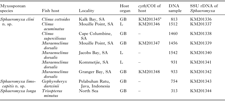
New sequence data on Sphaeromyxa species from this study

a cytb sequence, the rest of sequences in the row are host COI sequences; SA, South Africa; GB, gall bladder; L, biliary ducts in liver.
Processing of samples
Gall bladders and livers of fishes were examined for the presence of myxosporean infections by light microscopy (LM) on Leica DM750 and Olympus BX51 microscope. Plasmodia and spore morphology were documented with Leica DM750 and Olympus DP70 digital camera using differential interference contrast. The spores (n = 20) were measured according to the guidelines of Lom and Arthur (Reference Lom and Arthur1989) using ImageJ v.1_44p software (Wayne Rasband, http://imagej.nih.gov/ij/). Samples were stored in 400 μL of TNES urea buffer (10 mm Tris-HCl with pH 8, 125 mm NaCl, 10 mm EDTA, 0·5% SDS and 4 m urea) or in 90% ethanol for subsequent DNA extraction.
For histological examination, approximately 8 mm3 of liver tissue of M. dorsalis (Mouille Point) was fixed for 24 h in Davidson fixative and stored in 70% ethanol. Samples were routinely dehydrated and embedded into paraffin. Sections were stained by haematoxylin and eosin (H&E) and Giemsa. For examination of fine structure of spores and plasmodia by transmission electron microscopy (TEM), 6 mm3 of liver tissue from C. acuminatus (Mouille Point) was fixed in cacodylate buffered 3% glutaraldehyde at 4 °C, rinsed in 0·1 m cacodylate buffer and postfixed in 1% osmium tetroxide. After graded acetone dehydration series, the samples were embedded in Spurr's resin. Semi-thin sections (1–2 μ m) were stained with toluidine blue and used for additional histological observation. Ultrathin sections (70 nm) were stained with uranyl acetate and lead citrate and examined in a JEOL JEM 1010 electron microscope operating at 80 kV. Images were collected with Megaview II soft paging system using analySIS software.
Host species identification
Most of the fish species were identified using morphological features (Froese and Pauly, Reference Froese and Pauly2014). The morphology-based identification of clinid fish hosts, that all have very similar morphology, was verified by sequencing the taxonomically informative genes cytochrome c oxidase I (COI) or cytochrome b (cytb) using previously described primers (Folmer et al. Reference Folmer, Black, Hoeh, Lutz and Vrijenhoek1994; Boore and Brown, Reference Boore and Brown2000).
DNA extraction, PCR amplification, cloning and sequencing
Total DNA was extracted using a standard phenol–chloroform protocol, after an overnight digestion with proteinase K (50 μg mL−1; Serva, Germany) at 55 °C. DNA was re-suspended in 50–100 μL−1 of DNAse-free water and left to dissolve overnight at 4 °C.
Almost complete SSU rDNA sequences of a Sphaeromyxa species from C. cottoides (Kalk Bay) as well as a Sphaeromyxa species from Gephyroberyx darwinii were obtained using universal eukaryotic ERIB1–ERIB10 primers in the first PCR. Two overlapping fragments of the parasite SSU rDNA were amplified in nested PCR by a combination of ERIB1–ACT1r and Myxgen4F–ERIB10 for a Sphaeromyxa species from C. cottoides or MyxGP2F–ERIB10 for a Sphaeromyxa species from G. darwinii (Barta et al. Reference Barta, Martin, Liberator, Dashkevicz, Anderson, Feighner, Elbrecht, Perkins-Barrow, Jenkins, Danforth, Ruff and Profous-Juchelka1997; Kent et al. Reference Kent, Khattra, Hervio and Devlin1998; Hallett and Diamant, Reference Hallett and Diamant2001; Diamant et al. Reference Diamant, Whipps and Kent2004). The composition of PCR reaction mixture was identical as for the Taq-Purple DNA polymerase protocol described by Bartošová et al. (Reference Bartošová, Fiala, Jirků, Cinková, Caffara, Fioravanti, Atkinson, Bartholomew and Holzer2013). Following PCR cycling conditions were used for the first and nested PCR, respectively: 95 °C 3 min, (95 °C 1 min, 48 °C 1 min, 72 °C 1 min 30 s) 30×, 72 °C 10 min or 95 °C 3 min, (95 °C 1 min, 50 °C 1 min, 72 °C 1 min) 30×, 72 °C 10 min.
Approximately 500 bp of the SSU rRNA gene of additional Sphaeromyxa samples from other clinid fish (Table 1) were amplified by a combination of primers newly designed in this study, i.e. SphmyxSSU520F (5′-AGGTCGTTCCCAMAAGGATTC-3′)–SphmyxSSU1140R (5′-ATCACCCGAAGACAGCAACC-3′). The composition of PCR reaction mixture was identical as for the TITANIUM Taq DNA polymerase protocol described by Bartošová et al. (Reference Bartošová, Fiala, Jirků, Cinková, Caffara, Fioravanti, Atkinson, Bartholomew and Holzer2013). Following PCR cycling conditions were used: 95 °C 3 min, (94 °C 40 s, 57 °C 40 s, 68 °C 50 s) 35×, 68 °C 8 min.
The missing 3′end of SSU rRNA gene sequence of S. longa was amplified from the identical DNA sample (isolate M0313) for which partial SSU and almost complete LSU rDNA data exist in GenBank (NCBI: DQ377691, FJ417072). For this purpose, Myxgen4F–ERIB10 primers were used. The composition of PCR reaction mixture was identical as for the TITANIUM Taq DNA polymerase protocol described by Bartošová et al. (Reference Bartošová, Fiala, Jirků, Cinková, Caffara, Fioravanti, Atkinson, Bartholomew and Holzer2013). Following PCR cycling conditions were used: 95 °C 3 min, (95 °C 50 s, 55 °C 50 s, 68 °C 1 min 40 s) 35×, 68 °C 10 min. The product, which was weakly amplified most probably due to low DNA concentration of parasite in the sample, was re-amplified using identical primers and PCR cycling parameters as in previous PCR.
PCR products were purified using a Gel/PCR DNA Fragments Extraction Kit (Geneaid Biotech Ltd, USA) and sequenced directly. As a mixed sequence was detected for S. longa in the chromatogram of the direct sequencing product, the amplified fragments were cloned into the pDrive Vector with a PCR Cloning Kit (Qiagen, Germany) and transformed into TOP10 chemically competent Escherichia coli cells (Life Technologies, Czech Republic). Plasmid DNA was isolated using a High Pure Plasmid Isolation Kit (Roche Applied Science, Germany) and two colonies were sequenced on ABI PRISM 3130 × 1 automatic sequencer (Applied Biosystems, Czech Republic) in the sequencing facility of the Faculty of Science and BC ASCR.
Alignments and phylogenetic analyses
The SSU rDNA sequences were aligned in MAFFT v6·864b (Katoh et al. Reference Katoh, Misawa, Kuma and Miyata2002) using the L-INS-i multiple alignment method, with a default gap opening penalty (–op = 1·53) and gap extension penalty (–ep = 0·0). The alignment was manually edited in BioEdit v7·0·5·2 (Hall, Reference Hall1999) and the ambiguous sections were removed manually.
The SSU rDNA-based alignment included newly obtained Sphaeromyxa sequences as well data from all Sphaeromyxa species available in GenBank and 12 non-sphaeromyxid members of the hepatic biliary group sensu Kristmundsson and Freeman (Reference Kristmundsson and Freeman2013). Chloromyxum cristatum (NCBI: AY604198; under the synonym Chloromyxum cyprini in GenBank) and Chloromyxum fluviatile (NCBI: GU471264) were used as outgroups.
Maximum parsimony (MP) analysis was performed in PAUP* v4.b10 (Swofford, Reference Swofford2003), using a heuristic search with random taxa addition, the ACCTRAN option, TBR swapping algorithm, all characters treated as unordered, a Ts/Tv ratio of 1:2, and gaps treated as missing data. Maximum likelihood (ML) analysis was performed in RAxML v7·0·3 (Stamatakis, Reference Stamatakis2006) using the GTR + Γ model. Bootstraps were based on 1000 replicates for both analyses. Bayesian inference (BI) analysis was performed in PhyloBayes v3·3.f. (Lartillot et al. Reference Lartillot, Lepage and Blanquart2009) using the CAT-GTR model of evolution. Two intertwined chains were run in parallel for a minimum of 500 cycles. Then, every 100 cycles, the bipartition frequencies and the summary variables were automatically done and the run stopped once all the discrepancies were lower or equal to 0·3 and all effective sizes were larger than 50. Burnin period was equal to one fifth of the total length of the chain.
RESULTS
Four out of the six clinid fish species examined, i.e. C. acuminatus, C. cottoides, C. superciliosus and M. dorsalis were found to be infected with a Sphaeromyxa which we describe as a new species using light microscopical, ultrastructural, histological and molecular data. The morphology of a Sphaeromyxa found in the gall bladder of G. darwinii also did not match any existing nominal Sphaeromyxa species and is described as new species of Sphaeromyxa in this study.
We obtained a novel cytb sequence of C. cottoides (NCBI: KM201345), the host of a Sphaeromyxa species from South Africa, which showed 81% sequence similarity with Abudefduf sparoides (Perciformes: Pomacentridae; NCBI: JQ707166; cytb of clinids missing in GenBank). We also obtained three COI sequences of C. acuminatus (NCBI: KM201346) and M. dorsalis (NCBI: KM201347, KM201348), additional hosts of the abovementioned Sphaeromyxa species, which showed 100% and 98% sequence similarity with their nucleotide GenBank reference data (NCBI: JF493211, JF493914, respectively) and 100 % sequence similarity with their amino acid GenBank reference data (NCBI: AEB16730, AEB17430), respectively (Table 1).
Species descriptions.
Phylum: Cnidaria Hatschek, 1888
Unranked subphylum: Myxozoa Grassé, 1970
Class: Myxosporea Bütschli, 1881
Order: Bivalvulida Schulman, 1959
Suborder: Variisporina Lom et Noble, 1984
Family: Sphaeromyxidae Lom et Noble, 1984
Genus: Sphaeromyxa Thélohan, 1892
Sphaeromyxa clini n. sp.
Type host: Clinus acuminatus, sad klipfish.
Other hosts: Clinus cottoides, bluntnose klipfish; C. superciliosus, super klipfish; M. dorsalis, nosestripe klipfish (Perciformes: Clinidae).
Type locality: South Africa: Mouille Point (Fig. 1).
Other localities: South Africa: Cape Columbine; Jacobs Bay; Kalk Bay; Kommetjie (Fig. 1).
Description of sporogonic stages: Flat polysporic plasmodia with irregular margins, pansporoblasts producing one or predominantly two spores (Fig. 2A and B). Ectoplasm composed of a compact 2·5–3·5 μm thick outer layer and a finely granular 4·5–7·5 μm thick inner layer. Endoplasm vacuolated and loosely connected (Fig. 2B). Plasmodia with small projections on their surface (Fig. 3D and E; more detailed description in ultrastructure section).
Light microscopy pictures and line drawings of Sphaeromyxa spores and plasmodia as seen in Nomarski differential interference contrast. (A–E) Sphaeromyxa clini n. sp. stages from (A–C) M. dorsalis and (D) C. acuminatus. (A) Plasmodium with spores in pansporoblasts; (B) Plasmodium with focus on the ectoplasmic and endoplasmic layers; (C) Myxospore in frontal view; (D) Myxospore in sutural view; (E) Myxospore in frontal (left) and sutural view (right). Myxospores of Sphaeromyxa longa in (F) frontal and (G) sutural view. Sphaeromyxa limocapitis n. sp. myxospores in (H, I) frontal and (H) sutural view. (J) Myxospore in frontal (left) and sutural view (right). Scale bar 5 μ m.

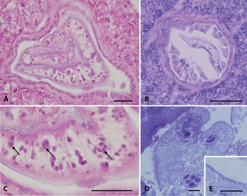
Histological and semi-thin sections of Sphaeromyxa clini n. sp. in the bile ducts of (A–C) M. dorsalis and (D–E) C. acuminatus. (A) H&E stained section of infected bile duct of M. dorsalis completely occluded with the sporogonic plasmodium of S. clini n. sp.; (B) Giemsa stained bile duct of M. dorsalis co-infected by the sporogonic stages of S. clini n. sp. (centrally) and Ceratomyxa sp. (peripherally); (C) Picture detail of the plasmodium with sporogonic stages of S. clini n. sp. (arrows); (D) Sporogonic plasmodium of S. clini n. sp. in the bile duct of C. acuminatus (E) with the detail on plasmodial surface with small projections covered with mucus layer. Scale bars 20 μ m (A–C) and 5 μ m (D–E).

Description of myxospores (measurements from C. acuminatus, C. superciliosus and M. dorsalis): Spores arcuate in frontal view tapering towards the ends (Fig. 2A–E) and slightly sigmoid in sutural view (Fig. 2D and E). Spore length 17·4–20·6 μ m (mean ± s.d. = 18·8 ± 0·9), spore width 4·5–6·1 μ m (5·3 ± 0·4), spore thickness 4·0–6·0 μ m (5·0 ± 0·7). Sutural line relatively indistinct, sigmoid (Fig. 2E). Two elongate oval polar capsules (PCs) with ribbon-like polar filament irregularly folded twice to three times inside each PC (Fig. 2A and C–E). PC length 5·3–6·6 μ m (5·9 ± 0·3), PC width 2·1–3·1 μ m (2·5 ± 0·2). Single binucleated sporoplasm occupying the space between PCs (Fig. 2D and E). Spore ridges not observed by LM. Mature spores were observed from March to April and from October to November.
Localization of sporogonic stages: Coelozoic, gall bladder, liver–bile ducts (Table 1).
Prevalence: 9% of all fish individuals (n = 140); detailed data: C. acuminatus: Mouille Point 60 % (n = 5); C. cottoides: Kalk Bay 6% (n = 18); C. superciliosus: Cape Columbine 14% (n = 7), Jacobs Bay 29% (n = 7); M. dorsalis: Mouille Point 100% (n = 5), Kommetjie 33% (n = 3); the rest of fish examined negative.
Pathology: In histology sections, the biliary ducts completely occluded by plasmodia causing dilatation (Fig. 3A and C).
Materials deposited: DNA samples (Table 1) stored in −80 °C, paraffin block No. 363 and block in resin nr. AK005 stored in the Laboratory of Fish Protistology, Institute of Parasitology, BC ASCR.
Molecular data: Seven SSU rDNA sequences (NCBI: KM201336–KM201342; Table 1) with 100% sequence identity in the overlapping region. Intragenomic variability: one polymorphic site (A/G) at position 895 (related to the almost complete sequence NCBI: KM201336). The almost complete sequence of S. clini n. sp. (NCBI: KM201336) was used for the phylogenetic analyses. Clusters within the ‘incurvata’ group of the Sphaeromyxa clade.
Ultrastructure: Surface of the plasmodium extends in fine projections ranging about 130–350 nm (Fig. 4A, B and D–F), covered with a double cell membrane (Fig. 4E). Cell membrane frequently invaginated to form small (approximately 100 nm) pinocytotic vesicles which were observed also deeper in the ectoplasm (Fig. 4B and C). Network of mucus up to 450 nm thick layer covering the plasmodial projections (Fig. 4A, B, D, and F). Approximately 1 μ m thick ectoplasmic layer (Fig. 4B and F) containing small pinocytotic vesicles (50–80 nm). Endoplasm additionally filled with electron-dense granules of variable size (0·5–1·0 μ m), lamellar structures, mitochondria and sporoblasts (Fig. 4F). Two secondary cells in a primary cell observed as the earliest developmental stages of sporogony (Fig. 4F and G). Pansporoblasts harbouring a single developing spore (Fig 4I). During capsulogenesis, PC primordium filled with granular substance and attached to the transversely striated external tube (Fig. 4H). Apex of the maturing polar capsule covered with an electron-dense cap (stopper-like structure) in the preexisting channel for polar filament discharge; three folds of the polar filament observed in the longitudinal section of PC (Fig. 4I). Polar filament with dark lumen and bright surface (Fig. 4I). Shell valves of the maturing spore having much darker cytoplasm than the cytoplasm of surrounding sporoblast. Spore most probably possessing ridges – these could not be clearly distinguished in our material due to the absence of mature spores in the examined plasmodium. Desmosome-like junction of the suture discernible between valve cells (Fig. 4I).
Ultrastructure of Sphaeromyxa clini n. sp. infection in the bile ducts of C. acuminatus. (A) Contact zone of the plasmodium with host bile duct endothelium; (B) Plasmodium containing pinocytotic vesicles and dense granules with (C) detail of pinocytotic vesicles in the membrane and outer layer of the endoplasm; (D) Projections and mucus layer on the surface of plasmodium with (E) detail of double cell membrane of plasmodium; (F) Plasmodium with projections and mucus layer on its surface; dense granules and early sporoblasts present in the endoplasm; (G) Early sporogonic stage with two secondary cells in the primary cell; (H) Immature spore with polar capsule primordium filled with granular substance and transversely striated external tube; (I) Monosporic pansporoblast containing maturing spore with polar capsule covered with an electron-dense cap and filled with polar filament folded three times and connected to the discharge canal; valve cells connected by desmosome cell junction. Abbreviations: DC, electron-dense cap; CD, channel for polar filament discharge; EN, endoplasm; EC, ectoplasm; ET, external tube; EB, endothelium of bile duct; M, mucus layer; PCa, polar capsule; PCe, primary cell; PF, polar filament; PP, plasmodial projections; PV, pinocytotic vesicle; SC, secondary cell; SP, Sphaeromyxa plasmodium; VJ, valve cell junction. Scale bar 1 μ m (A, B, D) and 2 μ m (F–I).

Etymology: clini refers to the name ‘Clinidae’ of fish family where both of its fish host genera belong.
Differential diagnosis: South African S. clini n. sp. is most similar to S. noblei described from a different clinid fish H. whiteleggii from Australia (Lom, Reference Lom2004). Besides host species and locality, they differ by a number of morphological features, i.e. spore shape (less arcuate in S. clini n. sp.) and mean length (18·8 μ m in S. noblei vs 20 μ m in S. clini n. sp.), thickness and structure of mucus layer on the plasmodial surface (thicker and ramified in S. noblei vs thinner and less branched in S. clini n. sp.), and thickness of plasmodial ectoplasm (15 μ m in S. noblei vs 1 μ m and 7–11 μ m in S. clini n. sp. as observed by TEM and LM, respectively) (Table 2). The comparison of the thickness of ectoplasm layer has to be taken with caution, as it may vary with plasmodial size and development, i.e. the small unsporulated plasmodia from the liver ducts may not be readily comparable with large plasmodia from the gall bladder. Moreover, as deduced from the TEM figures in Lom (Reference Lom2004), the thickness of ectoplasm of S. noblei appears to be 5 μ m rather than 15 μ m as referred in the text. Sphaeromyxa clini n. sp. has several additional characteristics which most probably remained unnoticed or are absent in S. noblei; more specifically wider tissue specificity (bile ducts additionally to gall bladder), double-layer plasmodial membrane and formation of pinocytotic vesicles.
Comparison of Sphaeromyxa clini n. sp. with morphologically similar members of the ‘incurvata’ group

NG, spore thickness not given in the literature.
Sphaeromyxa clini n. sp. differs considerably from Sphaeromyxa sp. from the South African clinid Pavoclinus graminis by more arcuate longer spores with longer PCs (Reed et al. Reference Reed, Basson and Van As2009). Spores of another species from intertidal blennies, S. tripterygii, are comparable in their length with S. clini n. sp.; however, they are relatively slender with truncate ends and narrower PCs (Laird, Reference Laird1953). Other two Sphaeromyxa species from the tidal blennies, S. gibbonsia and S. incurvata, have longer spores and larger ovoid or pyriform PCs than S. clini n. sp. (Doflein, Reference Doflein1898; Kudo, Reference Kudo1919; Noble Reference Noble1939) (Table 2).
The phylogenetically most closely related species, Sphaeromyxa hellandi, resembles to S. clini n. sp. in spore shape but differs by longer spores and longer PCs, host specificity and geographic distribution (Kalavati and MacKenzie, Reference Kalavati and MacKenzie1999). Other species of the ‘incurvata’ clade, Sphaeromyxa lycodi Kristmundsson et Freeman, 2013 and Sphaeromyxa kenti Whipps et Font, 2013, differ from S. clini n. sp. by possessing more arcuate spores, longer PCs, host species spectrum and geographic distribution. Sphaeromyxa kenti has similar spore dimensions as S. clini n. sp. whereas spores of S. lycodi are larger (Kristmundsson and Freeman, Reference Kristmundsson and Freeman2013; Whipps and Font, Reference Whipps and Font2013). Other arcuate species, Sphaeromyxa arcuata Fantham, Reference Fantham1930, Sphaeromyxa elegini Dogiel, 1948, Sphaeromyxa exneri Awerinzew, 1913, Sphaeromyxa curvula Fantham, Reference Fantham1930, S. incurvata Doflein, Reference Doflein1898, Sphaeromyxa maiyai Morrison et Pratt, 1973, and Sphaeromyxa sabrazesi Laveran et Mesnil, 1900 differ from S. clini n. sp. by their geographic distribution, host species, spore size and shape of PCs (Doflein, Reference Doflein1898; Kudo, Reference Kudo1919; Fantham, Reference Fantham1930; Shulman, Reference Shulman1966; Morrison and Pratt, Reference Morrison and Pratt1973; Aseeva, Reference Aseeva2002). Moreover, some of them possess specific features atypical for Sphaeromyxa species, i.e. disporic plasmodia in S. elegini and absence of mucus on the plasmodial membrane in S. maiyai (Shulman, Reference Shulman1966; Morrison and Pratt, Reference Morrison and Pratt1973; Aseeva Reference Aseeva2002) (Table 2).
Other remarks: Co-infections of S. clini n. sp. and Ceratomyxa sp. sporogonic stages observed in the infected tubules (Fig. 3B).
Sphaeromyxa limocapitis n. sp.
Type host: G. darwinii, Darwin's slimehead (Beryciformes: Trachichthyidae).
Type locality: Fish market at Pelabuhan Ratu, Java, Indonesia (6°59′02″N, 106°32′37″E).
Description of sporogonic stages: Not observed.
Description of myxospores: Spores straight to slightly curved in frontal view (straight on one side and slightly convex on another side) tapering towards the pointed ends (Fig. 2H–J); spore straight in sutural view (Fig. 2H and J). Spore length 26·7–30·1 μ m (27·7 ± 1·3), spore width 5·6–6·7 μ m (6·1 ± 0·4), spore thickness 4·1–5·7 μ m (5·1 ± 0·7). Sutural line slightly sigmoid (Fig. 2J). Two elongate oval PCs with ribbon-like polar filament irregularly folded four times inside each PC (Fig. 2H–J). PC length 6·0–7·8 μ m (6·8 ± 0·6), PC width 2·7–3·5 μ m (3·2 ± 0·2). Single binucleated sporoplasm occupying the space between PCs (Fig. 2I and J). Spore ridges not observed.
Localization of sporogonic stages: Coelozoic, gall bladder.
Prevalence: 100% (n = 1).
Materials deposited: DNA samples (Table 1) stored in −80 °C in the Laboratory of Fish Protistology, Institute of Parasitology, BC ASCR.
Molecular data: SSU rDNA sequence (NCBI: KM201343; Table 1). Clusters in a separate lineage of the Sphaeromyxa clade.
Etymology: limocapitis refers to the English name ‘slimehead’ of the family Trachichthyidae where G. darwinii belongs.
Differential diagnosis: Sphaeromyxa limocapitis n. sp. described from the Central Indo-Pacific and possessing straight/slightly curved spores with pointed ends has an uncommon spore shape rather similar to certain marine Myxidium spp. than to most of sphaeromyxids. From the ten Sphaeromyxa spp. reported from the Western Indo-Pacific (Bay of Bengal) and Central Indo-Pacific (the coast of Borneo Island), only Sphaeromyxa bengalensis Sarkar, Reference Sarkar2010, Sphaeromyxa diacanthusa Sarkar, Reference Sarkar2004 and Sphaeromyxa lomi Moser et Noble, Reference Moser and Noble1977 have similar fusiform spores as S. limocapitis n. sp. However, they differ from the new sphaeromyxid by host species, locality and by having (i) smaller striated spores with bluntly rounded ends (S. diacanthusa) or rounded/truncated spore ends (S. lomi) or (ii) spores with moderately truncated ends and spirally coiled polar filament hence with overall morphology similar to Myxidium morphotype (S. bengalensis) (Sarkar Reference Sarkar2004; Sarkar, Reference Sarkar2010 and others reviewed in Sarkar, Reference Sarkar1999 and Lom, Reference Lom2004). Sphaeromyxa magna Zhukov, 1964 and Sphaeromyxa reinhardti Jameson, Reference Jameson1929 having similar morphology as S. limocapitis n. sp. but described from different marine realm (i.e. Arctic and Temperate Northern Pacific, respectively), differ from the new species by smaller spores with truncated ends, host specificity and distribution (Jameson Reference Jameson1929; Shulman Reference Shulman1966).
Phylogenetic analyses
The strongly supported Sphaeromyxa clade (node A in Fig. 5A) clustering within the freshwater myxosporean lineage split into (i) the ‘incurvata’ group, (ii) the ‘balbianii’ group and (iii) S. limocapitis n. sp. lineage. Sphaeromyxa clini n. sp. clustered with high nodal support sister to the clade composed of S. hellandi sequences within the ‘incurvata’ group. Sphaeromyxa longa (Fig. 2F and G), for which additional 765 bp at its 3′end were newly sequenced in this study (NCBI: KM201344), clustered with strong nodal support with Sphaeromyxa artedielli Karlsbakk, Einen et Bartošová, 2013 within the ‘balbianii’ group (Fig. 5A). The new species S. limocapitis n. sp. created a separate lineage which was (i) positioned at the base of the Sphaeromyxa clade in the ML analysis (Fig. 5A and B left), (ii) unresolved within the Sphaeromyxa clade in the MP consensus tree (Fig. 5B middle) or (iii) positioned sister to the ‘balbianii’ group in the BI analysis (Fig. 5B right).
Phylogeny of myxosporeans focused on the Sphaeromyxa clade. (A) Maximum likelihood (ML) tree showing the clustering of Sphaeromyxa species with new sequence data within the Sphaeromyxa clade. Chloromyxum fluviatile and Chloromyxum cristatum were selected as outgroups, their common branch shortened to 50% of original length. Newly sequenced taxa in bold. Bold branches lead to a node with a 100% ML/maximum parsimony (MP) bootstrap support and 1·00 Bayesian inference (BI) posterior probability. Dashes indicate bootstrap values <50 or node not present in the MP or BI tree. PCs, polar capsules; (B) Variable positioning of S. limocapitis n. sp. within the Sphaeromyxa clade in the ML, MP and BI analyses. Nodal supports (even <50) shown for nodes A and B (defined in Fig. 5A). IG, ‘incurvata’ group; BG, ‘balbianii’ group; LL, ‘limocapitis’ lineage.

DISCUSSION
Sphaeromyxa clini n. sp. from South African C. acuminatus, C. cottoides, C. superciliosus and M. dorsalis (this study) was found to be most similar to S. noblei described from H. whiteleggii from Australia (Lom, Reference Lom2004) by overall shape of their spores and plasmodia. However, both species possess several differences in their morphology, host species and geographic distribution. Future sequencing of S. noblei from its type host and type locality and its subsequent comparison with the sequence data obtained for S. clini n. sp. in this study can help to ascertain if the observed differences are expressed as species-specific features or are due to variability in one species (S. noblei). The second option would be reasoned by low host specificity and a wide distribution reported for a number of Sphaeromyxa spp. (Lom, Reference Lom2004) and by evolutionary implications. More specifically, even though the abovementioned clinid fish hosts of S. clini n. sp. and S. noblei are restricted to different biogeographic realms (temperate Southern Africa vs temperate Australasia; Spalding et al. Reference Spalding, Fox, Allen, Davidson, Ferdaña, Finlayson, Halpern, Jorge, Lombana, Lourie, Martin, McManus, Molnar, Recchia and Robertson2007), they are closely related phylogenetically (Lin and Hastings, Reference Lin and Hastings2013). Clinids have an ancestral distribution in the temperate region and successfully crossed intervening tropical regions during their evolutionary history (Lin and Hastings, Reference Lin and Hastings2013). The common ancestor of clinids lived some 20 Ma ago (Lin and Hastings, Reference Lin and Hastings2013) when continents had almost the same position as at present, i.e. Australia was placed a bit more south from its current position. Therefore, we assume these clinid fish may possibly share a similar parasite fauna inherited from their ancestor.
Regarding the intraspecies variability, no sequence differences were observed in S. clini n. sp. isolates from different fish species and localities even though oceanographic barriers do affect the population structuring of South African intertidal fishes (Von der Heyden et al. Reference von der Heyden, Bowie, Prochazka, Bloomer, Crane and Bernardi2011) along with their parasites, as for example shown for a myxosporean Ceratomyxa cottoidi closely associated with its host C. cottoides (Reed et al. Reference Reed, Basson, Van As and Dyková2007, Reference Reed, Bartošová, Fiala and Dyková2011).
As for the fish host identification of S. clini n. sp., the 2% sequence divergence observed between the COI nucleotide sequence of M. dorsalis from Moullie Point and GenBank reference data of M. dorsalis from Haga Haga and Wooleys Pool (both South Africa) is due to the cryptic species nature of the fish host resulting from phylogeographic breaks (Von der Heyden et al. Reference von der Heyden, Bowie, Prochazka, Bloomer, Crane and Bernardi2011).
The Sphaeromyxa species, which are all parasites of marine fish, cluster with marine Myxidium species, i.e. Myxidium coryphaenoidium Noble, Reference Noble1966 and Myxidium baueri Kovaljova & Gaevskaya, 1982, within the freshwater myxosporean lineage (Fiala, Reference Fiala2006; Kalavati et al. Reference Kalavati, Mackenzie, Collins, Hemmingsen and Brickle2013). Sphaeromyxa species, especially S. limocapitis n. sp. forming a separate lineage of the Sphaeromyxa clade, share many morphological similarities with phylogenetically closely related M. coryphaenoidium and M. baueri as well as with other morphologically similar myxidiids without sequence data in GenBank i.e. Myxidium bajacalifornium Noble, Reference Noble1966, Myxidium melanocetum Noble, Reference Noble1966, Myxidium melanostigmum Noble, Reference Noble1966 and Myxidium noblei Konovalov, 1966 (Noble, Reference Noble1966; Zubchenko and Krasin, Reference Zubchenko and Krasin1980). Besides similar fusiform/spindle shaped spores (straight with tapering ends) and infecting the gall bladder of marine fish, the abovementioned Myxidium spp. possess rough polar filament (Zubchenko and Krasin, Reference Zubchenko and Krasin1980) which is heavy at its proximal end and becoming thinner and more tightly coiled at distal end (Noble, Reference Noble1966). Sphaeromyxa species also have a thick polar filament with a broad base, however being zig-zag folded and not spirally coiled as in Myxidium species. This morphological, biological and molecular evidence strongly corresponds with the hypothesis that Sphaeromyxa species had evolved by a change in the character of polar filament from the spindle-shaped Myxidium ancestor, which then returned to the marine environment (Fiala and Bartošová, Reference Fiala and Bartošová2010; Kristmundsson and Freeman, Reference Kristmundsson and Freeman2013). Our phylogenetic analyses indicate that the sphaeromyxids then diverged into species still possessing ancestral characters (the ‘limocapitis’ lineage with straight/slightly curved fusiform spores, pointed spore ends and elongate oval PCs) or expressing evolutionary novelties (the ‘incurvata’ group with arcuate or slightly curved spores, rounded spore ends, smooth or finely striated twisted valves and elongate oval or pyriform PCs; the ‘balbianii’ group with straight fusiform or ovoid spores, truncate spore ends, striated valves and ovoid PCs). Thus, S. limocapitis n. sp. may represent a missing link in the evolution of sphaeromyxids.
Herein, we describe a Sphaeromyxa species parasitizing the deep sea trachichthyid fish, S. limocapitis n. sp., based on new morphological and sequence data. We also provide morphological and sequence data for a newly described species S. clini n. sp. from clinid fishes. Further sequencing of additional Sphaeromyxa species, especially of those with unique spore morphologies, may reveal the existence of other phylogenetic clades within this myxosporean group and thus provide more insights into Sphaeromyxa radiation. Moreover, future study of the life cycles of Sphaeromyxa representatives, for which the invertebrate host remains unknown, may help to clarify why these parasites of marine fish cluster within the clade encompassing parasites which mostly alternate between freshwater fish and oligochaetes.
ACKNOWLEDGEMENTS
The authors would like to thank Drs Ivan Fiala, Miloslav Jirků and Astrid S. Holzer (Institute of Parasitology, Biology Centre of the ASCR, České Budějovice, Czech Republic) for the help with manuscript corrections. The authors thank Dr Ivan Fiala for providing DNA sample and pictures of S. longa and to Dr Milan Říha (Institute of Hydrobiology, Biology Centre of the ASCR, České Budějovice, Czech Republic) and Dr Harry Palm (University of Rostock, Rostock, Germany) for help with sampling of the fish in Indonesia. The authors also thank the three anonymous referees for their valuable comments.
FINANCIAL SUPPORT
This work was supported by the Czech Science Foundation (Centre of Excellence 505/12/G112), The National Research Foundation (Thuthuka) of South Africa (TTK200705020002008) and the University of Cape Town Research Committee.