Introduction
Cholesteatoma is an accumulation of squamous epithelium and keratin debris within the middle ear and mastoid bone. Multiple theories exist as to its aetiology and pathogenesis. One of the more widely accepted theories is that in the acquired form, the disease arises due to retraction of the tympanic membrane, which is predisposed by altered middle ear ventilation and chronic otitis media (COM), or can result from iatrogenic insertion following otological surgery.Reference Kennedy and Singh1 Symptoms include otorrhoea, tinnitus, hearing loss, vertigo and, if more progressive, facial weakness and intracranial infections.Reference Basa, Levi, Field and O’Reilly2 The progression of cholesteatoma results in an inflammatory process and subsequent injury to surrounding structures, including the ossicles, lateral semicircular canal (LSCC), tegmen tympani and facial nerve canal. Failure to control related infection can lead to fatal complications, such as meningitis, abscesses and cavernous sinus thrombosis.Reference Kuo, Shiao, Yung, Sakagami, Sudhoff and Wang3
The mainstay of definitive management is surgical excision with a form of tympanomastoid procedure. Surgeons are assisted through pre-operative imaging modalities, including high-resolution computed tomography (HRCT), to guide their approach to the procedure and estimate the extent of the disease. However, in recent years, the question of whether the use of HRCT is essential in the pre-surgical assessment of uncomplicated cholesteatoma has been questioned.Reference Stefanescu, Balica, Motoi, Grigorita, Georgescu and Iovanescu4 High-resolution computed tomography has limitations; for example, it does not reliably differentiate between cholesteatoma and other soft tissue structures, and therefore, some studies suggest it cannot accurately demonstrate the extent of disease. Furthermore, there have been reports that it overestimates cholesteatoma growth.Reference Schwartz, Lane, Bolster and Neff5, Reference Razek, Ghonim and Ashraf6
This systematic review aims to examine the currently available evidence as to the reliability of HRCT to detect cholesteatoma extension and damage to the ossicles, LSCC, tegmen tympani and the facial nerve.
Materials and methods
Registration and databases searched
This systematic review was registered on PROSPERO (ID: CRD42023409051). An initial search of the Medline, Embase and Cochrane databases was conducted on 2 October 2023 and repeated on 17 December 2024 using the following search terms: CT or Computed tomography or HRCT or High-resolution computed tomography AND Cholesteatoma or Chronic squamous otitis media or CSOM or attico-antral disease or surgical findings or middle ear disease.
Inclusion and exclusion criteria
Inclusion criteria were observational studies of human adults aged over 18 which directly compared CT/HRCT findings to intra-operative findings in those diagnosed with cholesteatoma. Exclusion criteria included any other imaging modality, studies using artificial intelligence, paediatric populations, congenital cholesteatomas, reviews or animal studies. The publication date was limited to studies published after 2013 to limit the variation in CT resolution and quality.
Outcomes
The primary outcome measure was concordance between pre-operative CT findings and intra-operative surgical findings. Secondary outcome measures were whether the timing between CT and surgery, the age of the patient, the severity of disease and the severity of clinical symptoms for the patient had any impact on this.
Data extraction and synthesis
Study results were reviewed by GF and SP. Studies were assessed against the inclusion and exclusion criteria listed above. Data extraction was performed in Excel (Microsoft, Redmond, Washington, USA). Both qualitative and quantitative analyses were used due to the varied reporting of outcomes. All studies were assessed for bias using the JBI bias tools.Reference Aromataris, Lockwood, Porritt, Pilla and Jordan7
Results
Study selection
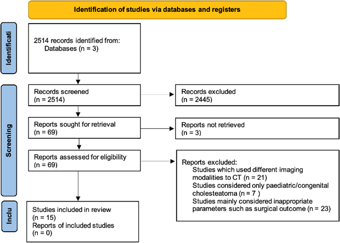
Figure 1 shows the PRISMA flowchart for the selection of the papers for this review. The initial search identified 2514 abstracts, which were screened; only 69 studies were considered to contain data relevant to the aim of this review and were therefore reviewed in full. Of these, 14 studies met the inclusion criteria and were selected for analysis. One additional study was selected for inclusion from a reference list.
PRISMA flowchart.

Study characteristics
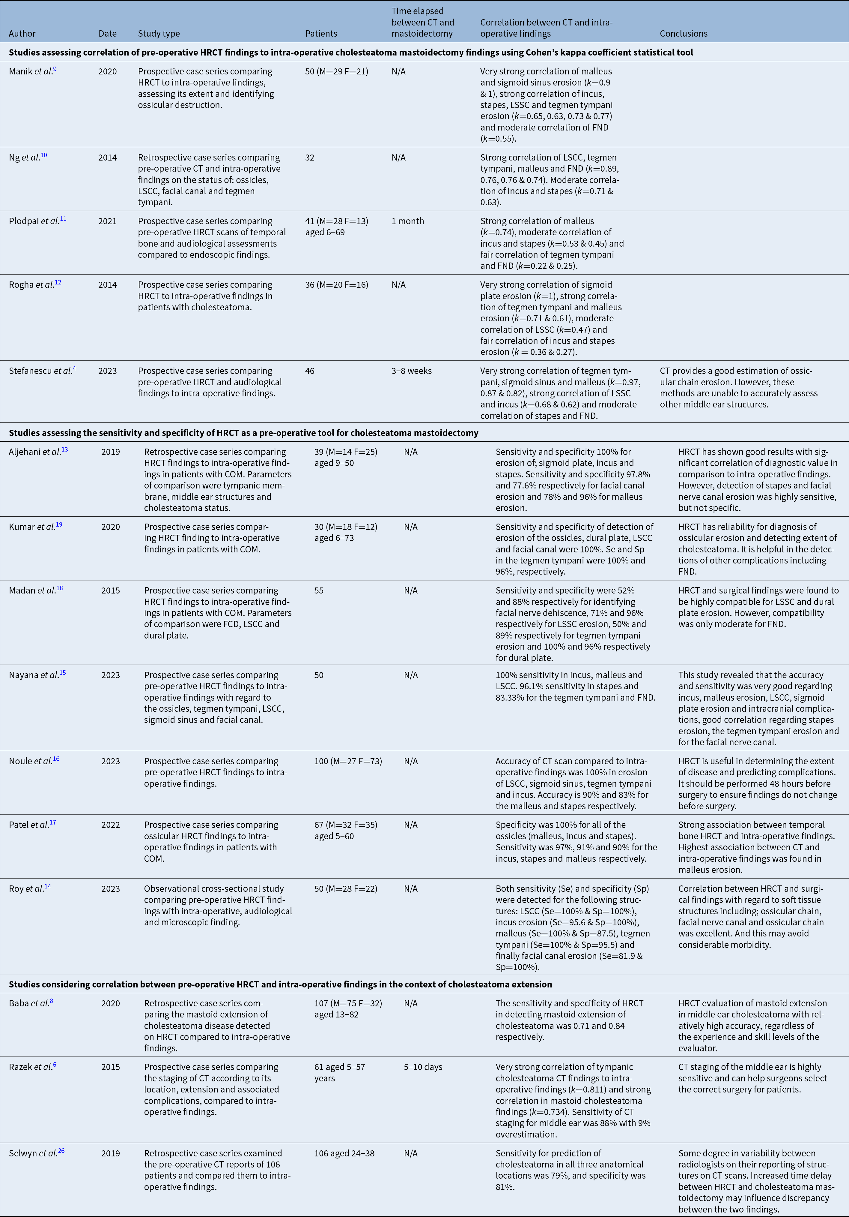
A summary of study characteristics can be found in Table 1. Overall, 15 studies which considered the correlation between HRCT findings and intra-operative findings of patients with cholesteatoma were included. Fourteen studies were case series – nine were prospective, five were retrospective. A single study was an observational cross-sectional study. All studies evaluated the accuracy of HRCT in detecting damage to middle ear structures by using statistical tools, including Cohen’s kappa coefficient, AC1 or measuring sensitivity and specificity, except Baba et al.,Reference Baba, Kurihara, Ogihara, Matsushima, Yamauchi and Ikeda8 which considered the extension of the cholesteatoma instead.
Study characteristics and outcomes; CT = computed tomography; HRCT = high-resolution computed tomography; FND = facial nerve dehiscence; LSCC = lateral semicircular canal; COM = chronic otitis media; Se = sensitivity; Sp = specificity

The ossicles
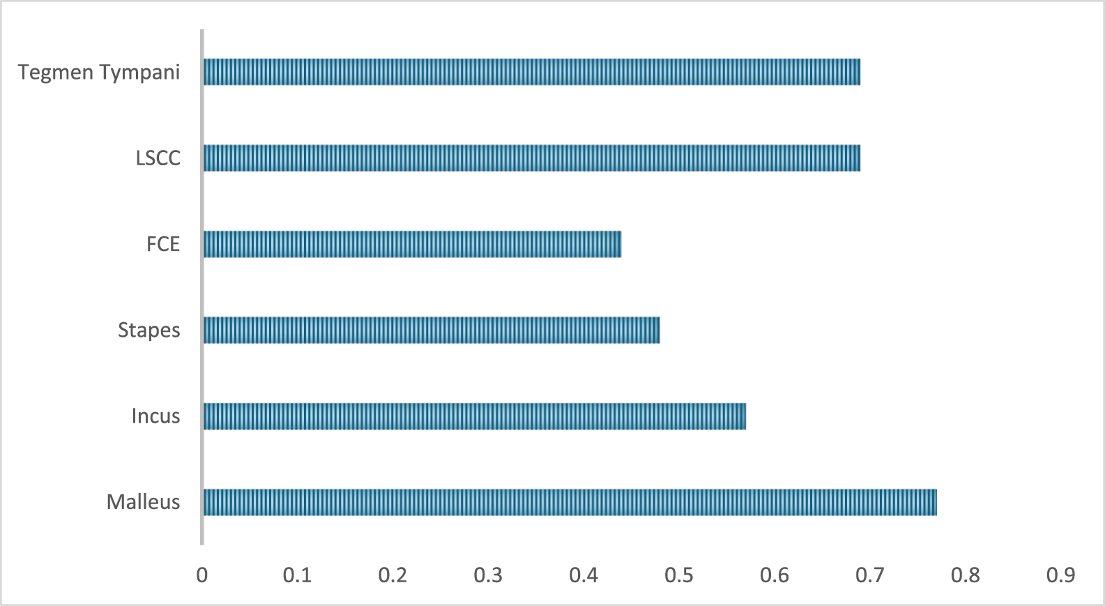
Five articles considered the correlation between ossicular erosion reported in pre-operative HRCT and ossicular erosion observed intra-operatively.Reference Stefanescu, Balica, Motoi, Grigorita, Georgescu and Iovanescu4, Reference Manik, Dabholkar, Bhalekar, Velankar, Chordia and Saberwal9–Reference Rogha, Hashemi, Mokhtarinejad, Eshaghian and Dadgostar12 These studies used Cohen’s kappa coefficient to assess the level of correlation between the two parameters, with the average being 0.76 (Strong). The average coefficient for each anatomical area can be seen in Figure 2, with study-level coefficients tabulated in Supplementary Table 1.
Bar graph showing the average kappa correlation value for middle ear structures from five studies.

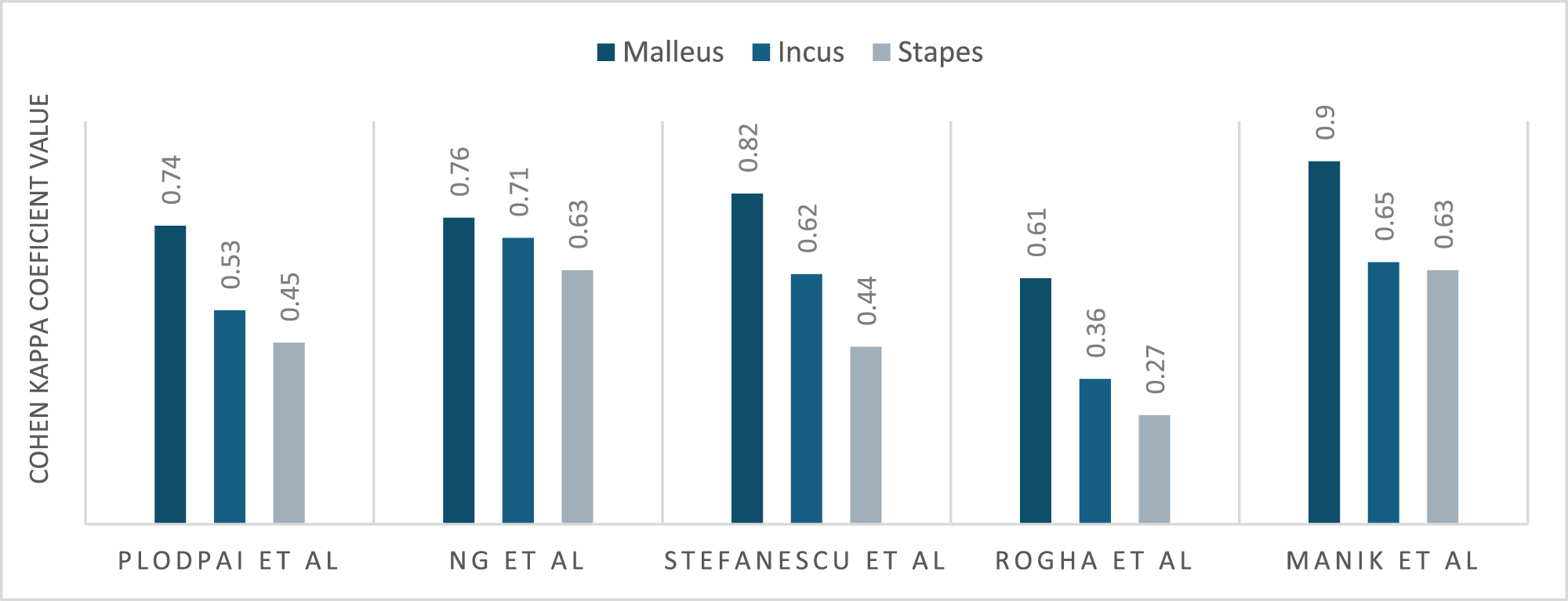
Each study found the correlation between malleus erosion in HRCT compared to intra-operative findings to be either very strong or strong. Figure 3 shows the grouped bar graph showing the correlation of ossicular erosion on HRCT compared to intra-operative findings at mastoidectomy. The extent of the erosion of the malleus showed the highest level of agreement in all studies compared to the other ossicles. In contrast, the extent of the erosion of the stapes showed the lowest level of agreement in all studies, ranging from fair to strong agreement.
Grouped bar graph illustrates the correlation of ossicular erosion on HRCT compared to intraoperative findings in cholesteatoma mastoidectomy.

Seven studies considered the sensitivity and specificity of HRCT as an imaging modality for assessing erosion of the ossicles. These studies demonstrated that HRCT has a high level of sensitivity for detecting erosion of the ossicles, with variation between individual ossicles. These studies demonstrated an average (weighted for the number of study participants) sensitivity for malleus erosion of 95 per cent, incus erosion of 99 per cent and stapes erosion of 96 per cent.Reference Aljehani and Alhussini13–Reference Kumar, Vudumula and Maghiben19
Facial nerve dehiscence
Five studies considered the correlation between facial nerve dehiscence (FND) reported in pre-operative HRCT and FND observed intra-operatively.Reference Stefanescu, Balica, Motoi, Grigorita, Georgescu and Iovanescu4, Reference Manik, Dabholkar, Bhalekar, Velankar, Chordia and Saberwal9, Reference Plodpai, Jongthitinon, Atchariyasathian, Pitathawatchai, Hirunpat and Kirtsreesakul11, Reference Rogha, Hashemi, Mokhtarinejad, Eshaghian and Dadgostar12 The level of agreement between these two parameters was between fair and strong, with an average coefficient of 0.44 (Moderate). Five studies considered the sensitivity and specificity of HRCT as an imaging modality for assessing erosion of the facial canal. These studies demonstrated an average sensitivity and specificity of 83 per cent and 92 per cent respectively, weighted for the number of study participants per study.Reference Aljehani and Alhussini13–Reference Nayana, Anusha, Apoorva and Khan15, Reference Madan, Turamanlar, Bucak, Acay, Gönül and Yıldız18, Reference Kumar, Vudumula and Maghiben19
Lateral semicircular canal
Four studies considered the correlation between LSCC erosion reported in pre-operative HRCT and LSCC erosion observed intra-operatively.Reference Stefanescu, Balica, Motoi, Grigorita, Georgescu and Iovanescu4, Reference Manik, Dabholkar, Bhalekar, Velankar, Chordia and Saberwal9, Reference Ng, Zhang, Soon, Tan, Tan and Mok10, Reference Rogha, Hashemi, Mokhtarinejad, Eshaghian and Dadgostar12 The agreement between these two parameters was between moderate and very strong, with an average coefficient of 0.69 (Strong). Five studies considered the sensitivity and specificity of HRCT as an imaging modality for assessing erosion of the LSCC.Reference Roy, Maji, Dhir and K14–Reference Noule, Samala, Yenganti, Thejas and Ejanthkar16, Reference Madan, Turamanlar, Bucak, Acay, Gönül and Yıldız18, Reference Kumar, Vudumula and Maghiben19 The average sensitivity and specificity were 93 per cent and 99 per cent respectively, weighted for the number of participants per study.
Tegmen tympani
Five studies considered the correlation between tegmen tympani erosion reported in pre-operative HRCT and tegmen erosion observed intra-operatively.Reference Stefanescu, Balica, Motoi, Grigorita, Georgescu and Iovanescu4, Reference Manik, Dabholkar, Bhalekar, Velankar, Chordia and Saberwal9–Reference Rogha, Hashemi, Mokhtarinejad, Eshaghian and Dadgostar12 The level of agreement between the two parameters was between fair and very strong, with an average coefficient of 0.68 (Strong). Five studies considered the sensitivity of HRCT as an imaging modality for assessing erosion of the tegmen tympani.Reference Roy, Maji, Dhir and K14–Reference Noule, Samala, Yenganti, Thejas and Ejanthkar16, Reference Madan, Turamanlar, Bucak, Acay, Gönül and Yıldız18, Reference Kumar, Vudumula and Maghiben19 These studies demonstrated an average sensitivity and specificity of 83 per cent and 95 per cent, respectively.
Cholesteatoma extension
Baba et al. examined the correlation between cholesteatoma extension detected with HRCT and intra-operative findings.Reference Baba, Kurihara, Ogihara, Matsushima, Yamauchi and Ikeda8 They compared the distance of cholesteatoma extension between two groups: surgically mastoid positive for cholesteatoma and surgically mastoid negative for cholesteatoma. The distance of cholesteatoma extension on HRCT in the positive group was significantly longer than in the negative group. They found HRCT can determine the extension of cholesteatoma with moderate sensitivity (71%) and specificity (84%). Razek et al. compared the correlation between HRCT findings and intra-operative findings in cases of mastoid cholesteatoma and tympanic cavity cholesteatoma.Reference Razek, Ghonim and Ashraf6 The correlation was stronger for tympanic cavity cholesteatoma (k = 0.811), than for mastoid cholesteatoma (k = 0.734).
Risk of bias
The risk of bias assessment is presented in Supplemental Tables Two and Three. The studies had a low risk of bias. In most cases, studies were prospective with well-defined inclusion and exclusion criteria, with participants entering at a similar stage in their disease progression. Another element that increased the risk of bias was variation in the quality of HRCT, with each study using different manufacturers of imaging devices and different HRCT slice thicknesses.
Discussion
To the best of our knowledge, previous studies have not synthesised data comparing temporal bone CT findings and intra-operative findings in cholesteatoma mastoidectomy. The studies included in this review looked at the correlation between these two parameters in the context of the erosion of different middle ear structures, including the ossicles, facial canal, LSSC and tegmen tympani. In addition, Baba et al. and Razek et al. considered this with regard to the extension and staging of the cholesteatoma, respectively and discussed the establishment of a new classification system for cholesteatoma extension and severity.Reference Razek, Ghonim and Ashraf6, Reference Baba, Kurihara, Ogihara, Matsushima, Yamauchi and Ikeda8
The ossicles are commonly damaged by the disease processes of cholesteatoma.Reference Gaurano and Joharjy20 Malleus erosion showed the highest average level of correlation (k = 0.77) between CT and intra-operative findings in five studies which used Cohen’s kappa coefficient.Reference Stefanescu, Balica, Motoi, Grigorita, Georgescu and Iovanescu4, Reference Manik, Dabholkar, Bhalekar, Velankar, Chordia and Saberwal9, Reference Plodpai, Jongthitinon, Atchariyasathian, Pitathawatchai, Hirunpat and Kirtsreesakul11, Reference Rogha, Hashemi, Mokhtarinejad, Eshaghian and Dadgostar12 Stapes erosion had the lowest level of correlation in all studies (k = 0.48). The stapes, being the smallest of the ossicles, may be masked by the surrounding soft tissue density of cholesteatoma, making erosion of the bone harder to detect on HRCT in comparison to the malleus and incus.Reference Gaurano and Joharjy20 Therefore, erosive detection on CT may correlate with the size of the ossicular bone, with the malleus being the largest, followed by the incus and the stapes. Despite variations between the ossicles, other studies included in this review demonstrated that HRCT detected erosion at an average sensitivity of 94 per cent, 99 per cent and 96 per cent for the malleus, incus and stapes, respectively. This demonstrates that HRCT is effective in identifying ossicular erosion.Reference Razek, Ghonim and Ashraf6
Five studies also considered the correlation of HRCT findings to facial nerve erosion.Reference Stefanescu, Balica, Motoi, Grigorita, Georgescu and Iovanescu4, Reference Manik, Dabholkar, Bhalekar, Velankar, Chordia and Saberwal9–Reference Rogha, Hashemi, Mokhtarinejad, Eshaghian and Dadgostar12 With an average kappa value of 0.55, the correlation of facial canal erosion on HRCT to intra-operative findings was lower than any other middle ear structure studied (Figure 3). Sensitivity and specificity were 82 per cent and 93 per cent, respectively, for FND detection. A proposed cause for this is the challenge in identifying FND in the vertical part of the facial canal due to the close proximity of the inflamed mastoid air cells.Reference Manik, Dabholkar, Bhalekar, Velankar, Chordia and Saberwal9 In addition, Stefanescu et al. argue that HRCT cannot reliably differentiate between cholesteatoma and granulation tissue, meaning detection of cholesteatoma may be unreliable.Reference Stefanescu, Balica, Motoi, Grigorita, Georgescu and Iovanescu4 It is useful to pre-operatively be aware of episodes of FND, as the nerve is at increased risk of injury either directly or indirectly through the use of local anaesthetic agents or displaced implants within the middle ear. Moody et al. showed that in 416 cases of cholesteatoma, 80 per cent had confirmed FND based on intra-operative findings. Identifying FND on HRCT may influence the approach to the tympanomastoidectomy and would influence the urgency of the surgery, particularly with any prior history of facial weakness.Reference Moody and Lambert21 The incidence of FND as a result of cholesteatoma disease has decreased from 27.7 per cent in 1998 to 17.7 per cent in 2018, likely due to the earlier detection of cholesteatoma disease and its subsequent management.Reference Gulotta, Pace, Iannella and Visconti22 If required, magnetic resonance imaging (MRI) can be used as an adjunct to HRCT in the identification of dehiscence of the facial nerve.Reference Gupta, Mends, Hagiwara, Fatterpekar and Roehm23
The LSCC resides within the medial wall of the tympanic cavity and can be subject to damage through bony erosions from cholesteatoma. Injury to this structure may result in symptoms of vertigo, autophony and hearing loss. Therefore, detecting damage is very important to improve patient outcomes and morbidity.Reference Liu, Semaan, Rayess and Megerian24 Another boundary of the tympanic cavity that is susceptible to erosion from cholesteatoma is the tegmen tympani. This forms the roof of the tympanic cavity, and erosion may result in meningoencephalocele formation or cause intracranial sepsis.Reference Egilmez, Hanege, Kalcioglu, Kaner and Kokten25 Five studies examined the correlation between LSCC and tegmen tympani HRCT findings and intra-operative findings (except Plodpai et al. who only considered LSCC).Reference Manik, Dabholkar, Bhalekar, Velankar, Chordia and Saberwal9–Reference Nayana, Anusha, Apoorva and Khan15 They found an identical average level of correlation in both the LSCC and tegmen tympani (k = 0.69). This value represents a strong correlation between CT and intra-operative findings. Therefore, it can be inferred that erosion of the LSCC and tegmen tympani can reliably be detected with HRCT. However, studies considering the sensitivity of HRCT in detecting erosion of these structures found values of 93 per cent and 83 per cent for LSCC and tegmen tympani, respectively. This highlights limitations in HRCT when detecting erosive disease of cholesteatoma at the tegmen tympani. A low specificity of 83 per cent indicated HRCT may overestimate LSCC and tegmen tympani erosion.
HRCT is also used to understand the extent of cholesteatoma disease, particularly into the petrous apex; however, estimating disease extension, particularly into the mastoid, can be challenging because inflammatory tissue may have similar appearances to cholesteatoma disease.Reference Manik, Dabholkar, Bhalekar, Velankar, Chordia and Saberwal9 Supporting this further, Razek et al. found a stronger correlation in tympanic findings (k = 0.811) in comparison to the findings within the mastoid air cells (k = 0.734).Reference Razek, Ghonim and Ashraf6 They developed a classification system outlining tympanic involvement, mastoid involvement and complications, yet this has not become widespread use.
Of the nine prospective studies included in this review, only three limited the time between HRCT and cholesteatoma mastoidectomy: Plodpai et al., Stefanescu et al. and Razek et al., who limited this time period to 1 month, 3 to 8 weeks and 5 to 10 days, respectively.Reference Stefanescu, Balica, Motoi, Grigorita, Georgescu and Iovanescu4, Reference Razek, Ghonim and Ashraf6, Reference Plodpai, Jongthitinon, Atchariyasathian, Pitathawatchai, Hirunpat and Kirtsreesakul11 Many of the other studies included were retrospective studies and included patients who underwent HRCT within a certain period, but data regarding individual time delay between CT and surgery were not available. Therefore, few conclusions can be drawn from these studies.
Selwyn et al. conducted a retrospective cohort study in 106 patients who had HRCT at a median time delay of 215 days before cholesteatoma mastoidectomy. This study aimed to ascertain the impact of increased time elapsed between pre-operative HRCT and cholesteatoma mastoidectomy.Reference Selwyn, Howard and Cuddihy26 They observed prediction of non-diseased middle ear detection was lower than the prediction of cholesteatoma (65% and 90%, respectively) with HRCT. They demonstrated that HRCT identified a high rate of false negatives; however, they concluded it is likely that this was due to the growth of cholesteatoma during the time between HRCT and cholesteatoma mastoidectomy. Cholesteatoma can be a rapidly progressive pathology, and therefore, it is likely that this increased duration between HRCT and surgery will reduce the accuracy of imaging, and efforts should be made to limit this time lag to improve HRCT accuracy.
Other imaging modalities have previously been considered for pre-operative imaging of cholesteatoma. These include contrast-enhanced CT, 3-dimensional CT, CT histogram analysis, subtraction CT and MRI. These are not commonly undertaken before initial tympanomastoid surgery for cholesteatoma in the UK. Diffusion-weighted MR imaging is employed to assess for recidivism. There is a large scope for future research and a review of the accuracy of these methods. This also provides scope for a future review. This review was limited by the large amount of statistical heterogeneity between selected studies, with variation in correlational tools used. In addition, there was heterogeneity between study designs. There was also variation in the quality of HRCT, with each study using different manufacturers of imaging devices and different HRCT slice thicknesses.
Despite the above results demonstrating strong correlations between pre-operative HRCT and intra-operative findings, it must be acknowledged that no pre-operative imaging modality has a 100 per cent sensitivity and specificity for disease extent and presence of anatomical abnormalities. Indeed, routine use of HRCT can only be justified if it can be shown to influence clinical management, and some who are sceptical of its use may suggest they approach every case as though there were abnormalities present, as though every step of dissection was fraught with danger. However, most surgeons would approach these ‘danger areas’, such as the LSCC, carefully as a matter of course, regardless of whether an HRCT was obtained. Pre-operative HRCT has other advantages, alongside diagnosing the extent of disease. It can act as a visual aid to facilitate consent, it can enable planning of the most appropriate approach and can prevent unnecessarily extensive surgeries when middle ear disease is excluded.Reference Yates, Flood, Banerjee and Clifford27
Other imaging techniques may offer complementary information. MRI has been shown to have a sensitivity of between 12.5–100 per cent and a specificity between 55–100 per cent.Reference Vercruysse, De Foer, Somers, Casselman and Offeciers28 A study by Sun et al. showed that diffusion-weighted and T1-weighted MRI combined with HRCT had a sensitivity of 92 per cent and specificity of 90 per cent for detecting cholesteatoma and an overall predictive value of 84 per cent for detecting the extent of disease.Reference Sun, Fan and Huang29 However, utilising MRI technologies has additional cost implications and is less sensitive for bony disease.
The main limitation of this study was the small population from which each study was drawn, potentially increasing the selection bias within the review. There was variation in the quality of HRCT, with each study using different manufacturers of imaging devices and different HRCT slice thicknesses.
Conclusions
Cholesteatoma is a destructive process which causes damage to the middle ear and mastoid air cells, which can be detected on HRCT. There is variation in the accuracy of HRCT in the detection of damage to different middle ear structures. HRCT can reliably assess erosion of the malleus, tegmen tympani, LSCC and cholesteatoma extension. However, the detection of stapes erosion and FND is less reliable. Alternative imaging modalities, such as MRI, are more effective in detecting the soft tissue extent in cholesteatoma. Increased time between HRCT and mastoidectomy could potentially reduce the accuracy of the pre-operative findings with progression of the cholesteatoma.
Supplementary material
The supplementary material for this article can be found at https://doi.org/10.1017/S0022215125102995.
Financial support
The research received no specific grant from any fundraising agency, commercial or not-for-profit sectors.
Competing interest
The authors declare none.
Ethical statement and informed consent
The study was conducted in accordance with the Declaration of Helsinki.