Introduction
Cryo-electron microscopy (cryo-EM) is flourishing as a popular method of choice in structural biology and is rapidly developing as the key approach to determining the three-dimensional (3D) structures of large macromolecular assemblies. The great potential of this technology was recognized with the award of the Nobel Prize in Chemistry in 2017 to J. Dubochet, J. Frank, and R. Henderson,1 who pioneered key early advances in the cryo-EM field. The purpose of this article is to review some of the developments in instrumentation and methods that have led to the rise of cryo-EM in the life science community and consider how researchers in the materials community might benefit from these advances. In particular, we will compare transmission electron microscopy (TEM) methods with those associated with scanning transmission electron microscopy (STEM) for cryogenic imaging in both biological science and materials science. In many ways, the development of new detector technologies paved the way for the breakthrough developments in cryo-EM. We briefly discuss these developments in detector technologies, as well as possible future directions in this field.
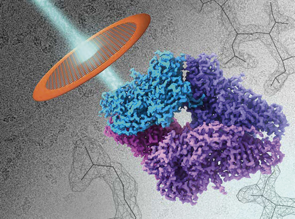
A notable aspect in the development of TEM instrumentation is that through the various revolutions in electron microscopy over much of the last century, which have led to so many exciting advances in our understanding of the structures of inorganic materials, semiconductors, and biological specimens, the fundamental design of electron optical columns has been largely preserved. This seeming constancy of design, however, has also laid the foundation for achieving extraordinary levels of precision in central elements of modern microscopes such as image and probe correctors, specimen stages, vacuum levels, and automation of specimen delivery. Improvements in the electron microscope have been matched by great advances in technology for ancillary equipment such as image and probe correctors, spectrometers, and detectors, which now allow the recovery of unprecedented levels of information on the 3D structures of proteins and nucleic acids in biological applications (see schematic in Figure 1).
Composite illustration of three layers of information obtained in modern cryo-electron microscopy imaging. (Upper left) The raw, low-dose projection images of proteins; (center) the 3D density map obtained by averaging tens of thousands of projection images of the tetrameric protein β-galactosidase, with each protomer colored separately. (Lower right background) The interpretation of the density map in terms of an atomic model, which allows unambiguous tracing of the path of the polypeptide and individual amino acids.

The central challenge in biological TEM imaging is radiation damage.Reference Henderson2 The revolution in the use of transmission electron imaging in biology has at its core three basic advances, each of which provides a way to partially overcome the effects of damage to organic matter from electron irradiation. First, the development of microscope stages maintained at temperatures close to that of liquid nitrogen provides the means to reduce the damage resulting from irradiation with electron beams. Compared to imaging at room temperature, there is a five- to tenfold reduction in radiation damage at cryogenic temperatures.Reference Hayward and M Glaeser3 Imaging at cryogenic temperatures allows the generation of images from vitrified biological specimens with higher signal-to-noise ratios than at room temperature for the same dose, and has been instrumental in driving the growth of the field of cryo-EM.
A second, and game-changing, advance that has dramatically increased the quality of information recovered from electron microscope images is the development of direct electron detectors.Reference Li, Mooney, Zheng, Booth, Braunfeld, Gubbens, Agard and Cheng4 Unlike the earlier generation of charge-coupled device (CCD) detectors, direct electron detectors do not involve an intermediate stage where incident electrons are converted into photons before detection. In addition to their greatly improved detective quantum efficiency, direct electron detectors can be used to collect a large number of images during a single exposure. Each image is a movie, distributed over many frames (typically 30–60 in current applications). The individual frames in a movie allow the application of powerful image-processing tools to correct for beam-induced motion of biological specimens during exposure by coherent averaging of the individual frames or selected subregions of frames.
A third and critical development is the explosion in methods to average the information from tens of thousands of images, each collected at low doses to minimize radiation damage.Reference Frank5 Images recorded in a transmission electron microscope are projection images, and by assigning the correct relative orientation to each image, and combining the information in large numbers of images, structures of radiation-sensitive specimens such as proteins and viruses can be visualized in three dimensions. The combination of these three advances has led to a dramatic growth in the use of TEM in structural biology and has made determination of protein structures at near-atomic resolution possible.Reference Bartesaghi, Merk, Banerjee, Matthies, Wu, Milne and Subramaniam6 The number of entries deposited in the Electron Microscopy Data Bank7 and the number of users in the life science community are both growing exponentially.
Among the technical advances in instrumentation, one notable feature is the transition from conventional side-entry stages, where TEM specimens were mounted at the end of a long and bulky stage, to one in which specimens are delivered into the column as a cartridge, greatly reducing instabilities in the stage arising from thermal and vibrational sources. This innovation, first introduced by the FEI Company in their Polara microscopes, is now a standard feature of advanced microscopes such as the Titan Krios8 and the CryoARM.9 Combined with the development of multispecimen holders, where as many as 12 specimens can be parked and sequentially delivered into the microscope column, these cartridge delivery systems are at the heart of the increased throughput and automation in biological TEM.
Cryo-EM for materials science applications
Although much of the progress in life science applications over the last decade has come from the use of TEM, there are developments in scanning transmission electron microscopy (STEM) imaging that should not be overlooked.Reference Sousa and Leapman10 TEM is the norm for cryo-EM imaging, where the specimen thickness is low (typically <100 nm for most applications). However, numerous biological applications require imaging of thick specimens, with thicknesses in the range of ∼1 µm, which are essentially at the limit of useful TEM imaging, even in 300 kV electron microscopes. For these thicker specimens, users generally apply electron tomographic methods to collect images over a wide range of tilt angles and combine the resulting images to obtain a 3D image. For these types of applications, which are usually at a resolution about an order of magnitude lower than those obtained with protein cryo-EM, STEM tomography offers distinct advantages.Reference Wolf, Elbaum, Müller-Reichert and Pigino11 STEM tomography allows imaging thicker samples than those for TEM because of the incoherent mode of contrast, and the high amplitude contrast at low spatial frequencies. STEM imaging with the probe beam focused on the specimen plane eliminates the need for corrections from the inversions arising from variations in contrast transfer function. The future of STEM imaging lies in exploiting the quantitative information it can generate because the scattering correlates directly with atomic density, providing valuable information on elemental composition in complex specimens such as cells and tissue.
Arguably, the most efficient cryo-EM instruments for structural biology also offer the fewest options in terms of multifunctionality. In part, this reduction in functionality leads to the stability and reproducibility that allows experiments to be conducted over several days in a semi-automated manner. In contrast, TEM or STEM instruments used for materials science applications are often advanced analytical electron microscopes that provide a plethora of methods to study the microstructure, crystallography, chemistry, and electronic properties of a wide range of materials. In materials science applications, it is often desirable to obtain imaging and diffraction information from the same region and then to supplement it with chemical information from x-ray energy-dispersive spectroscopy (XEDS) or electron energy-loss spectroscopy (EELS). The cartridge delivery systems central to the advances in life sciences provide one major limitation for materials science applications, since they only allow the specimen to be tilted on one axis. The question that arises is, can we have it all? Is it possible, now or in the future, to have the stability of the dedicated cryo-EM instrument in a multifunctional transmission electron microscope/scanning transmission electron microscope for materials science applications?
As discussed earlier, stage stability in dedicated cryo-EM instruments is achieved by using a cartridge system, which means the specimen is almost isolated from the external environment once it is inserted into the microscope stage. The sample can be moved in all directions, but can only be tilted on one axis. In cryo-EM for materials science, it is critical to have a five-axis stage to be able to move the specimen in x, y, and z directions, as well as tilt the specimen on two orthogonal axes so a specific crystallographic direction is aligned along the electron-optical axis (i.e., the viewing direction). Currently, this requires the use of a side-entry specimen rod with double-tilt functionality. These specimen rods are stable at room temperature, as evidenced by the numerous publications that show atomic-resolution images with exceptional precision.Reference Pennycook and Nellist12
In most current designs, low-temperature side-entry rods have a bulky liquid nitrogen dewar mounted on the end of the rod with a “cold finger” that connects through the body of the holder to the specimen position. While this allows cooling of the sample to low temperatures, it also transmits vibrations from the external environment (e.g., convection currents in the nitrogen) to the specimen. Ultimately, this compromises the stability of the specimen, which means that atomic resolution at low temperature is compromised.Reference Savitzky, El Baggari, Clement, Waite, Goodge, Baek, Sheckelton, Pasco, Nair, Schreiber, Hoffman, Admasu, Kim, Cheong, Bhattacharya, Schlom, McQueen, Hovden and Kourkoutis13 While some researchers are exploring ways around this, from combining multiple fast acquisitions to mitigating drift and other effects, it is clear that specimen holders, with improved stability and development of a five-axis stage based on the cartridge designs used in dedicated cryo-EM instruments, are critical steps to realize cryo-EM for materials applications.Reference Goodge, Bianco, Zandbergen and Kourkoutis14 It is also worth emphasizing another key difference between the needs of the life science and materials science cryo-EM communities. In life sciences, the goal is to maintain the specimen in its vitrified state at liquid-nitrogen or liquid-helium temperatures. In materials science, researchers often wish to study properties or phenomena as a function of temperature, so it is important to be able to vary the temperature of the specimen in the microscope column. In this issue, Minor et al.Reference Minor, Denes and Muller15 address the issue of whether the new quantum science would be enabled by a variable temperature cryo-EM that could operate from low temperatures, below 77 K, to room temperature and higher.
In materials science applications, the chemical composition of the specimen may be spatially inhomogeneous, and electron microscopy has become the key tool to investigate chemical heterogeneity with high spatial resolution. The most common technique used to achieve this in the TEM is XEDS, where the incident electron transfers energy to an electron in the specimen, exciting it to a higher energy state. Upon relaxing to the ground state, the specimen emits energy, often in the form of an x-ray; the energy of these x-rays are characteristic of the element that is excited. X-ray emission from the specimen is isotropic and subtends a full solid angle around the excitation site. Due to engineering constraints, XEDS detectors span a finite solid angle in the vicinity of the specimen, and the spectral quality is related to size of the solid angle subtended.
For cryo-EM, it is essential to maintain a good vacuum, ideally ultrahigh vacuum (UHV, <10–9 mbar [1 × 10⁻⁷ Pa]), in the specimen area, to minimize contamination and to have minimal temperature drift. This is achieved by the introduction of cryo-shields—cooled plates that surround the specimen area, improving the vacuum and ensuring the specimen is not exposed to any hot surfaces. These cryo-shields prevent any useful x-ray signal from reaching the detectors and effectively eliminate the use of XEDS in cryo-EM. While alternative techniques, such as EELS can also be used for chemical analysis, the loss of XEDS as a spectroscopic option in the microscope is significant. Solutions such as retractable cryo-blades circumvent this specific issue, but ultimately, a new type of UHV stage that delivers temperature stability while still enabling XEDS will likely be important for the effective use of cryo-EM in materials science.Reference Homo, Booy, Labouesse, Lepault and Dubochet16
Technological improvements such as image and probe aberration correctors, specimen stages, vacuum levels, and automation of specimen delivery have delivered significant benefits to electron microscopists in both physical and life sciences. However, to realize further improvements in spatial resolution and stability, improvements in electron detectors are necessary. For the past four decades, the best method for digitally collecting TEM images has been the use of radiation-sensitive scintillators coupled to CCD or complementary metal oxide semiconductor (CMOS) devices. Both of these technologies have advantages and disadvantages for a given application, and both use an optically coupled scintillator to convert the incoming electrons into a photon. This conversion to photons has plagued both biological and materials researchers by effectively blurring the incoming signal, referred to as the point-spread function (PSF) of the detector, and limiting the resolution gains possible from the improvements in electron optics. This problem is eliminated in direct electron detectors, which are composed of radiation-hardened CMOS architectures that can tolerate direct exposure to high-energy electrons and then convert the electrons directly, to read out the image electronically with single electron sensitivity, at frame rates from 400 to greater than 1000 fps.Reference Tate, Purohit, Chamberlain and Nguyen17
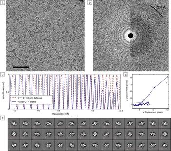
The improved detection quantum efficiency as well as the ability to collect a large number of images during a single exposure were critical for realizing gains in the life science community. Some of the recent revolutions seen in biological research come from direct electron detectors that typically have 4 k × 4 K pixels for real-space TEM imaging, where single electron sensitivity with low noise is paramount due to the low electron dose imaging conditions used for sensitive biological specimens. The fast frame rates of these detectors are excellent for capturing sharper and cleaner biological images by correcting for beam-induced motion (Figure 2).Reference Jiang, Chen, Han, Deb, Gao, Xie, Purohit, Tate, Park, Gruner, Elser and Muller18
Demonstration of the quality of low-dose images recorded using a Gatan K2 detector, illustrated using E. coli β-galactosidase, the test protein first used to demonstrate that atomic-resolution structures could be obtained using single-particle cryo-electron microscopy (as presented in Reference Bartesaghi, Merk, Banerjee, Matthies, Wu, Milne and SubramaniamReference 6). (a) Average of 38 aligned movie frames acquired over a 7.6 s exposure window (45 e/ Å2 total accumulated dose). Images were recorded at 300 kV and –1 μm defocus in super-resolution mode using a physical pixel size of 0.637 Å. Scale bar = 50 nm. (b) Fast Fourier transformations of image in (a) showing the extent of Thon rings that are visible to resolutions beyond ∼3 Å, obtained using periodogram averaging (left panel) from the average of aligned frames and (right panel) from the raw individual frames. (c) One-dimensional power spectrum profile obtained by radially averaging the Fourier transform obtained from the individual frames (right panel in [b]) with peaks visible at 2.5 Å resolution. The solid blue line shows the profile of the contrast transfer function (CTF) derived from the data, while the red dashed line shows the theoretical profile obtained using the computed CTF value, illustrating the excellent fit between the experimental and theoretical profiles. (d) Trajectory of movement for each of the 38 frames during the 7.6 s exposure used to correct the drift in the image shown in (a). Displacements are measured in multiples of the super-resolution pixel size and numbered in sequence, with only the 1st, 19th, and 38th frames shown for the sake of clarity. The plot shows that the displacements are largest in the first 5–6 frames. (e) Two-dimensional reference-free class averages showing that the data set is composed of images of characteristic β-galactosidase projection images that span a range of orientations, including top, side, and tilted views of the protein. A wide distribution of views enables more complete sampling of orientations, resulting in a more isotropic resolution.

In materials research, imaging of beam-sensitive materials such as polymers or metal–organic frameworks (MOFs), or, in situ experimentation, require similar low-dose TEM imaging conditions as those implemented in biological imaging. As discussed previously, STEM characterization is much more commonly used in materials science, and technological improvements in probe aberration correction have unlocked the theorized potential of this reciprocal-space imaging method, if electron detectors can be optimized for high-dynamic range with single electron sensitivity. The dilemma for STEM acquisition lies in the ability to collect the large number of electrons that pass through the sample unaffected, while simultaneously detecting the small fraction of electrons scattered from the crystal.Reference Courtland19
While some researchers are attempting to use the fast readout time of direct electron detectors used for biological research to achieve a high dynamic range, others are working on hybrid pixel array detectors, with large bit depth, to provide a 1,000,000:1 dynamic range within a single pixel, allowing for direct electron-beam imaging while also maintaining single electron sensitivity. A high-speed, high-sensitivity, and high-dynamic-range pixel array detector could provide researchers the opportunity to characterize electric and magnetic fields with unprecedented accuracy and spatial resolution, even down to the atomic scale,Reference Tate, Purohit, Chamberlain and Nguyen17 with applications to characterize devices such as next-generation photovoltaics and solid-state magnetic storage.
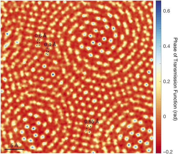
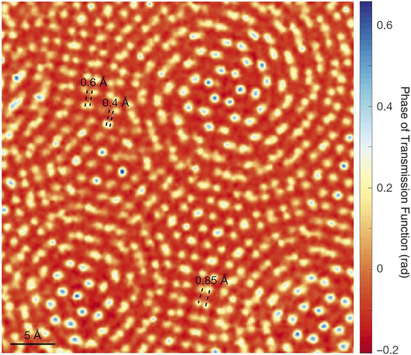
Classically, STEM imaging has been performed using multiple CCDs or solid-state detectors, capable of detecting diffracted or transmitted intensities over some angular range and limited by fixed geometries defined by the detector location relative to the sample. An alternative to this approach is capturing the complete, unsaturated diffraction pattern at every pixel while in scanning mode. This produces a 4D-STEM image containing spatially resolved diffraction patterns in each pixel, and allows virtual aperture post-processing to extract bright-field, dark-field, center-of-mass, and differential phase-contrast information from a single acquisition. The ability to extract this quantity of information represents a significant expansion in the characterization methods possible for electron scattered images and could produce transformative results for materials research. Such detectors may even have an impact on the use of cryo-STEM for life sciences in the area of cryo-electron tomography by allowing post-processing adjustment of contrast by selecting optimum scattering angles. In the past, technological limitations with cameras have prevented researchers from collecting complete diffraction patterns. If the unsaturated electron intensities on an absolute scale can be collected, information such as local sample thickness can be determined and additional characterization techniques such as nanodiffraction, fluctuation electron microscopy, holography, and ptychography, a method to recover phase information from the full phase space to increase image resolution, become possible.Reference Courtland19 Pixel array detectors have been recently used to show short-range order in bulk metallic glasses and to break the diffraction resolution limit in MoS2 by achieving 0.39 Å spatial resolution, as shown in Figure 3.Reference Jiang, Chen, Han, Deb, Gao, Xie, Purohit, Tate, Park, Gruner, Elser and Muller18
One of the advantages of the use of direct electron detectors for cryo-electron microscopy applications in materials research will be the capability to increase the maximum attainable resolution using full-field ptychography. Recently, researchers have reported 0.39 Å resolution in a MoS2 twisted bilayer using an electron microscope pixel array detector and full-field ptychography reconstruction to effectively “break” the diffraction limit. The Rayleigh resolution for the annular dark-field scanning transmission electron microscope is 1.2 Å for these imaging conditions.Reference Jiang, Chen, Han, Deb, Gao, Xie, Purohit, Tate, Park, Gruner, Elser and Muller18
Going forward, detector improvements that have the potential for further advancements for materials research will depend on increasing the number of pixels on pixel array detectors while maintaining single electron sensitivity and providing enough dynamic range to collect the direct electron beam. Data storage and transfer speed, as well as processing software to handle large file sizes may well become critical limiting factors, but there is every reason to hope that they will keep pace with instrument development in the coming years.
Summary
In closing, while we have covered mostly instrumentation advances, the practical utility of these advances in hardware design would be minimal without the extraordinary advances in operating software for microscopes. The vast majority of tuning and alignment operations that would have typically been carried out by highly trained experts can now be carried out by users with relatively modest amounts of training. This reduction in skill required to operate an electron microscope, combined with all of the advances that have led to improvements in data quality, are undeniably a major part of the story of the remarkable growth of modern electron microscopy, especially in the life sciences.
Cryo-EM in materials science is at an exciting stage of development and the developments necessary to make it as impactful are already under way. We predict that the life science and materials science cryo-EM communities will increasingly overlap and benefit significantly from shared experiences and knowledge. The challenges of data acquisition, manipulation, and storage, as well as the developing role of artificial intelligence leading to “turnkey” operation of even the most complex instruments, are common themes in both communities. With collaboration and communication, we anticipate great progress in the next decade in this field.
Acknowledgments
D.W.M. is supported by the State of Ohio Third Frontier Program as an Ohio Research Scholar. S.S. was supported by a Canada Excellence Research Chair Award from the Government of Canada.

Robert E.A. Williams is currently the assistant director for Research and Development at the Center for Electron Microscopy and Analysis at The Ohio State University. He received his BS degree from Virginia Tech in 2001, and his MS degree in 2006 and his PhD degree in 2010 from The Ohio State University, both in materials science and engineering. His research interests include inorganic and organic materials, with a focus on advanced materials characterization. His current research focuses on high-resolution electron microscopy and advanced characterization techniques. Williams can be reached by email at williams.2156@osu.edu.

David William McComb is a professor of materials science and engineering and director of the Center for Electron Microscopy and Analysis at The Ohio State University. He received his BS degree in chemistry in 1987 from the University of Glasgow, Scotland, and his PhD degree in physics in 1990 from the University of Cambridge, UK. His research focuses on the development and application of scanning transmission electron microscopy and electron energy-loss spectroscopy as a subnanometer-scale probe of chemistry, structure, and bonding. He is a Fellow of the Royal Society of Chemistry, the Institute of Materials, and the Microscopy Society of America. McComb can be reached by email at mccomb.29@osu.edu.

Sriram Subramaniam is a professor and the Gobind Khorana Canada Excellence Research Chair in Precision Cancer Drug Design at The University of British Columbia, Canada. He received his PhD degree in physical chemistry in 1987 from Stanford University and completed postdoctoral training at the Massachusetts Institute of Technology. From 1998 to 2018, he served as a senior investigator at the National Cancer Institute, National Institutes of Health. He is also the founding director of the National Cryo-electron microscopy Facility at the National Cancer Institute, currently serving as their scientific advisor. His research focuses on single-particle cryo-electron microscopy methods that determine protein and protein-bound drug structures at atomic resolution. Subramaniam can be reached by email at sriram.subramaniam@ubc.ca.