Introduction
Fertilization is pivotal for organisms of sexual reproduction and, beyond doubt, sperm motility is essential for its success. Sea urchins reproduce by external fertilization and produce large amounts of gametes; these two features make the sea urchin an excellent model to study general mechanisms of fertilization. Sea urchin sperm are immotile in the gonads due to the high CO2 tension and K+ concentration that provide an acidic pH environment (Christen et al., Reference Christen, Schackmann and Shapiro1982, Reference Christen, Schackmann and Shapiro1983; Lee et al., Reference Lee, Johnson and Epel1983). Under these conditions, the dyneins (ATPases, whose activity is regulated by phosphorylation), the molecular motors of the axoneme that propel the flagella, are inactive. When sea urchin spawn into the seawater, sperm motility initiates as intracellular pH increases, activating the dyneins (Lee, Reference Lee1984). Sperm motility is also regulated by diffusible sperm-activating peptides (SAPs) from the outer egg layer (Suzuki, Reference Suzuki1995). Upon binding to its receptor in the sperm flagella, speract, the first purified and structurally identified SAP from S. purpuratus eggs (Hansbrough and Garbers, Reference Hansbrough and Garbers1980; Suzuki et al., Reference Suzuki, Nomura, Ohtake and Isaka1981), elicits rapid physiological changes in cyclic nucleotide levels (cGMP and cAMP), membrane potential (Em), intracellular pH (pHi), and Ca2+ ([Ca2+]i), as well as depolarization of the mitochondrial membrane potential (ΔΨ), increases in the NADH levels and changes in the protein phosphorylation patterns in sperm (Darszon et al., Reference Darszon, Guerrero, Galindo, Nishigaki and Wood2008; Kaupp et al., Reference Kaupp, Kashikar and Weyand2008; Loza-Huerta et al., Reference Loza-Huerta, Vera-Estrella, Darszon and Beltrán2013; García-Rincón et al., Reference García-Rincón, Darszon and Beltrán2016). As mature sperm are unable to synthesize new proteins, their need to alter their functions through protein phosphorylation/dephosphorylation is higher than in other cell types. Protein phosphorylation plays an important role during the activation and regulation of sperm motility (Tash and Means, Reference Tash and Means1983; Murofushi et al., Reference Murofushi, Ishiguro, Takahashi, Ikeda and Sakai1986; Tash, Reference Tash1989; Bracho et al., Reference Bracho, Fritch and Tash1998; Nakajima et al., Reference Nakajima, Morita, Takemura, Kamimura and Okuno2005; Su et al., Reference Su, Chen, Zhou and Vacquier2005). The cAMP increases induced by SAPs activate PKA, which phosphorylates sperm proteins (Su et al., Reference Su, Chen, Zhou and Vacquier2005). In vitro experiments in demembranated sperm indicated that cAMP-dependent changes in protein phosphorylation are essential for sperm motility activation (Brokaw, Reference Brokaw1987; Inaba, Reference Inaba2003) and that a cAMP-independent protein phosphorylation is also involved, suggesting the participation of other kinases and changes (Hayashi et al., Reference Hayashi, Yamamoto, Yonekawa and Morisawa1987; Dey and Brokaw, Reference Dey and Brokaw1991; Morita et al., Reference Morita, Takemura and Okuno2004; Nakajima et al., Reference Nakajima, Morita, Takemura, Kamimura and Okuno2005). However, in demembranated sperm, the fundamental ion permeability changes involved in the regulation of motility are eliminated, limiting the usefulness of this model. There has been only one report in intact Lytechinus pictus sperm showing that PKC is implicated in motility initiation and its maintenance (White et al., Reference White, de Lamirande and Gagnon2007).
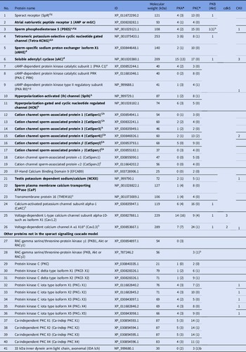
Several proteins of the proposed model for the speract signalling cascade (Table 1) contain phosphorylation sites for PKA and/or PKC (Ward et al., Reference Ward, Moy and Vacquier1986; Hanoune and Defer, Reference Hanoune and Defer2001; Su and Vacquier, Reference Su and Vacquier2006; Darszon et al., Reference Darszon, Guerrero, Galindo, Nishigaki and Wood2008; Vacquier et al., Reference Vacquier, Loza-Huerta, García-Rincón, Darszon and Beltrán2014; Guerrero et al., Reference Guerrero, Gadêlha, Ramírez-Gómez, Ramírez, Beltrán and Tuval2020). Furthermore, although initially it was reported that the S. purpuratus kinome contained four different PKC genes (PKC β, δ, ϵ and ι) (Bradham et al., Reference Bradham, Foltz, Beane, Arnone, Rizzo, Coffman, Mushegian, Goel, Morales, Geneviere, Lapraz, Robertson, Kelkar, Loza-Coll, Townley, Raisch, Roux, Lepage, Gache, McClay and Manning2006) we found that the isoenzymes present in the S. purpuratus genome are: PKC [regulated by Ca2+ and diacylglycerol (DAG)], PKCδ with isoforms X1 and X2 (regulated by DAG), PKCι X1–X5 (regulated by phosphatidylserine as all the PKC isoenzymes) (Newton, Reference Newton1995, Reference Newton2018), and four isoforms of the Ca2+-independent PKC (X1–X4) (Table 1). As the antibodies used as a tool to follow the phosphorylation changes associated to motility, recognize motifs also in the RAC-gamma serine/threonine-protein kinase (PKB or Akt) and there are seven proteins in the speract signalling cascade with putative PKB phosphorylation sites, Table 1 also includes the two PKB isoenzymes present in the S. purpuratus genome. As shown in Table 1, all the proteins in the list seem to be regulated by phosphorylation. How the phosphorylation changes regulate sperm motility is not fully understood.
Proteins from the speract signalling cascade with predicted phosphorylation sites by different kinases

Only predicted phosphorylation sites with scores > 0.7 are indicated.
a (#) Number of phosphorylated motifs recognized by anti-PKA substrates and anti-PKC substrates, respectively.
b In the PKB column indicates the recognized motif by anti-PKA substrates.
Proteins detected by proteomic analysis in sperm of S. purpuratus.
c Su et al., Reference Su, Chen, Zhou and Vacquier2005.
d Nomura and Vacquier, Reference Nomura and Vacquier2006.
e Loza-Huerta et al., Reference Loza-Huerta, Vera-Estrella, Darszon and Beltrán2013.
f Espinal-Enríquez et al., Reference Espinal-Enríquez, Priego-Espinosa, Darszon, Beltrán and Martínez-Mekler2017.
g Beltrán Núñez, Reference Beltrán Núñez, Arenas Rios and Fuentes Mascorro2019 and in sperm of Arbacia punctulata in bold (Trötschel et al., Reference Trötschel, Hamzeh, Alvarez, Pascal, Lavryk, Bönigk, Körschen, Müller, Poetsch, Rennhack, Gui, Nicastro, Strünker, Seifert and Kaupp2020).
Our results using swimming intact S. purpuratus sea urchin sperm indicate that both PKA and PKC are involved in basal and speract-stimulated sperm motility through a signalling pathway that comprises a crosstalk between both enzymes, as described for bovine sperm capacitation (Cohen et al., Reference Cohen, Rubinstein, Gur and Breitbart2004; Alonso et al., Reference Alonso, Osycka-Salut, Castellano, Cesari, Siervi Di, Mutto, Johannisson, Morrell, Davio and Perez-Martinez2017) and the human sperm acrosome reaction (Doherty et al., Reference Doherty, Tarchala, Radwanska and Jonge de1995). This proposal is supported by the fact that PKA inhibition modifies the phosphorylation levels of both PKA and PKC substrates in the flagellum of sea urchin sperm and that PKC inhibition has a similar effect.
Materials and methods
Materials
S. purpuratus sea urchins were collected in Ensenada B.C. Mexico and maintained in commercial sea water (Oceanic, Grupo Acuario Lomas, S.A. de C.V.) at 14–15°C. H89 dihydrochloride, Rp-adenosine-3′,5′-monophosphorothionate (Rp-cAMPS) and bisindolylmaleimide (BIM) were purchased from Calbiochem, EMD Biosciences (San Diego, California). Sodium orthovanadate (Na3VO4), sodium fluoride (NaF), DNase and Poly-HEME (poly(2-hydroxyethylmethacrylate)) were purchased from Sigma-Aldrich (St. Louis, MO). Chelerythrine chloride was purchased from Alomone Labs (Jerusalem, Israel). PhosphoSafe™ Extraction Reagent was purchased from Novagent, Merck Biosciences (Darmstadt, Germany). Complete EDTA-free and the calpain inhibitors I and II, from Roche Applied Sciences (Indianapolis, IN, USA). Speract was synthesized in Lourival Possani’s laboratory (Instituto de Biotecnología-UNAM, México). Immobilon-P, PVDF membranes (IPVH00010) for western blot (WB), 0.45 µm, were from Millipore (Suwanee, GA, USA). Primary rabbit antibodies, were obtained from Cell Signalling Technology (Danvers, MA, USA): phospho-(Ser/Thr) PKA substrates antibody (anti-PKA substrates, no. 9621) that recognizes the general substrate sequence –R–x–x–pS/T–, of the AGC family kinases [PKA, PKB (Akt) and PKC], and phospho-(Ser) PKC substrates antibody (anti-PKC substrates, no. 2261) that recognizes the sequence (R/K)XS(hydrophobic)(R/K). The specificity of both antibodies, anti-PKA substrates (Su et al., Reference Su, Chen, Zhou and Vacquier2005) and anti-PKC substrates (White et al., Reference White, de Lamirande and Gagnon2007) for sea urchin sperm proteins, has been previously proven (Loza-Huerta et al., Reference Loza-Huerta, Vera-Estrella, Darszon and Beltrán2013). Secondary HRP-labelled antibody was obtained from Santa Cruz Biotechnology, Inc. (Santa Cruz, CA, USA). Super Signal West Pico ECL detection kit was purchased from Pierce (Rockford, IL, USA).
Gametes
S. purpuratus sea urchin sperm were obtained by intracoelomic injection of 0.5 M KCl, and were collected and kept on ice as dry sperm until use. Artificial sea water (ASW) contained the following, in mM: 486 NaCl, 10 CaCl2, 26 MgCl2, 10 KCl, 30 MgSO4, 2.5 NaHCO3, 0.1 EDTA and 10 HEPES (∼1000 mOsm, pH 8). Seawater with low Ca2+ (1CaASW) was ASW pH 7.5 containing only 1 mM CaCl2. Sea urchin sperm were made immotile, suspending them in ASW pH 6 (buffered with 5 mM MES), containing 50 mM KCl (Bracho et al., Reference Bracho, Fritch and Tash1997) instead of 10 mM. We confirmed that, as reported, sperm under these conditions (ISW, pH 6) had no motility that they recovered when diluted in ASW pH 8.0.
Sperm motility analysis
Diluted sperm (1:4000 in 1CaASW 7.5) were further diluted (1:100) in ASW pH 8 (∼2 × 10⁶ cells ml −1) with or without PKA (H89 or Rp-cAMPS) or PKC (chelerythrine or BIM) inhibitors in a Petri dish [treated with 0.1% (w/v) poly-HEME in ethanol] and maintained at 14°C on a microscope stage (Eclipse TE300; Nikon; Melville, New York, USA) throughout the experiment. Sperm motility was evaluated using dark-field illumination at ×40 magnification and was recorded on an EMCCD Andor camera (DU888, Andor iXon; Oxford, UK). Images were collected with the Andor iQ 1.8 software (Andor).
Bioimaging
Bioimaging (NC) at 5 fps during five-time intervals: 1–2, 5–6, 10–11, 20–21 and 30–31 min of incubation with or without the different inhibitors. Sperm head trajectories were tracked using ImageJ software 1.47c (Waine Rasband, NIH, USA) with the function ‘M Track J’ to calculate circular speed (µm/s). Controls were the sperm in ASW pH 8 containing <0.1% dimethyl sulfoxide (DMSO), the dilution medium of the inhibitors.
SDS-PAGE and western blot
Dry sperm (∼8 × 10¹⁰/ml) were diluted (1:400) in ISW or ASW in the absence or presence of inhibitors for PKA or PKC. After incubation at 14°C for different times, the proteins were precipitated with trichloroacetic acid (10% final) for 30 min on ice and centrifuged (23,100 g for 5 min at 4°C). The sperm pellet was cleaned with cold acetone four times (23,000 g for 5 min at 4°C) and the proteins resuspended in a cocktail of phosphatase (PhosphoSafe Extraction® 1×, 1 mM Na2VO4 and 50 mM NaF) and protease (2× Complete EDTA-free, 4 µg/ml calpain I and II and 1 mM EGTA) inhibitors and 10 µg/ml DNase. The proteins were solubilized at 70°C/30 min with 1/5 of 5× concentrated sample buffer (10% SDS, 50% glycerol, 8 mM EDTA, 0.5 M Trizma base pH 6.8, 0.001% bromophenol blue and 5% β-mercaptoethanol). Sperm proteins were resolved on 10% SDS-PAGE according to Laemmli (Reference Laemmli1970) and transferred to Immobilon-P membranes for WB experiments with anti-PKA substrates or anti-PKC substrates diluted 1:500 in TBS (50 mM Tris–HCl and 150 mM NaCl pH 7.6) containing 0.05% Tween 20 (TBS-T). Primary antibodies were detected with an HRP-conjugated goat anti-rabbit IgG secondary antibody (diluted 1:1000 in TBS-T) and revealed by chemiluminescence with Super Signal West Pico ECL substrate, as described (Loza-Huerta et al., Reference Loza-Huerta, Vera-Estrella, Darszon and Beltrán2013).
Predicted phosphorylation sites and protein amphipathicity
Analysis of the predicted phosphorylation protein sites was performed using the NetPhos-3.1 server. TMpred from Expasy was used to determine protein amphipathicity to eliminate extracellular and transmembrane predicted phosphorylation sites of the proteins in Table 1.
Statistical analysis
Densitometry analysis of the PKA substrates or PKC substrates phosphorylation levels (WB) and protein abundance (amido black-stained Immobilon-P membrane after the WB), was carried out using the ImageJ program. The intensity of each PKA substrate or PKC substrate peak was divided by the intensity of the internal loading control (amido black-stained protein band in the motile condition) in the corresponding line. To compare the results between different experiments we used Image Studio™ Lite software by LI-COR and normalizing taking as ‘1’ the motile sperm condition in each independent experiment. Data for Figs 1, 2, 4 and 5 were analyzed in GraphPad Prism 7 software with unpaired two-tailed t-test, or analysis of variance (ANOVA) when comparing the effect of different concentrations of inhibitors on sperm motility (Fig. 3).
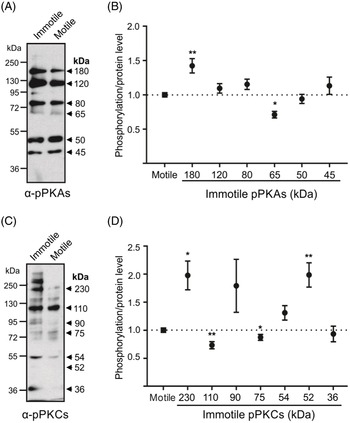
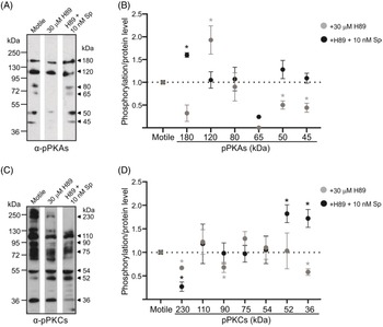
Phosphorylation levels of some phospho-(S/T) PKA substrates and PKC substrates change with the activation of motility. Strongylocentrotus purpuratus spermatozoa were incubated in ISW (immotile), or ASW (motile) and WB experiments with ∼15 µg (A, C) of the precipitated and solubilized proteins (see Materials and methods) were resolved in 10% SDS-PAGE. Phospho-(S/T) PKA substrates (A) and PKC substrates (C) phosphorylation levels were detected by western blot (WB) with α-phospho-(S/T) PKA substrates and α-PKCs antibodies, respectively. Values on the left and right of the WB indicate molecular weight (MW) of protein standards and motility-associated substrates respectively. Blots are representative of independent experiments. (B, D) Phosphorylation levels were normalized considering as ‘1’ the corresponding band of the motile condition, obtaining the ratio of phosphorylation:protein levels for each independent experiment. Asterisks indicate significant changes in n = 3–8 independent experiments. *, P ≤ 0.05); **, P ≤ 0.01, using a two-tailed one-sample t-test.

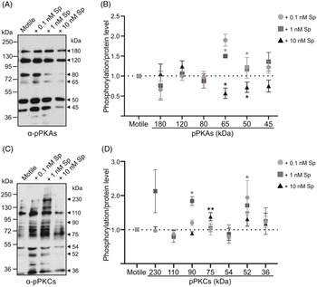
Stimulation of sperm with speract modifies PKA substrates and PKC substrates phosphorylation levels. Strongylocentrotus purpuratus sperm were incubated in ASW in the absence (motile) or the presence of different speract concentrations (0.1, 1 and 10 nM). The phosphorylation levels of phospho-(S/T) PKA substrates (A, B) and PKC substrates (C, D) were detected and evaluated as in Fig. 1. (A, C) Arrowheads indicate the molecular weight of motility-associated substrates and blots are representative of independent experiments. Normalization in (B, D) as in Fig. 1. n = 3–8 independent experiments. *, P ≤ 0.05; **, P ≤ 0.01 using a two-tailed one-sample t-test.

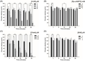
PKA and PKC are involved in the regulation of the motility of sea urchin sperm. Strongylocentrotus purpuratus sperm were diluted 1:4000 in ASW and incubated for 1, 5, 10, 20 and 30 min with different concentrations of PKA inhibitors: (A) 0, 2, 5, 10 and 30 µM H89; (B) 0, 0.1, 0.2, 0.5; or 1 mM Rp-cAMPS and PKC inhibitors: (C) 0, 2.5, 5 and 7.5 µM chelerythrine (Chele); (D) 0, 0.1, 1 and 5 µM bisindolylmaleimide I (BIM) and the velocity of sperm was measured. Values are means ± SEM of 10 independent experiments made in triplicate. The motility control (0) was performed in sperm suspensions containing the same volume (<0.1% v/v) of DMSO, the solvent for the inhibitors. ***, P ≤ 0.001; **, P ≤ 0.01; *, P ≤ 0.05; by ANOVA.

PKA regulates both basal and the speract-stimulated motility by changing the phosphorylation levels of specific proteins in sperm that includes PKC substrates. Strongylocentrotus purpuratus sperm were incubated for 10 min in ASW (motile) in the absence or presence of 30 µM H89 (PKA inhibitor) and then stimulated or not with speract for 10 s. Western blot of PKA substrates (A) and PKC substrates (C) of lysates of whole sperm exposed to the different conditions are shown. Arrowheads indicate the molecular weight of motility-associated substrates. (B, D) Normalization as in Figure 1. Blots are representative of three independent experiments. *, P ≤ 0.05; **, P ≤ 0.01; using two-tailed one-sample t-test.

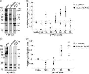
PKC modulates sea urchin sperm motility through protein phosphorylation by PKA. Strongylocentrotus purpuratus sperm were incubated for 10 min in ASW (motile), in the absence or presence of 5 µM chelerythrine (PKC inhibitor) and then stimulated or not with speract for 10 s. Western blot of PKC substrates (A) or PKA substrates (C) of lysates of whole sperm exposed to the different conditions are shown. Arrowheads indicate the molecular weight of motility-associated substrates. (B, D) Normalization as in Figure 1. Blots are representative of three independent experiments. *, P ≤ 0.05 using two-tailed one-sample t-test.

Results
In intact sea urchin sperm, the phosphorylation patterns of PKA substrates and PKC substrates change with motility
In sea urchin as in most species, sperm remain in a quiescent state in the gonads due to a low pHi maintained by a high CO2 tension and initiate to beat their flagella upon spawning (Johnson et al., Reference Johnson, Clapper, Winkler, Lee and Epel1983). It is clear that sperm motility depends on pH and on the external ionic composition (Christen et al., Reference Christen, Schackmann and Shapiro1982). In this work we considered both conditions to keep sperm in an immotile state (Bracho et al., Reference Bracho, Fritch and Tash1997, see Methods).
Previously we have shown by immunofluorescence experiments that PKA substrates and PKC substrates are distributed along the S. purpuratus sperm (Loza-Huerta et al., Reference Loza-Huerta, Vera-Estrella, Darszon and Beltrán2013). We confirmed the presence of some of these substrates by tandem mass spectrometry (MS/MS), in Low Density Detergent Insoluble Membranes (LD-DIM) isolated from whole sperm, as well as from their flagella (Beltrán Núñez, Reference Beltrán Núñez, Arenas Rios and Fuentes Mascorro2019). In addition, we reported that the phosphorylation levels of PKA substrates and PKC substrates associated to LD-DIM derived from intact sperm changed with their motility condition (Loza-Huerta et al., Reference Loza-Huerta, Vera-Estrella, Darszon and Beltrán2013).
To evaluate the participation of PKA and PKC in intact sperm, initially we used specific antibodies (see Materials and methods) that recognized substrates phosphorylated by PKA (anti-PKA substrates; Su et al., Reference Su, Chen, Zhou and Vacquier2005; Loza-Huerta et al., Reference Loza-Huerta, Vera-Estrella, Darszon and Beltrán2013) (Fig. 1A) or by PKC (anti-PKC substrates; White et al., Reference White, de Lamirande and Gagnon2007; Loza-Huerta et al., Reference Loza-Huerta, Vera-Estrella, Darszon and Beltrán2013) in immotile and motile S. purpuratus sperm. Because, as we mentioned before, anti-PKA substrates also recognize PKB (Akt) and PKC, we analyzed in silico the predicted phosphorylation sites by different kinases (NetPhos program; score > 0.7) in the proteins from the proposed speract-triggered signalling cascade (Guerrero et al., Reference Guerrero, Gadêlha, Ramírez-Gómez, Ramírez, Beltrán and Tuval2020) in S. purpuratus sperm. The results are shown in Table 1 that also includes the PKA subunits identified by proteomic analysis in membrane microdomains isolated from S. purpuratus sperm exposed to different motility conditions (Loza-Huerta et al., Reference Loza-Huerta, Vera-Estrella, Darszon and Beltrán2013), as well as isoenzymes of PKB and PKC found in the S. purpuratus genome. In this analysis, the sites located within the membrane as well as the extracellular ones were eliminated using the TMpred program that predicts membrane-spanning regions and protein orientation. As can be seen in Table 1, all the proteins from the cascade triggered by speract had predicted sites for phosphorylation by PKA and PKC, seven by PKB (akt), two by cdk5 and 11 by CKII. Furthermore, the number of motifs recognized by anti-PKA substrates (–R–x–x–pS/T–), and anti-PKC substrates ((R/K)XS(hydrophobic)(R/K)) in each protein are also shown. It is interesting that, while anti-PKA substrates recognize most (35; 85%) of the 41 proteins in the list (Table 1), anti-PKCs only detected 16 proteins (37%); 15 proteins (36%) can be detected by both antibodies and only five (CatSperβ, CatSperϵ1,2, EFCAB9 and PKC) cannot be detected by either antibody. Fig. 1A shows that immotile sperm contained six main PKA substrates with relative mobility (Mr) of ∼180, 120, 80, 65, 50 and 45 kDa. Fig. 1B summarizes the normalized values obtained as in Fig. 1A, quantified by the Image Studio™ Lite software taking as ‘1’ the ratio phosphorylation (WB):protein level (amido black staining after WB, not shown). As we can see (Fig. 1B) there were significant changes in the phosphorylation levels of two proteins detected by anti-PKA substrates, an increase in the one of 180 kDa (1→1.43 ± 0.07) and a decrease in the 65 kDa (1→0.71 ± 0.05), when comparing the motile with the immotile condition.
Similarly, the anti-PKC substrates (Fig. 1C) detected different proteins [Mr of ∼230, 110, 90, 75, 54, 52 (barely seen) and 36 kDa]. Comparison of immotile vs motile sperm showed that, under immotile conditions, the phosphorylation levels of both the 230 kDa and 52 kDa PKC substrates were significantly higher (1→1.98 ± 0.26 and to 1.99 ± 0.0.22, respectively), while the ones at 110 kDa were lower (1→0.73 ± 0.06) (Fig. 1D).
Speract stimulation modifies the phosphorylation levels of PKA substrates and PKC substrates in sea urchin sperm
The speract receptor in the sperm flagella detected a concentration gradient of the peptide as sea urchin sperm approached the homologous egg (Darszon et al., Reference Darszon, Guerrero, Galindo, Nishigaki and Wood2008; Kaupp et al., Reference Kaupp, Kashikar and Weyand2008). In an attempt to understand the dynamic changes that could be occurring within a speract gradient, in Fig. 2 we show that distinct speract concentrations alter differentially the phosphorylation levels of some of the PKA substrates and PKC substrates detected in motile sperm. Speract concentrations of 0.1, 1 and 10 nM, changed the phosphorylation levels of two PKA substrates-detected proteins significantly, in comparison with the motile condition. Speract 0.1 nM (1.9→1.5 ± 0.15) and 1 nM (1→1.5 ± 0.04) increased the phosphorylation levels of the 65 kDa and 50 kDa (1→1.19 ± 0.07) proteins, while 10 nM decreased them (65 kDa, 1→0.57 ± 0.13; 50 kDa, 1→0.72 ± 0.13) (Fig. 2B). Under the same conditions, we observed that the phosphorylation levels of the proteins detected by anti-PKC substrates responded differently to the rise in speract concentrations (Fig. 2C,D). Therefore, 1 nM speract increased the phosphorylation levels of the 90 kDa (1→1.84 ± 0.13) and 52 kDa (1→1.71 ± 0.22) proteins and 10 nM enhanced significantly the phosphorylation level of the one of 75 kDa (1→1.38 ± 0.12) (Fig. 2D). It is important to note that a PKC substrate of 40 kDa is elevated only when sperm are stimulated with 0.1 nM and 1 nM speract (Fig. 2C) and that there were no significant changes in the proteins detected by anti-PKC substrates of 230, 110, 54 and 36 kDa (Fig. 2C).
Both PKA and PKC inhibitors decrease sea urchin sperm motility
As our results and previous reports suggested that both PKA and PKC are involved in sea urchin sperm motility regulation (White et al., Reference White, de Lamirande and Gagnon2007; Loza-Huerta et al., Reference Loza-Huerta, Vera-Estrella, Darszon and Beltrán2013), we evaluated how both kinases affected the circular sperm motility (µm/s). For this, we exposed the cells to different concentrations and times of a pair of PKA [(H89; Chijiwa et al., Reference Chijiwa, Mishima, Hagiwara, Sano, Hayashi, Inoue, Naito, Toshioka and Hidaka1990) and (Rp-cAMPS; Yokozaki et al., Reference Yokozaki, Tortora, Pepe, Maronde, Genieser, Jastorff and Cho-Chung1992)] or PKC [chelerythrine (Chele); Herbert et al., Reference Herbert, Augereau, Gleye and Maffrand1990] and bisindolylmaleimide I (BIM; Toullec et al., Reference Toullec, Pianetti, Coste, Bellevergue, Grand-Perret, Ajakane, Baudet, Boissin, Boursier and Loriolle1991) inhibitors, and measured their circular velocity. Fig. 3A shows that H89 inhibited the circular velocity of sperm in a time-dependent (0–30 min) and concentration-dependent (0–30 µM) manner. However, the inhibition achieved with Rp-cAMPS was small (0–1 mM; Fig. 3B) similar to that observed in L. pictus sperm (White et al., Reference White, de Lamirande and Gagnon2007). The lack of inhibition of sperm motility with Rp-cAMP could be due to a low affinity of the compound for the cAMP-dependent protein kinase type II regulatory subunit (PKA RII; Yokozaki et al., Reference Yokozaki, Tortora, Pepe, Maronde, Genieser, Jastorff and Cho-Chung1992). Similarly, there was a clear inhibition of the circular velocity with the PKC inhibitor chelerythrine (Chele; 0–7.5 µM; Fig. 3C), but almost none with bisindolylmaleimide (BIM; 0–5 µM; Fig. 3D). Although this last result was unexpected, it is in line with the fact that the S. purpuratus genome lacks the conventional isoenzymes PKC-α, -β, -γ that BIM inhibits (Newton, Reference Newton2018) and suggests the participation of BIM-insensitive isoenzymes of PKC in the regulation of proteins involved in the circular swimming of sperm. Table 2 summarizes the concentration range tested, as reported for other cell types [(H89; Chijiwa et al., Reference Chijiwa, Mishima, Hagiwara, Sano, Hayashi, Inoue, Naito, Toshioka and Hidaka1990); (Rp-cAMPS; Yokozaki et al., Reference Yokozaki, Tortora, Pepe, Maronde, Genieser, Jastorff and Cho-Chung1992); (Chele; Herbert et al., Reference Herbert, Augereau, Gleye and Maffrand1990) and (BIM; Toullec et al., Reference Toullec, Pianetti, Coste, Bellevergue, Grand-Perret, Ajakane, Baudet, Boissin, Boursier and Loriolle1991)], the optimal conditions to get the maximum motility inhibition in sea urchin sperm, as well as the type of action for each inhibitor.
Effect of inhibitors of PKA and PKC on the motility of sea urchin sperm

Maximum motility inhibition indicates: inhibitor concentration/preincubation time at 14°C (% inhibition).
References: aChijiwa et al., Reference Chijiwa, Mishima, Hagiwara, Sano, Hayashi, Inoue, Naito, Toshioka and Hidaka1990; bYokozaki et al., Reference Yokozaki, Tortora, Pepe, Maronde, Genieser, Jastorff and Cho-Chung1992; cHerbert et al., Reference Herbert, Augereau, Gleye and Maffrand1990; dToullec et al., Reference Toullec, Pianetti, Coste, Bellevergue, Grand-Perret, Ajakane, Baudet, Boissin, Boursier and Loriolle1991.
Protein phosphorylation by PKA and PKC influences motility
Considering our previous results and as several proteins implicated in sea urchin sperm motility have putative phosphorylation sites in serine or threonine by PKA and/or PKC (Loza-Huerta et al., Reference Loza-Huerta, Vera-Estrella, Darszon and Beltrán2013), we evaluated by WB the phosphorylation patterns of proteins detected by anti-PKA substrates and anti-PKC substrates in sperm pre-exposed to PKA and PKC inhibitors under conditions that inhibited circular motility and then stimulated with speract. Fig. 4A shows a representative WB experiment with whole sperm lysates (pre-exposed 10 min to 30 µM H89 that inhibited sperm circular velocity ∼70%) immunodecorated with anti-PKA substrates, and Fig. 4B shows its normalized values. As anticipated (Fig. 4B), the PKA inhibition diminished significantly with the phosphorylation level of the 50 (1→0.50 ± 0.09), and 45 (1→0.44 ± 0.10) kDa) proteins. Fig. 4B also shows that stimulation with 10 nM speract of sperm exposed to H89 returns the phosphorylation levels of three proteins (120, 50 and 45 kDa), to values close to those of motile sperm. The changes induced by speract, might be inhibiting the activity of some phosphatases. Surprisingly, H89 increased the phosphorylation level of the PKA substrates of 120 kDa (1→1,93 ± 0.31) and after speract stimulation of the 180 kDa protein (1→1.60 ± 0.05). Similar results were obtained in HEK293A cells, in which H89 increased the phosphorylation of the ribosomal protein S6 kinase (S6K1) and PKB (Akt) kinases. The authors proposed that H89 could either inhibit a phosphatase or promote the phosphorylation of S6K1 and PKB through an unknown target, such as a kinase (Melick and Jewell, Reference Melick and Jewell2020). It is worth mentioning that, although the sea urchin genome contains S6Kβ1 (XP_030848310.1; Table 1), it seems not to have a target in the signalling cascade of speract. However it does have two isoenzynes of PKB. In addition, from seven proteins with predicted phosphorylation sites by PKB, only PDE5 has a consensus motif for anti-PKA substrates. So it is possible that H89 is inhibiting PKB. Indeed, PDE5 is one of the substrates of PKA identified by proteomic analysis in microdomains of isolated flagella from S. purpuratus sperm, whose phosphorylation pattern changed with motility (Beltrán Núñez, Reference Beltrán Núñez, Arenas Rios and Fuentes Mascorro2019). Notably, when we evaluated the involvement of PKA in the phosphorylation levels of PKC substrates (Fig. 4C,D), we observed a significant decrease in the phosphorylation of two PKC substrates [230 (1→0.67 ± 0.03) and 36 (1→0.58 ± 0.07) kDa]. Finally, when sperm were preincubated with H89 and then stimulated with 10 nM speract, only three proteins detected by anti-PKC substrates changed significantly, the 230 kDa that disappeared, and the 52 kDa (1→1.82 ± 0.19) and 36 kDa (1→1,72 ± 0.19) that increased their phosphorylation degree (Fig. 4D), suggesting the probable participation of PKA in the regulation of these proteins.
Similarly, to evaluate the participation of PKC in the phosphorylation patterns of proteins detected by anti-PKC substrates (Fig. 5A) and anti-PKA substrates (Fig. 5C), we incubated (10 min) sperm with the PKC inhibitor chelerythrine (Chele; 5 µM), which inhibited the sperm circular velocity by ∼90% (Fig. 3 and Table 2). Contrary to what was expected, chelerythrine increased the phosphorylation levels of the 75 kDa protein (1→1.73 ± 0.15) detected by anti-PKC substrates and chelerythrine + speract, the ones at 54 kDa (1→2.09 ± 0.33) revealed in motile sperm (Fig. 5B). At this point, it is worth noting that we looked at the balance between phosphorylation (by kinases) and dephosphorylation (by phosphatases), and we could not rule out the possibility that chelerythrine may be inhibiting a phosphatase. Under the same conditions (Chele; 5 µM) we detected a significant decrease in the phosphorylation of the120 kDa protein (1→0.38 ± 0.2) and an increase in the protein 45 kDa (1→1.41 ± 0.04) detected by anti-PKA substrates (Fig. 5D). Stimulation by speract of the chelerythrine pre-treated sperm showed a decrease in the phosphorylation level of the 80 kDa protein (1→0.52 ± 0.14), and an increase in the phosphorylation level of the 45 kDa protein detected by anti-PKA substrates (1→2.53 ± 0.30) (Fig. 5D). To aid comprehensive imaging of the results, these are summarized in Table 3.
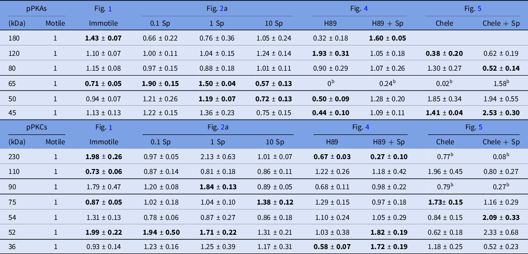
Changes in the phosphorylation levels of PKA substrates and PKC substrates in sperm exposed to different motility conditions

Numbers in bold are the significative changes indicated in the figures (*), comparing with the motile condition.
aSp: is nM [Speract].
b Averages of two experiments.
Discussion
The information shown in Table 1 suggests that the signalling cascade triggered by speract is mainly regulated by kinases of the AGC family that comprises cAMP-dependent protein kinase (PKA), PKC and PKB [activated by the phosphoinositide 3-kinase (PI3K); Manning and Toker, Reference Manning and Toker2017]. In turn PKC is divided into three isotypes: PKC conventional [α, β, γ; regulated by Ca2+ and diacylglycerol (DAG)], PKC novel (δ, ϵ, η, θ; regulated by DAG) and PKC atypical (ζ, ι; regulated by phosphatidylserine as all the PKC isoenzymes) (Newton, Reference Newton2018). Doing a search in the S. purpuratus genome, we found PKC, PKCδ, PKCι and the Ca2+-independent PKC. The fact that calphostin C (a DAG competitor) inhibited L. pictus sperm motility supported the presence of DAG-regulated PKC activity in sperm of sea urchin (White et al., Reference White, de Lamirande and Gagnon2007).
The fact that the different compounds used to evaluate the participation of PKA and PKC in sperm motility did not inhibit with the same efficiency suggested a different mechanism of action for PKA and the participation of distinct isoenzymes of PKC.
By using WB experiments with antibodies against phosphorylated PKA or PKC substrates and inhibitors to both enzymes in motile and speract-stimulated intact S. purpuratus sea urchin sperm (Table 3), we confirmed the participation of PKA (Loza-Huerta et al., Reference Loza-Huerta, Vera-Estrella, Darszon and Beltrán2013), as proposed earlier for demembranated sperm (Tash et al., Reference Tash, Hidaka and Means1986; Su et al., Reference Su, Chen, Zhou and Vacquier2005), and PKC, as suggested for S. purpuratus (Loza-Huerta et al., Reference Loza-Huerta, Vera-Estrella, Darszon and Beltrán2013) and L. pictus (White et al., Reference White, de Lamirande and Gagnon2007), in modulating their phosphorylation levels. The finding that the inhibition of PKA (Figs 3A, 4A,B) altered the phosphorylation levels of PKC substrates (Fig. 4C, D) and that PKC inhibition (Figs 3C, 5A,B) modified PKA substrates (Fig. 5C,D) suggested that a crosstalk mechanism between these two enzymes regulated S. purpuratus sperm motility.
Our results also indicated that the participation of PKC in the motility of S. purpuratus sperm was as important as that of PKA. As we mentioned before, the binding of speract to its receptor triggered several ionic changes that included an increase in the [Ca2+]i (Wood et al., Reference Wood, Nishigaki, Furuta, Baba and Darszon2005) that could participate in the regulation of Ca2+-dependent kinases/phosphatases. Indeed, the [Ca2+]i was different depending on the motility condition of the sperm (immotile, motile or stimulated by speract). Although in S. purpuratus sperm, several isoforms of transmembrane (tm) adenylyl cyclases (AC) (tmAC1, tmAC2, tmAC5, and tmAC9; Beltrán et al., Reference Beltrán, Vacquier, Moy, Chen, Buck, Levin and Darszon2007; Vacquier et al., Reference Vacquier, Loza-Huerta, García-Rincón, Darszon and Beltrán2014) are expressed, more than 90% of the cAMP is produced by soluble AC (sAC; Vacquier et al., Reference Vacquier, Loza-Huerta, García-Rincón, Darszon and Beltrán2014), activated by alkaline pHi (Nomura et al., Reference Nomura, Beltrán, Darszon and Vacquier2005; Körschen et al., Reference Körschen, Hamzeh, Pascal, Alvarez, Bönigk, Kaur, Levin, Buck, Kambach, Michino, Jennings, Sato, Seifert, Strünker, Steegborn and Kaupp2021) and by bicarbonate (Beltrán et al., Reference Beltrán, Vacquier, Moy, Chen, Buck, Levin and Darszon2007). In addition, Table 1 shows that all the proteins from the signalling cascade of speract (Guerrero et al., Reference Guerrero, Gadêlha, Ramírez-Gómez, Ramírez, Beltrán and Tuval2020) had predicted sites for phosphorylation by PKA and PKC and all but five (Catsperβ, Catsperϵ1, Catsperϵ2, EFCAB9 and PKC) contained at least one motif recognized by anti-PKA substrates. Table 1 also showed that, from the 41 proteins included, there were seven [SpR (121 kDa), PDE5 (108 kDa), Tetra-KCNG (253 kDa), sAC (209 kDa), CaKC (119 kDa), Cav1.2 (α1D subunit, 229 kDa) and Cav2.3 (A α1 X18 (289kDa)] that contained, in addition to PKA and PKC phosphorylation sites, one predicted site for PKB. Furthermore, 13 proteins (Tetra-KCNG, PKA RII, SpIh, CatSper4, KNCE, Cav1.2, Cav2.3, PKCδ X1, X2 and Ca-indep PKC X1–X5) contained a motif recognized by the anti-PKC substrates. Additionally, Cav1.2 and Cav2.3 had three and two Cdk5 predicted phosphorylation sites, respectively, and there were 11 proteins (PDE5, sAC, PKA RII, CatSper4, KNCE, Cav2.3 and PKCι X1–X5) with at least one predicted phosphorylation site for CKII. This suggests that all the proteins in the cascade triggered by the speract may be regulated by phosphorylation.
Unlike previous results with demembranated sperm, our findings using intact sea urchin sperm from S. purpuratus indicated that both PKA and PKC participated in the regulation of motility. Moreover, White et al. (Reference White, de Lamirande and Gagnon2007) detected four PKC substrates (200, 100, 65 and 28 kDa) in immotile L. pictus sperm whose phosphorylation level increased when reactivating the sperm motility by diluting the cells in ASW pH 8, in which all but the 65 kDa protein were associated with the flagella (White et al., Reference White, de Lamirande and Gagnon2007).
Although we used the same antibody to detect PKC substrates in immotile S. purpuratus sperm in our WB experiments, the Mr of the bands detected in this work (230, 110, 90, 75, 54, 52 and 36 kDa) was different. In addition to the fact that the sperm in these two studies were from distinct species, the differences could also be due to variations in the experimental conditions and medium used to immobilize the sperm, which led to changes at least in [Ca2+]i (activating or inactivating kinases/phosphatases) and [K+]i (ΔEm) (Supporting information Table S1).
As in mouse, in S. purpuratus sperm, a calmodulin (CaM)-dependent protein phosphatase involved in the Ca2+-dependent regulation of flagellar motility has been described (Tash et al., Reference Tash, Krinks, Patel, Means, Klee and Means1988). Furthermore, human sperm contains a Ca2+/CaM-dependent protein kinase type IV involved in motility (Marín-Briggiler et al., Reference Marín-Briggiler, Jha, Chertihin, Buffone, Herr, Vazquez-Levin and Visconti2005) that is also in the S. purpuratus genome (XP_030832817.1). It will be important to determine if that enzyme is also present in the sea urchin sperm, as well as the phosphatases PP1A (gi|72045279) and PPA2 (gi|115675671), and PKA RII that were identified by proteomic analysis in samples of flagella from S. purpuratus, among other proteins from the speract signalling cascade (reviewed in Beltrán Núñez, Reference Beltrán Núñez, Arenas Rios and Fuentes Mascorro2019; Guerrero et al., Reference Guerrero, Gadêlha, Ramírez-Gómez, Ramírez, Beltrán and Tuval2020). In the flagella of mouse sperm, CatSper is located in four linear nanodomains colocalizing with a Ca2+/CaM-dependent kinase II (CaMKII) and the phosphatase calcineurin (PP2B-Aγ) forming a microdomain important for motility (Chung et al., Reference Chung, Shim, Everley, Gygi, Zhuang and Clapham2014). A Ca2+-dependent PP2B is bound to the axoneme in sea urchin sperm, regulating dynein phosphorylation (Tash et al., Reference Tash, Krinks, Patel, Means, Klee and Means1988).
The involvement of PKA and PKC in the regulation of both the initiation and the speract-stimulated motility, as well as the crosstalk between the two enzymes, is clear. The fact that different concentrations of speract modified the phosphorylation patterns of sperm differentially could suggest that, while the cells are swimming towards the egg, the speract gradient detection system modifies the phosphorylation levels of various proteins to regulate sperm motility, and that both PKA and PKC are involved.
For possible identity of some of the PKA substrates and PKC substrates PDE5 (NP_001029121.1; 108 kDa), Table 1 contains predicted phosphorylation sites for PKA (four), PKB (one), PKC (15), and CKII (one) and three motifs (two in PKA and one in PKB) recognized by anti-PKA substrates. This protein was detected previously in sea urchin sperm by proteomic analysis as well as by WB experiments with anti-PKA substrates (Su et al., Reference Su, Chen, Zhou and Vacquier2005; Nomura and Vacquier, Reference Nomura and Vacquier2006; Beltrán Núñez, Reference Beltrán Núñez, Arenas Rios and Fuentes Mascorro2019; Trötschel et al., Reference Trötschel, Hamzeh, Alvarez, Pascal, Lavryk, Bönigk, Körschen, Müller, Poetsch, Rennhack, Gui, Nicastro, Strünker, Seifert and Kaupp2020). As we mentioned in the results section, when sperm are preincubated with H89, the phosphorylation degree of the 120 kDa protein revealed with anti-PKA substrates increases (1→1.93 ± 0.31). As H89 raises the level of phosphorylation of PKB (Melick and Jewell, Reference Melick and Jewell2020), it is possible that PKB could in turn elevate the phosphorylation level of the 120 kDa protein. All that together suggests that the 120 kDa protein might correspond to PDE5.
Sea urchin sperm sAC could be detected by WB with anti-sAC as a full-length protein (190 kDa) or as a truncated form of 65 kDa (Beltrán et al., Reference Beltrán, Vacquier, Moy, Chen, Buck, Levin and Darszon2007). It is highly probable that the 65 kDa protein detected by anti-PKA substrates corresponded to sAC. This protein has 15 putative phosphorylation sites by PKA and one by PKB, in addition to 13 recognition motifs by anti-PKA substrates (Table 1). As can be seen in Table 3, the phosphorylation degree of this protein is very much regulated by the motility condition and by speract. The degree of phosphorylation of this protein increased with motility (0.71 ± 0.05→1) and even more with 0.1 nM speract (1→1.90 ± 0.15), to start diminishing with 1 nM speract (1.90 ± 0.15→1.50 ± 0.04) and further with 10 nM speract (1.50 ± 0.04→0.57 ± 0.13). The levels of phosphorylation of the 65 kDa protein were inhibited by H89 (1→0) even when stimulated by speract (0→0.24) (Table 3 and Fig. 4) and by chelerythrine (1→0.02), which reverted when the cells pre-exposed to chelerythrine were stimulated by speract (Figure 5 and Table 3). These results could suggest that the sAC in addition to being regulated by pHi and bicarbonate (Nomura et al., Reference Nomura, Beltrán, Darszon and Vacquier2005; Beltrán et al., Reference Beltrán, Vacquier, Moy, Chen, Buck, Levin and Darszon2007, Vacquier et al., Reference Vacquier, Loza-Huerta, García-Rincón, Darszon and Beltrán2014) is also regulated by phosphorylation at least by PKA and that there is a crosstalk between PKA and PKC. As indicated earlier, sAC is responsible for synthesizing more than 90% of the cAMP in S. purpuratus sperm (Vacquier et al., Reference Vacquier, Loza-Huerta, García-Rincón, Darszon and Beltrán2014).
The 45 kDa PKA substrate could correspond to the S. purpuratus cAMP-dependent protein kinase type II regulatory subunit (PKA RII; NP_999688.1; 42 kDa) detected with the anti-phospho-(Ser/Thr) PKA substrate by Loza-Huerta et al. (Reference Loza-Huerta, Vera-Estrella, Darszon and Beltrán2013) in membrane microdomains (LD-DIM) from whole S. purpuratus sperm or isolated flagella. This is considering that it contains three motifs recognized by the antibody against PKA substrates and one PKA predicted site by the NetPhos-3.1 Server (Table 1). In addition, the phosphorylation levels of this substrate are lowered (1→0.44± 0.1) by H89 and reverted to basal levels (1.09 ± 0.1) when stimulated with speract. As the 36 kDa protein detected by the anti-PKC substrates had a similar behaviour (H89, 1→0.58 ± 0.07 and H89+Sp, 1→1.72 ± 0.19), it could correspond to PKA that has three predicted sites for PKC and one motif recognized by anti-PKC substrates. This strengthens the proposed cross-regulation between PKA and PKC.
As for the 90 kDa PKC substrate, it may correspond to the reported K+-dependent Na+/Ca2+ exchanger (suNCKX; NP_999790.1 with calculated molecular mass of 72 kDa, five putative N-linked glycosylation sites and two PKA sites), cloned from S. purpuratus testis. This exchanger was identified in the flagella of sea urchin sperm and proposed to play a major role in keeping low levels of [Ca2+]i in swimming sperm. In SDS-PAGE, this protein runs with an Mr of 90 kDa (Ward et al., Reference Ward, Moy and Vacquier1986; Su and Vacquier, Reference Su and Vacquier2002). When analyzing the suNCKX sequence with NetPhos-3.1, we found that the exchanger contains, in addition to two reported predicted PKA sites, five for PKC. Furthermore, suNCKX also contained one motif recognized by anti-PKA substrates and another recognized by the anti-PKC substrates, confirming that it is a PKA substrate and PKC substrate, and the possibility that the 90 kDa protein corresponds to the suNCKX exchanger involved in the speract signalling cascade (Espinal-Enríquez et al., Reference Espinal-Enríquez, Priego-Espinosa, Darszon, Beltrán and Martínez-Mekler2017; Guerrero et al., Reference Guerrero, Gadêlha, Ramírez-Gómez, Ramírez, Beltrán and Tuval2020). It is important to note that speract 1 nM promotes the increase (Sp 1 nM; 1→1.84 ± 0.13) in phosphorylation of the 90 kDa PKC substrate, while it diminishes the phosphorylation of the 80 kDa protein detected by anti-PKA substrates. Probably both proteins correspond to the suNCKX. Indeed, our proteomic analysis of LD-DIM from S. purpuratus sperm resolved in SDS-PAGE indicated that suNCKX migrates within a molecular weight (MW) range 75–100 kDa (C. Beltrán, unpublished).
Finally, the 36 kDa PKC substrate (Fig. 4) may correspond to the axonemal p30 important for the motility of S. purpuratus sperm. This protein was cloned and sequenced from sea urchin (Gingras et al., Reference Gingras, White, Garin, Multigner, Job, Cosson, Huitorel, Zingg, Dumas and Gagnon1996) and corresponds to the inner dynein arm light chain (NP_999680.1; 33 kDa) in the S. purpuratus genome. It has three predicted sites for PKC according to the NetPhos-3.1 Server.
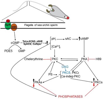
In conclusion, this is the first work that shows that the changes (Fig. 6) induced by the chemoattractant and motility regulator peptide speract activate at least PKA and PKC in intact sea urchin sperm, and that there is cross-communication between PKA and PKC in the onset of motility as stimulated by speract. The PKC isoforms involved in sperm motility as well as its subcellular localization remain to be determined. There is still a long way to go and experiments to be done to unravel the complexity of speract/PKA/PKC signalling in sperm.
PKA and PKC crosstalk model in sea urchin sperm. Binding of speract (Sp) to its receptor (SpR) activates a membrane guanylyl cyclase (mGC) and increasing cGMP levels. cGMP regulated by phosphodiesterase 5 (PDE5) binds to and opens the K+ channel; tetra-KCNG, and K+ efflux hyperpolarizes the cell, activating the sperm-specific Na+/H+ exchanger (sNHE) and alkalizing pHi. The alkaline pHi regulates the sAC elevating the cAMP levels that activate PKA, and modulate the Na+ channel (SpHCN; also stimulated by hyperpolarization), and the sperm-specific Ca2+ channel, CatSper (Hwang et al., Reference Hwang, Mannowetz, Yongdeng Zhang, Robert, Everley, Steven, Gygi, Bewersdorf, Lishko and Jean-Ju Chung2019; Loyo-Celis et al., Reference Loyo-Celis, Orta, Beltrán and Darszon2021). The increases in Ca2+ and diacylglycerol (DAG), activate PKC. DAG also modulates PKCδ and phosphatidylserine (PS) regulates all PKC isoenzymes (Newton, Reference Newton2018). All these changes modulate kinases and phosphatases that in turn regulate the degree of phosphorylation/dephosphorylation of PKA substrates and PKC substrates involved in sperm motility (Guerrero et al., Reference Guerrero, Gadêlha, Ramírez-Gómez, Ramírez, Beltrán and Tuval2020). Chelerythrine (PKC; Herbert et al., Reference Herbert, Augereau, Gleye and Maffrand1990) and H89 (PKA; Chijiwa et al., Reference Chijiwa, Mishima, Hagiwara, Sano, Hayashi, Inoue, Naito, Toshioka and Hidaka1990) are inhibitors of kinases.

Supplementary material
To view supplementary material for this article, please visit https://doi.org/10.1017/S0967199421000915
Acknowledgements
We would like to thank JL. de la Vega-Beltrán and A. Blancas for technical support; S. Ainsworth and O. Arriaga for library services and J.M. Hurtado, R. Rodríguez, D. Santiago and A. Ocádiz for computer services.
Financial support
This work was supported by UNAM-DGAPA-PAPIIT (IN206016 and IN215519 to CB and IN205516 and IN200919 to AD) and CONACyT (Fronteras 71) to AD. AL-H was a PhD student of the Doctorado en Ciéncias Biomédicas-UNAM programme, supported by CONACyT and UNAM-DGAPA-PAPIIT. HP-C is a PhD student of the Doctorado en Ciencias Biomédicas-UNAM programme, supported by CONACyT.
Statement of interest
The authors declare that they have no conflict of interest in the research reported.
Ethical standards
The authors assert that all procedures contributing to this work complied with the ethical standards of the relevant national and institutional guidelines on the care and use of laboratory animals.