Introduction
Endocannabinoids (ECBs) and their receptors have been found throughout the central nervous system (CNS) and play an important role in the control of neuronal excitability. Cannabinoids modulate neuronal excitability by controlling neurotransmitter (NT) release. The ECBs N-arachidonoylethanolamine (anandamide or AEA) and 2-arachidonoyl glycerol are synthesized and released from postsynaptic neurons in response to depolarization (Kreitzer & Regehr, Reference Kreitzer and Regehr2001) or metabotropic receptor activation (Maejima et al., Reference Maejima, Hashimoto, Yoshida, Aiba and Kano2001a ,Reference Maejima, Ohno-Shosaku and Kano b ; Adermark et al., Reference Adermark, Talani and Lovinger2009). Once released, ECBs travel retrogradely to activate the presynaptic cannabinoid receptor 1 (CB1R) and hyperpolarize presynaptic terminals, thus reducing GABA and glutamate release (Diana & Marty, Reference Diana and Marty2004; Chevaleyre et al., Reference Chevaleyre, Takahashi and Castillo2006; De Petrocellis & Di Marzo, Reference De Petrocellis and Di Marzo2009; Heifets & Castillo, Reference Heifets and Castillo2009).
The activity of cannabinoids has been looked at in many areas of the brain including mouse cerebellar Purkinje cells (Kreitzer & Regehr, Reference Kreitzer and Regehr2001; Maejima et al., Reference Maejima, Hashimoto, Yoshida, Aiba and Kano2001a ,Reference Maejima, Ohno-Shosaku and Kano b ; Diana et al., Reference Diana, Levenes, Mackie and Marty2002; Galante & Diana, Reference Galante and Diana2004), pyramidal neurons in the hippocampus in mice and rats (Wang & Zucker, Reference Wang and Zucker2001; Trettel & Levine, Reference Trettel and Levine2003; Fortin et al., Reference Fortin, Trettel and Levine2004), and autaptic hippocampal neurons in culture (Straiker & Mackie, Reference Straiker and Mackie2005; Kellogg et al., Reference Kellogg, Mackie and Straiker2009) and in mouse visual cortex (Crozier et al., Reference Crozier, Wang, Liu and Bear2007; Huang et al., Reference Huang, Yasuda, Sarihi and Tsumoto2008). Recently, it was shown that the maturation of GABAergic signaling in mouse visual cortex depends on ECB-mediated long-term depression (LTD) (Jiang et al., Reference Jiang, Huang, de Pasquale, Millman, Song, Lee, Tsumoto and Kirkwood2010a ), sequentially developing from layers 4 to 2/3 during adolescence (Jiang et al., Reference Jiang, Sohya, Sarihi, Yanagawa and Tsumoto2010b ). The ECB system has also been localized to cells within the retina, suggesting a possible role in retinal processing (see Yazulla, Reference Yazulla2008, for review). It is speculated that disrupting the ECB system in the retina with marijuana use (in which the psychoactive component delta-9-tetrahydrocannabinol is a cannabinoid) can alter the visual signal improving night vision (Russo et al., Reference Russo, Merzouki, Mesa, Frey and Bach2004), further suggesting a role for cannabinoids in vision at an early stage of visual processing. The exact role of the ECB system in the retina and in vision, however, is poorly understood.
The mRNA for cannabinoid receptors has been detected in embryonic and adult rat retinas (Buckley et al., Reference Buckley, Hansson, Harta and Mezey1998; Lu et al., Reference Lu, Straiker and Maguire2000), and the receptors themselves have been localized to the anterior eye and retina of humans (Straiker et al., Reference Straiker, Maguire, Mackie and Lindsey1999a ). CB1Rs and their ligands have also been localized to the inner plexiform layer (IPL) and outer plexiform layer in rhesus monkey, mouse, rat, chick, goldfish, and tiger salamander (Straiker et al., Reference Straiker, Stella, Piomelli, Mackie, Karten and Maguire1999b ) and specifically found on horizontal cells and rod bipolar cells of the rat (Yazulla et al., Reference Yazulla, Studholme, McIntosh and Deutsch1999) as well as on Müller glial cells and bipolar cells in goldfish retina (Yazulla et al., Reference Yazulla, Studholme, McIntosh and Fan2000).
While the presence of the ECB system in the retina has been established, relatively few studies have looked at its physiological role. At the first synapse of the retina, a cannabinoid agonist has been shown to modulate potassium and calcium currents in rod and cone photoreceptor terminals in tiger salamander and goldfish retina (Fan & Yazulla, Reference Fan and Yazulla2003; Straiker & Sullivan, Reference Straiker and Sullivan2003). In addition, a cannabinoid agonist was shown to inhibit high–voltage-activated calcium channel currents in cultured retinal ganglion cells (RGCs) (Lalonde et al., Reference Lalonde, Jollimore, Stevens, Barnes and Kelly2006). More recently, a cannabinoid agonist was found to modulate spontaneous transmitter release in cultured embryonic amacrine cells of chick embryo proportionally to the initial frequency of minis (Warrier & Wilson, Reference Warrier and Wilson2007).
To investigate the potential role of ECBs as neuromodulators in retinal signaling, we examined the effects of a CB1R cannabinoid agonist WIN55212-2 on spontaneous synaptic activity. Voltage-clamp recordings were carried out from mouse RGCs in the whole-mount preparation. We hypothesized that a cannabinoid receptors (CB1) agonist would reduce the amount of spontaneous postsynaptic currents (SPSCs) affecting both inhibitory (IPSCs) and excitatory (EPSCs) currents, on the basis that CB1R activation leads to a reduction in NT release. Additionally, we investigated the role of ECBs at two different stages of retinal development, adolescent mice before the surge in spontaneous synaptic activity associated the critical period (P14–P21), and adult mice (older than 6 weeks). Overall, we found that a cannabinoid agonist reduced the frequency of both inhibitory and excitatory postsynaptic currents in RGCs without affecting their amplitude and kinetics, suggesting a presynaptic locus of action. These experiments support the role of ECBs as negative modulators of presynaptic release having a direct influence in retinal processing and hence vision.
Methods
All animal procedures had approval from the Animal Ethics Committee of the University of Sydney and followed the guidelines for animal experiments issued the Australian Code of Practice for the Care and Use of Animals for Scientific Purposes and the National Health and Medical Research Council of Australia.
Tissue preparation
Two groups of C57bl/6j mice were used for this study, classified as adolescent (between P14–P21 days old) and adult mice (older than 6 weeks old). Mice were euthanized by cervical dislocation, and the eyes were enucleated and dissected under carboxygenated Ames medium under white light, thus the tissue was light adapted. A hemiretina photoreceptor side down was mounted in a recording chamber, which was then mounted in an upright microscope (Axioskop 40; Zeiss, Carl Zeiss, Oberkochen, Germany). The tissue was continually perfused with carboxygenated Ames medium at 3–5 ml/min at 35°C. A small hole was torn in the inner limiting membrane to gain access to the ganglion cell layer where recordings were done in low ambient light conditions.
Recordings
Whole-cell patch-clamp recordings were obtained using borosilicate glass pipettes with a resistance of 6–8 MΩ. High resistance seals (>1 GΩ) were made on the cell body of large neurons (>15 μm diameter) in the ganglion cell layer and held at a holding potential of −65 mV (corrected for liquid junction potential). Spontaneous currents were recorded for 3–6 min periods using Pulse 8.67 (HEKA Electronik, Lambrecht, Germany) software.
Solutions
Intracellular solution containing (in millimolar) CsCl: 150, HEPES: 10, MgCl2: 4.6, ATP-Na: 4, GTP-Na: 0.4, tetrabutylammonium (TBA) (5 mM), and QX314 (5 mM) were also added to the intracellular solution to block voltage-gated potassium and sodium currents. This intracellular solution containing high CsCl was used to set E cl− to ∼0 mV to increase the driving force of GABA-mediated currents. Under these conditions, both spontaneous excitatory and inhibitory synaptic currents were recorded as inward currents when holding cells at −65 mV. Additionally, an intracellular solution containing Cesium Methanesulfonate containing (in millimolar) Cesium Methanesulfonate: 100, HEPES: 20, EGTA: 10, CaCl2: 1, MgCl2: 4.6, ATP-Na: 4, GTP-Na: 0.4, creatine phosphate: 20, creatine Ph-kinase: 250, TBA: 5 was used to determine the reversal potential of spontaneous synaptic currents in the presence of a GABAA antagonist.
Drug applications
Spontaneous synaptic activity was recorded under several pharmacological conditions. Ames medium containing 5 μm NBQX (AMPA/kainate receptor blocker) was used to block glutamatergic currents to specifically isolate and record inhibitory currents. Alternatively, 5 μm SR95531 (GABAA receptor blocker) was used to block GABAergic currents to specifically isolate and record excitatory currents. Recordings were taken before and after bath application of WIN55212-2 (5 μm), a cannabinoid (CB1) receptor agonist, and then washed out with fresh Ames medium to measure the recovery of activity.
Data analysis
Recordings were analyzed off-line using Mini Analysis 6.0.3 (Synaptosoft, Decatur, GA). Minimum detection thresholds were set at amplitude: 5 pA and area: 30 fC for EPSCs and amplitude: 30 pA and area: 50 fC for IPSCs. The rise time, decay time, area, and amplitude were recorded and analyzed as well as the frequency of currents. Kolmogorov–Smirnov (K-S) statistical analysis was used to quantify differences among distributions (cumulative plots) and Student’s t-test to test the equality or difference of mean frequency values.
Results
RGCs receive spontaneous and light-driven inputs from bipolar and amacrine cells. Pharmacological and biophysical characterization of these spontaneous EPSCs and IPSCs has revealed glutamatergic inputs as well as GABAergic and glycinergic inputs (Protti et al., Reference Protti, Gerschenfeld and Llano1997; Tian et al., Reference Tian, Hwang and Copenhagen1998; Flores-Herr et al., Reference Flores-Herr, Protti and Wassle2001). Our experiments consisted of whole-cell recordings of SPSCs from mouse RGCs. These recordings were taken in whole-mount retinas with cells held at −65 mV below activation threshold of high–voltage-activated Ca2+ currents, to prevent release of endogenous cannabinoids. Consistent with previous observations, all RGCs recorded from displayed spontaneous synaptic activity. The majority of SPSCs observed were attributed to GABAergic and glutamatergic currents. When both these currents were blocked, we observed minimal spontaneous currents (data not shown). This suggests that under our recording conditions, RGCs receive mostly GABAergic IPSCs rather than glycinergic input, as previously shown to be the more common scenario (Protti et al., Reference Protti, Gerschenfeld and Llano1997; Tian et al., Reference Tian, Hwang and Copenhagen1998).
Cannabinoids reduce SPSCs in RGCs
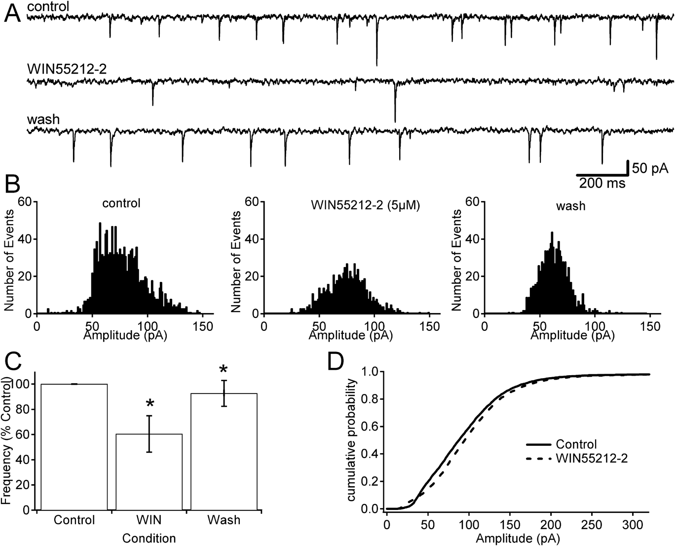
To investigate whether or not cannabinoids play a modulatory role on transmitter release in the retina, we studied the effect of the CB1R agonist WIN55212-2 on the frequency and amplitude of spontaneous synaptic currents in RGCs; bath application of 5 μM WIN55212-2 significantly reduced the frequency of SPSCs. Fig. 1A shows representative traces of spontaneous activity in a RGC in control conditions, in which SPSC frequency was 4.8 events/s. Bath application of 5 μM WIN55212-2 reduced spontaneous frequency to 1.1 events/s and this effect was reversible (SPSC frequency 4.2 events/s after wash out of the drug). Fig. 1B shows amplitude histograms for the same cell in the three experimental conditions. Across all cells tested, control SPSC frequency ranged from 1.3 to 9.4 events/s (mean = 6.4).
The effect of WIN55212-2 on SPSCs in RGCs. (A) Representative traces from a continuous recording from a RGC obtained using a CsCl intracellular solution at holding potential −65 mV. The frequency of SPSCs was reversibly reduced by bath application of 5 μM WIN55212-2. (B) Amplitude distributions of SPSCs in control condition (1907 events), in the presence of 5 μM WIN55212-2 (440 events), and after the drug was washed out (1666 events). The total sampling time was the same for each histogram, 400 s with 1 pA bin widths. (C) Averages of the effect of WIN55212-2 (5 μM) on SPSC frequency as percentage with respect to the initial control activity across all cells. On average, there was a significant reversible reduction in the frequency of currents (P < 0.05, n = 8). (D) Cumulative probability versus SPSC amplitude from the same cell showing that the amplitude distribution of currents was not altered in by the drug treatment (WIN55212-2, dotted trace) as compared to the control condition (solid trace, K-S: P > 0.05, 1000 currents for each condition), consistent with a presynaptic locus of action.

Across all eight cells tested, application of WIN55212-2 produced a reduction in seven, while one cell showed little change. The combined result of the effect of WIN55212-2 in all the cells (Fig. 1C) showed a significant reduction overall to approximately 60% of control (P < 0.05, n = 8). This reduction was reversible upon washing out the drug, leading to a recovery in SPSC activity to ∼93% of the control frequency (P < 0.05, n = 7). The reduction caused by the drug application ranged from 1.4 to 2.5 events/s (mean = 1.8) and was not proportional with the initial recorded frequency of the cell.
While WIN reduced the frequency of SPSCs, it had no effect on the amplitude of these currents as shown by the cumulative probability of SPSC amplitudes (Fig. 1D). The K-S test shows that these two distributions are not significantly different (P > 0.05), suggesting a presynaptic locus of action for WIN55212-2, which is consistent with the described mechanisms of action of the ECB system in other CNS areas.
Cannabinoids reduce IPSCs in RGCs
The observed reducing effect of a cannabinoid agonist on spontaneous frequency does not discriminate whether the modulatory effect of WIN on spontaneous synaptic activity was due to its action on the inputs originating from bipolar cells, amacrine cells, or both. Nevertheless, they demonstrate that this effect occurs in physiological conditions, in which transmitter release and the balance of excitation and inhibition have not been altered. To further characterize this effect, IPSCs were isolated using 5 μm NBQX, which blocks AMPA/KAINATE receptors and thus, spontaneous EPSCs from bipolar cells.
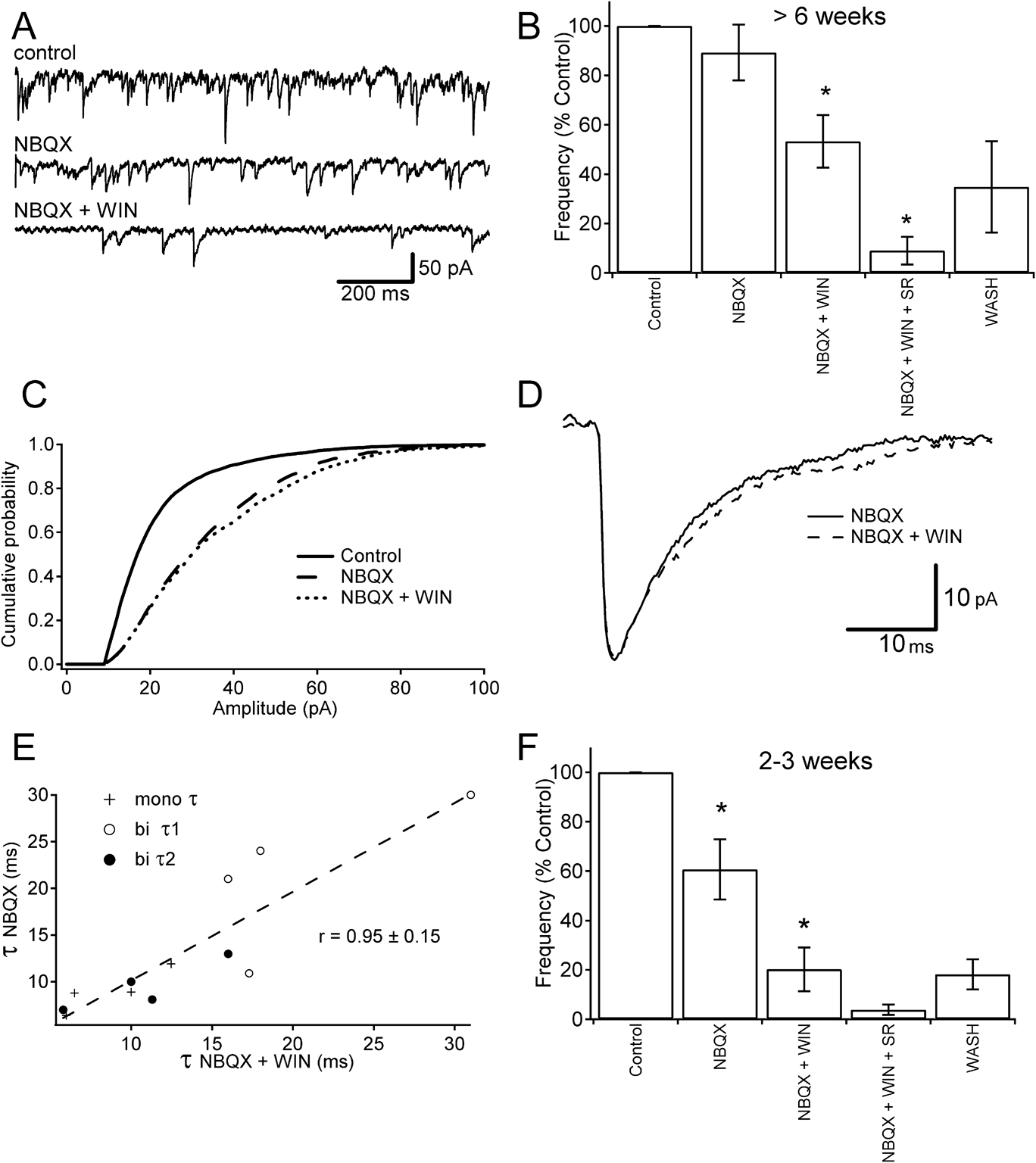
Fig. 2A shows representative traces of spontaneous activity in a RGC in successive pharmacological conditions in adult mice. In the control condition, both IPSCs and EPSCs were present; bath application of NBQX removed EPSCs by blocking AMPA/kainate receptors leading to a lower frequency of SPSCs, with the remaining events consisting of inhibitory currents (6.8 events/s). The frequency of IPSCs was further reduced by bath application of 5 μm WIN55212-2 (4.7 events/s).
The effect of WIN55212-2 on inhibitory PSCs in RGCs. (A) Representative traces from an adult mouse RGC of a continuous recording obtained using a CsCl intracellular solution at holding potential −65 mV In control conditions, both excitatory and inhibitory currents were present. The excitatory currents were removed by blocking glutamate receptors via bath application of NBQX (5 μm). The frequency of the remaining inhibitory currents was reduced after the addition of WIN55212-2 (5 μm) a CB1 receptor agonist. (B) Mean IPSCs frequency from all cells during successive drug applications in adult mice. Addition of WIN55212-2 significantly reduced the frequency of inhibitory currents (P < 0.05, n = 8). The majority of the remaining currents were blocked by bath application of SR95531, a GABAa receptor blocker. (C) Representative cumulative probability plot of current amplitude in adult mouse RGC (curves generated with 1000 currents in each recording condition). After excitatory currents were blocked with NBQX, there was a shift to the right in the amplitude distribution when compared to control conditions. Addition of WIN55212-2, however, did not cause any shift, indicating that the decrease in spontaneous synaptic transmission is mediated presynaptically. (D) Comparison of an average IPSC trace before and after the addition of WIN55212-2 (5 μm) in adult mouse (>300 currents for each condition), showing that the amplitude and decay time did not change between the NBQX condition (solid line) and the NBQX + WIN55212-2 condition (dashed line). (E) Average τ values before and after application of WIN55212-2 in adult mice. The average IPSCs were consisted of monoexponential (crosses) and biexponential decay times (τ1 = open circle, τ2 = closed circle). Pre-WIN application decay values had a correlation with post-WIN values of 0.95 ± 0.15 (n = 8 cells). (F) WIN55212-2 causes a similar reduction in frequency in adolescent mice as observed in adult (P < 0.05, n = 7). Unlike in adult mice, however, isolating IPSCs with 5 μM NBQX significantly reduced current frequency (P < 0.05, n = 7).

Fig. 2B summarizes the average effect of successive pharmacological treatments on SPSC frequency across all cells in adult mice. Bath application of NBQX caused a change in frequency in all cells, with either increases or decreases in synaptic activity observed at the individual cell level; however, the mean only showed an overall small decrease and this effect was not significant (see Discussion). Application of WIN caused a significant reduction in frequency of the remaining IPSCs of ∼42% (P < 0.05 n = 8). The remaining activity was reduced to near baseline (4%) after blocking GABAergic IPSCs with 5 μM SR9553. Washing out NBQX, WIN, and SR95531 from the tissue allowed some recovery (∼35%).
SPSCs recorded in control conditions, which include both EPSCs and IPSC, had a mean amplitude of −24.7 pA in eight cells. Application of 5 μM NBQX to remove AMPA/KA receptor-mediated events led to a nonsignificant increase to −33.7 pA (average of all events in all cells). Further application of WIN55212-2 (5 μM) did not produce any significant change in the mean amplitude of IPSCs (−29.6 pA, P > 0.05; n = 8 cells). Fig. 2C shows the distribution of current amplitude in one representative RGC from an adult mouse (1000 currents in each condition). Addition of NBQX alone shifted the cumulative distribution of amplitudes to the right as the smaller amplitude excitatory currents were blocked, thus leaving only larger amplitude inhibitory currents (Protti et al., Reference Protti, Gerschenfeld and Llano1997; Tian et al., Reference Tian, Hwang and Copenhagen1998). Further addition of WIN, however, did not cause any change in the cumulative distribution indicating that cannabinoids have minimal effect on the amplitude of the IPSCs and hence exert their effect presynaptically (Statistical analysis using K-S indicates P > 0.05). Average traces, each derived from over 300 IPSCs before and after the application of WIN55212-2 in a RGC from adult mouse retina, show that the mean amplitude and decay time were unchanged by the cannabinoid agonist (Fig. 2D).
To further assess whether or not WIN55212-2 had any postsynaptic effects, we analyzed its effect on the IPSCs kinetics. Similar decay times were observed before and after WIN application in adult mice. Decay time constants were calculated by generating an average IPSC from over 100 individual currents for each adult cell tested. The average decay times were monoexponential in 50% and biexponential in the remaining 50% of cells (n = 8). Fig. 2E shows that the average decay time constants (τ 1 and τ 2) before and after WIN have a correlation R = 0.95 ± 0.15, indicating that the kinetics of postsynaptic currents were not affected by WIN55212-2. These results are, again, consistent with a presynaptic locus of action, as we have previously observed for the overall spontaneous synaptic activity.
RGCs have been observed to exhibit frequent transient bursting activity prior to and during eye opening (Galli & Maffei, Reference Galli and Maffei1988; Wong et al., Reference Wong, Meister and Shatz1993) that is due to increased SPSCs (Tian & Copenhagen, Reference Tian and Copenhagen2001). Also, in other areas of the brain cannabinoids have been shown to have a differential effect between adolescent and adult mice (Kang-Park et al., Reference Kang-Park, Wilson, Kuhn, Moore and Swartzwelder2007). Of particular interest is the fact that the ECB system is involved in the maturation of GABAergic neurotransmission in visual cortex during the developmental critical period (Jiang et al., Reference Jiang, Huang, de Pasquale, Millman, Song, Lee, Tsumoto and Kirkwood2010a ). Given the reported dynamic of synaptic activity during retinal development, the action of cannabinoids may also change over time. Thus, the effects of WIN55212-2 on IPSCs were also examined in tissue from adolescent mice (P14–P21 days) to determine whether or not cannabinoids have a similar effect at earlier developmental stages in the retina.
The average frequencies in Fig. 2F show that isolating inhibitory currents with 5 μM NBQX produces a significant reduction in SPSCs to ∼60% of control frequency in adolescent mice (P < 0.05, n = 7). This blocking effect was much greater to what was observed in adult mice, in which isolating IPSCs caused no significant change in frequency. The addition of WIN caused a further decrease in frequency of ∼67% (P < 0.05, n = 7). The remaining activity was reduced to 2% after blocking GABAergic IPSCs with 5 μM SR95531. Washing out NBQX, WIN, and SR from the tissue allowed some recovery of approximately 20% of the control frequency.
Cannabinoids reduce EPSCs in RGCs
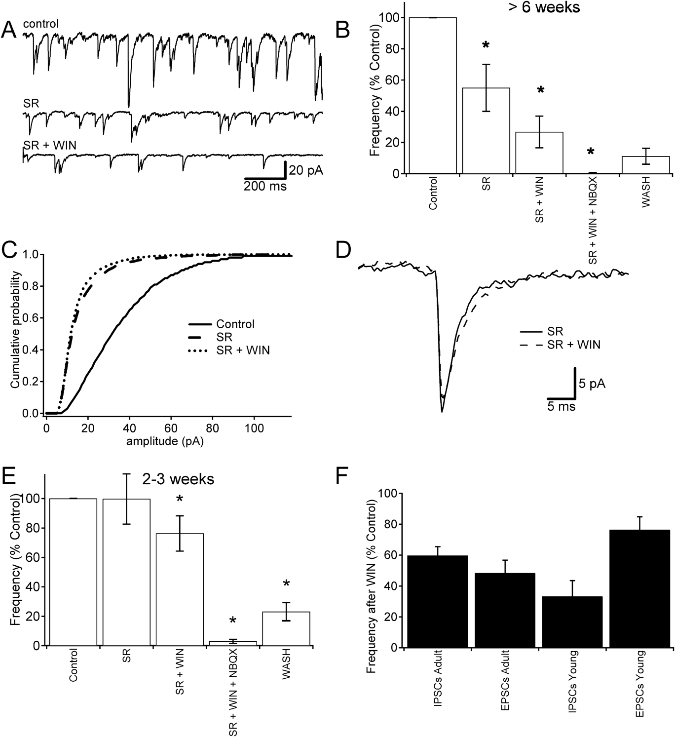
To determine whether or not WIN55212-2 also had an effect on excitatory inputs arising from bipolar cells, we isolated EPSCs by blocking GABAA receptors with 5 μm SR95531. Fig. 3A shows representative traces from an adult RGC in successive pharmacological conditions. In the control condition, both IPSCs and EPSCs were present; bath application of 5 μm SR95531 removed GABAergic currents, thus isolating EPSCs and resulting in a lower frequency of SPSCs (6.8 events/s). Addition of cannabinoids (5 μm WIN55212-2) further reduced the frequency of the remaining EPSCs (4.9 events/s). In a different set of experiments, we confirmed that the remaining spontaneous synaptic currents after SR95531 application were mediated by excitatory NTs by assessing their reversal potential using a Cesium Methanesulfonate intracellular solution. Under these conditions, SPSCs reversed on average at −5 mV (n = 8 cells; 1000 SPSCs; data not shown) consistent with nonselective cationic currents characteristic of AMPA/KA receptors and ruling out the presence of unblocked GABAA-mediated IPSCs and of glycine-mediated IPSCs in these recording conditions.
The effect of WIN55212-2 on excitatory PSCs in RGCs. (A) Representative traces from a continuous recording in adult mouse obtained using a CsCl intracellular solution. Holding potential −65 mV. Control condition contains both excitatory and inhibitory currents. Inhibitory currents were removed by blocking GABAa receptors via bath application of SR95531 (5 μm). The remaining excitatory currents were reduced after the addition of WIN55212-2 (5 μm), a CB1 receptor agonist. (B) Comparison of the mean frequency from all cells during successive drug application in adult mice. EPSCs were isolated by blocking GABAA receptors with 5 μm SR95531, which caused a significant reduction in the frequency of SPSCs. The remainder of the EPSCs was significantly reduced by the addition of WIN55212-2 (P < 0.5, n = 8). Remaining EPSCs were blocked (~1% activity left) with 5 μm NBQX, a glutamate receptor blocker, indicating that EPSCs were excitatory glutamate currents. (C) Representative cumulative probability plot of current amplitude in an adult mouse RGC. After blocking inhibitory currents with SR95531, a shift to the left in the distribution when compared to the control was observed. The addition of WIN55212-2, however, did not cause a significant shift (P > 0.05, n = 1015 currents) showing that the amplitude distribution of EPSCs was not altered, indicating that the observed decrease in spontaneous transmission is mediated presynaptically. (D) Comparison of average EPSC traces before and after the application of WIN55212-2 (5 μm) in adult mouse (>80 currents for each condition); amplitude and decay time did not change between the SR95531 condition (solid line) and the WIN55212-2 condition (dashed line). (E) The effect of WIN55212-2 on SPSC frequency is similar in adolescent to the one observed in adults. WIN55212-2 significantly reduced current frequency by ~24% (P < 0.05, n = 8). Isolating EPSCs with SR, however, did not lead to a significant drop in current frequency similar to that observed in adult mice. (F) The remaining frequency of spontaneous events after the addition of WIN55212-2 in all four conditions, IPSCs were reduced by ∼40 and ∼67% in adult (n = 8) and adolescent (n = 7) mice, respectively. EPSCs were reduced by ∼51 and ∼24% in adult (n = 8) and adolescent (n = 7) mice, respectively (mean 54.44%).

Fig. 3B shows the mean SPSC frequencies across eight cells in successive pharmacological conditions in adult mice. Blocking GABAergic currents caused a significant drop of approximately 55% in spontaneous activity (P < 0.05, n = 8). Application of a cannabinoid agonist caused a reduction in the frequency of the remaining EPSCs in seven of eight cells with an average of ∼49% reduced activity (P < 0.05, n = 8).
In control conditions, the mean amplitude of all events (EPSCs and IPSCs) was −38.5 pA for eight cells recorded in adult mice. Mean amplitude was reduced to −21.8 pA after inhibitory currents were removed with SR95531 (5 μM). After bath application of WIN55212-2 (5 μM), the mean amplitude of all remaining events was not significantly changed (−19.2 pA; P > 0.05; n = 7 cells, in one cell, the frequency of EPSCs after WIN application was zero; thus, this cell did not contribute towards the mean amplitude). Fig. 3C shows the cumulative amplitude distribution from one of the adult mouse RGCs. A shift to the left in the cumulative distribution of amplitudes compared to control was observed when GABA currents were blocked with SR95531, consistent with the removal of IPSCs that have larger amplitudes, but no change occurred when WIN was added. Statistical analysis using K-S indicates that amplitude distributions before and after WIN are not significantly different (P > 0.05). Fig. 3D displays average traces before and after WIN application in an adult mouse RGC (>80 EPSCs in each condition), showing that the currents have similar amplitudes and decay times and indicating negligible effects from the cannabinoid agonist on the postsynaptic properties of the EPSC. Across all cells, EPSCs had single exponential decays ranging from 3.3 to 4.4 ms (n = 7) and did not change after application of WIN55212-2 (data not shown). These results suggest that WIN does not affect the amplitude and time course of EPSCs, similar to the effects observed on IPSCs and further support a presynaptic locus of action.
Fig. 3E shows the changes in mean frequency that result from the same pharmacological treatments when performed on adolescent mice. Application of SR caused a change in frequency in all cells with either increases or decreases of synaptic activity observed at the individual cell level; the average, however, was not significantly changed from control (see Discussion). The addition of 5 μm WIN55212-2, however, caused a significant decrease in the remaining EPSC frequency of ∼24% (P < 0.05, n = 8). The remaining activity was reduced to near baseline (<1%) in both adolescent and adult mice after blocking glutamate EPSCs with 5 μM NBQX. Washing out the tissue of SR, WIN, and NBQX allowed some recovery of the spontaneous synaptic frequency in both conditions (∼11% in adult, ∼23% in adolescent).
Fig. 3F shows the remaining frequency of spontaneous synaptic activity after the application of a cannabinoid agonist to isolated IPSCs and EPSCs in adult and adolescent mice. With isolated IPSCs, cannabinoids had a smaller effect on adult mice reducing frequency by 40% compared to adolescent mice, in which they reduced by 67%. EPSCs, however, had the opposite trend with cannabinoids causing a 51% reduction in frequency in adult mice compared to a 23% reduction in adolescent mice, the smallest cannabinoid-induced reduction that we observed in all the conditions.
Discussion
We carried out experiments to determine the effects of cannabinoids on spontaneous transmitter release in the mouse retina and found that cannabinoids reduce the frequency of SPSCs in RGCs. While the frequency of spontaneous events was significantly reduced, there was no change in their amplitude and kinetics indicating that the drug had no effect on the postsynaptic receptors and consistent with a presynaptic locus of action. This suggests that ECBs may have a role as negative modulators of synaptic activity in the retina and are potentially involved in retinal processing.
As described in Results, at the individual cell level, application of SR or NBQX always modified spontaneous activity by either increasing or decreasing its frequency, although in some cases pooled data did not show significant differences. This apparent lack of effect, as observed in Figs. 2C and 3D, reflects the widespread action of these blockers on the retinal network, that is, SR95531 blocks not only GABAergic events on ganglion cells but also inhibitory inputs onto bipolar cell terminals, leading to increased bipolar cell output; conversely, NBQX does not only block excitatory events on ganglion cells but also on the networks of amacrine cells, which provide inhibitory inputs to ganglion cells, thus affecting also the frequency of inhibitory events. Consequently, in a complex scenario as in the retinal network, with serial synapses, the final results of these actions could be either increases or decreases in spontaneous activity. Regardless of the direction of the change in frequency induced by the antagonists, cannabinoid agonists always had a consistent reducing effect on the frequency of the remaining currents.
Our results are consistent with findings in other parts of the brain where CB1R activation has been shown to lead to a reduction of GABA and glutamate release (Kreitzer & Regehr, Reference Kreitzer and Regehr2001; Maejima et al., Reference Maejima, Hashimoto, Yoshida, Aiba and Kano2001a ,Reference Maejima, Ohno-Shosaku and Kano b ; Diana et al., Reference Diana, Levenes, Mackie and Marty2002). As previously shown (Protti et al., Reference Protti, Gerschenfeld and Llano1997; Tian et al., Reference Tian, Hwang and Copenhagen1998), our recordings from RGCs revealed that the majority of postsynaptic currents corresponded to GABAergic and glutamatergic events. Using specific GABAA or glutamate receptor blockers, we were able to isolate these currents, and we found that cannabinoids reduced the frequency of both GABA and glutamate currents presynaptically. Glycinergic currents have also been shown to be present in RGCs but only minimal levels of spontaneous glycinergic currents were observed; when GABA and glutamate currents were blocked, spontaneous activity was near baseline. Because of this, we can conclude that in our IPSC and EPSC experiments that cannabinoids exerted their effect on GABA and glutamate release, respectively. When glycine receptors were blocked with strychnine, baseline current became unstable and displayed strong oscillations making recordings and subsequent analysis of spontaneous currents difficult. Consequently, strychnine was not used in other recordings, and it is thus not possible to state, from our studies, whether cannabinoids had an effect on spontaneous glycine release.
CB1R expression is well preserved across species. Previous studies have shown extensive CB1R expression in the IPL on amacrine and bipolar dendrites in rhesus monkey, mouse, rat, chick, goldfish, and tiger salamander (Straiker et al., Reference Straiker, Stella, Piomelli, Mackie, Karten and Maguire1999 b; Yazulla et al., Reference Yazulla, Studholme, McIntosh and Fan2000). Although CB1Rs have been observed in RGCs as well (Lalonde et al., Reference Lalonde, Jollimore, Stevens, Barnes and Kelly2006), their level of expression is much lower compared to in the IPL.
The maturation of GABAergic release in mouse visual cortex depends on both visual experience and activation of cannabinoid receptors (Jiang et al., Reference Jiang, Huang, de Pasquale, Millman, Song, Lee, Tsumoto and Kirkwood2010a ). It has been shown that activation of CB1 receptors in visual cortex of adolescent mice reduces NT release probability, induces LTD, and reduces short-term depression as well as response variability. Based on this, it was proposed that visually induced ECB-mediated LTD causes a developmental decrease in the release probability of GABA sequentially from layers 4 to 2/3 during adolescence (Jiang et al., Reference Jiang, Huang, de Pasquale, Millman, Song, Lee, Tsumoto and Kirkwood2010a ,Reference Jiang, Sohya, Sarihi, Yanagawa and Tsumoto b ). Our experiments were conducted in both adolescent (P14–P21 days) and adult mice (>6 weeks). Interestingly, in our recordings from adolescent mice, IPSCs displayed a higher frequency and transient bursting activity as opposed to more stable and predictable release in adult mice, similar to the behavior described in mouse visual cortex (Jiang et al., Reference Jiang, Huang, de Pasquale, Millman, Song, Lee, Tsumoto and Kirkwood2010a ) and in mouse RGCs (Tian & Copenhagen, Reference Tian and Copenhagen2001). In our study, cannabinoids had the largest effect on IPSCs in young mice blocking 67% of activity remaining after drug application compared to 40% in adult, clearly indicating that IPSCs in adolescent mice are more susceptible to cannabinoids and suggesting a similar cannabinoid-induced maturation of GABAergic release in the retina as seen in the visual cortex. EPSCs showed the opposite trend, however, with EPSCs in adolescent mice being the least susceptible to cannabinoids blocking 24% of activity compared to 51% in adult mice.
Interpretation of the observed cannabinoid effects needs to take into account several factors. Experiments were done in whole-mount preparations of the mouse retina, in which drugs were bath applied affecting all retinal synapses. Thus, reduction in frequency of events could arise at several different stages. The largest contribution is likely due to the effect of CB1R activation at first-order synapses upon the RGC under recording, namely inhibitory inputs from amacrine cells and excitatory inputs from bipolar cells, which would be uniformly reduced regardless of their polarity. Alternatively, changes could occur earlier in the network at second-order and/or more distant synapses from RGCs. A reduction in NT release at these synapses might not necessarily cause a reduction of SPSCs at the RGC and hence are less predictable, that is, cannabinoid activity at amacrine and horizontal cells could reduce inhibition upon bipolar cells leading to more excitation and hence more NT release at downstream synapses upon RGCs. Such a network effect could explain why in some cases the cannabinoid agonist had little effect or increased synaptic currents.
Few studies have looked at the modulatory effects of cannabinoids on cellular activity in the retina and only one on RGCs in mammals. Lalonde et al. (Reference Lalonde, Jollimore, Stevens, Barnes and Kelly2006) found that activating CB1Rs in cultured rat RGCs inhibited high–voltage-activated Ca2+ channel currents and hence could modify the excitability of RGCs affecting retinal output. Our results in whole-mount mice retina show that cannabinoids dampen the inputs into RGCs by reducing presynaptic NT release. Taken together, these studies show that ECBs have a role in directly modulating RGC inputs and outputs.
A report on amacrine cells of the chicken retina showed a different effect of WIN55212-2, in which the cannabinoid agonist increased NT release. Warrier et al. (Reference Warrier and Wilson2007) showed in cultured embryonic chick retina that amacrines with an initial low frequency of events undergo a significant increase after addition of a cannabinoid agonist. These cells inputs originate from autapses and synapses from other amacrine cells, and despite all the SPSCs being inhibitory, the cannabinoids increased their frequency. From our experiments, it is unknown whether this scenario also occurs in the whole-mount retina but if so, it could possibly explain why some RGCs had no change or a small increase in postsynaptic currents after adding a cannabinoid agonist.
Cannabinoids have also been found to differentially modulate photoreceptors at the first synapse of retinal processing in tiger salamander and goldfish (Fan & Yazulla, Reference Fan and Yazulla2003; Straiker & Sullivan, Reference Straiker and Sullivan2003). Characterization of the ECB system in other brain areas has shown that CB1R activation lowers adenylate cyclase activity and hence cAMP levels, resulting in lowered protein kinase A activity (Ameri et al., Reference Ameri, Wilhelm and Simmet1999; Mukhopadhyay et al., Reference Mukhopadhyay, Shim, Assi, Norford and Howlett2002), which in turn increases K+ efflux and decreases Ca2+ influx causing hyperpolarization and consequently leading to reduced NT release. Some studies, however, show data contradictory to this mechanism; in tiger salamander, it was found that CB1 activation leads to an increase in inward calcium currents and a decrease in outward potassium currents leading to depolarization in rod photoreceptors (Straiker & Sullivan, Reference Straiker and Sullivan2003). In goldfish cones, it was also found that cannabinoids suppress outward potassium currents and inward calcium currents at concentrations >1 μm (Fan & Yazulla, Reference Fan and Yazulla2003). In our experiments, exogenous cannabinoids could cause photoreceptors to depolarize in this way, hence altering the photoreceptors NT release. This could be a contributing factor to the lower frequency of SPSCs observed downstream in RGCs.
Cannabinoids have also been shown to modulate NT release from bipolar cells. Straiker & Sullivan (Reference Straiker and Sullivan2003) found that a cannabinoid receptor agonist induced a dramatic reduction in voltage-gated L-type calcium channel currents in tiger salamander bipolar cells. Since these channels control glutamate release, it suggests that cannabinoids modulate glutamatergic synaptic transmission between bipolar cells onto amacrines and RGCs. Yazulla et al. (Reference Yazulla, Studholme, McIntosh and Fan2000) found that cannabinoids suppressed hyperpolarization due to the delayed rectifying potassium current in retinal bipolar cells and postulated that this would result in a more tonic ON response to increments of light. From these studies, it is apparent that cannabinoids are active at the synapse between bipolar cells and RGCs, and cannabinoid-mediated modulation of NT release from bipolar cells is likely a large contributing source of the reduction in frequency of postsynaptic currents on RGCs in our experiments.
The ECB system has several putative roles elsewhere in the CNS, some of which have been targeted pharmacologically including short- and long-term plasticity (Egashira et al., Reference Egashira, Ishigami, Mishima, Iwasaki, Oishi and Fujiwara2008); long-term potentiation and memory (Leweke et al., Reference Leweke, Kampmann, Radwan, Dietrich, Johannes, Emrich and Munte1998; Mallet & Beninger, Reference Mallet and Beninger1998), pain (Elikkottil et al., Reference Elikkottil, Gupta and Gupta2009; Kinsey et al., Reference Kinsey, Long, O’Neal, Abdullah, Poklis, Boger, Cravatt and Lichtman2009), and appetite control (Kirkham, Reference Kirkham2009). Cannabinoids have also been used medicinally as a treatment for glaucoma by lowering intraocular pressure and triggering neuroprotective mechanisms to reduce RGC death (Tomida et al., Reference Tomida, Pertwee and Azuara-Blanco2004; Nucci et al., Reference Nucci, Bari, Spano, Corasaniti, Bagetta, Maccarrone and Morrone2008). As a result, it becomes imperative to better understand the actions of exogenous cannabinoids in the retina when considering their use as a pharmacological treatment.
We have shown that cannabinoids reduce NT release from amacrine and bipolar cells onto RGCs. The extent of expression of cannabinoid receptors in the retina suggest a significant influence on all levels of processing and at the output of the retina and represents a large unexplored area in our understanding of the visual system. The precise roles of the retinal cannabinoids system are still unknown, although it appears to have similar behavior to that observed in other brain areas, suggesting putative roles in gain control as well as short- and long-term plasticity. Exogenous cannabinoids like those found in marijuana would disrupt these mechanisms and likely alter signal transmission in the visual system. Further research is needed to learn the exact role cannabinoids play in our visual system and to better understand the effects of manipulating this system.
Acknowledgments
We thank Craig Vonhoff, Stefano Di Marco, and Jin Huang for critical input and discussions. This work was supported by an Australian Research Council grant (DP0988227) to D.A.P. and an Australian Postgraduate award scholarship to T.M.