Implications
Proper development of adipose tissue is critically important for efficiency and quality of beef production. As a result, adipose tissue development in beef cattle has been extensively studied, but mechanistic exploration is hampered by the lack of a protocol to efficiently induce adipogenic differentiation of bovine cells in vitro. In this study, we describe a novel spheroid method, which differs from previous adipogenic methods based on rodent cells and results in highly efficient adipogenic differentiation of bovine stromal vascular cells (SVCs). This method will be useful for studying adipose development of ruminant animals in a three-dimensional setting mimicking in vivo adipose development.
Introduction
Although the accumulation of intramuscular fat, or marbling, in cattle is critically important for palatability and value of meat, subcutaneous and visceral fat accumulation is economically unfavourable (Smith et al., Reference Smith, Savell, Cross, Carpenter, Murphey, Davis, Abraham, Parrish and Berry1987). Therefore, a thorough understanding of adipocyte biology in livestock species bears implications for not only increasing the efficiency of livestock production, but also the quality of ultimate product, meat. Up to now, a large amount of our understanding of adipocytes has resulted from studies using the murine cell lines 3T3-L1 (Green and Kehinde, Reference Green and Kehinde1974) and C3H10T1/2 (Taylor and Jones, Reference Taylor and Jones1979; Tang et al., Reference Tang, Otto and Lane2004), along with some embryonic stem cell lines (Dani et al., Reference Dani, Smith, Dessolin, Leroy, Staccini, Villageois, Darimont and Ailhaud1997). Based on knowledge obtained through these studies, adipogenesis can be roughly separated into two steps: the adipogenic commitment of progenitor cells resulting in pre-adipocytes and the adipogenic differentiation of these pre-adipocytes into mature adipocytes.
Several molecular markers can be noted at the different stages of adipogenesis. Preadipocyte factor 1 is a marker in pre-adipocytes that inhibits terminal adipogenic differentiation (Villena et al., Reference Vishvanath, MacPherson, Hepler, Wang, Shao, Spurgin, Wang, Kusminski, Morley and Gupta2002) and works in coordination with zinc-finger protein 423 (Zfp423), a transcription factor, to control adipogenic lineage commitment. In addition, Zfp423 acts to further induce the peroxisome proliferator-activated receptor (PPAR)γ expression which promotes terminal adipogenic differentiation (Gupta et al., Reference Gupta, Arany, Seale, Mepani, Ye, Conroe, Roby, Kulaga, Reed and Spiegelman2010). Indeed, PPARγ is necessary and sufficient for terminal adipogenesis (Rosen and MacDougald, Reference Rosen and MacDougald2006). Furthermore, PPARγ can receive reinforcement from CCAAT-enhancer-binding protein (C/EBP)α to promote the terminal differentiation of pre-adipocytes into mature white adipocytes (Rosen et al., Reference Rosen, Hsu, Wang, Sakai, Freeman, Gonzalez and Spiegelman2002). The proteins C/EBPβ/δ are also expressed during the very early stages of adipogenesis triggering the expression of PPARγ (Fajas et al., Reference Fajas, Debril and Auwerx2001).
Differences in biomolecule metabolism exist between ruminant and non-ruminant species. One example is the difference in moieties used to generate lipid droplets within adipose tissue. For instance, ruminant adipocytes rely on volatile fatty acids, such as acetate, as major lipogenic precursors while non-ruminants mainly rely on long-chain fatty acids derived from circulation (Laliotis et al., Reference Laliotis, Bizelis and Rogdakis2010). For non-ruminant animals like swine, adipocytes can efficiently differentiate under the conventional adipogenic protocol used for rodent cells (Qu et al., Reference Qu, Donkin and Ajuwon2015). On the contrary, cells from ruminant animals such as cattle (Raoufi et al., Reference Raoufi, Tajik, Dehghan, Eini and Barin2011; Sun et al., Reference Sun, Yu, Gao, Zhao, Hua, Cai, Guan and Ma2014; Guan et al., Reference Guan, Hu, Liu, Xing, Zhou, Liang, Yang, Jin, Bao, Gao, Du, Li and Zhang2017) and sheep (Pu and Veiga-Lopez, Reference Pu and Veiga-Lopez2017) experience difficulty in the accumulation of lipids when rodent cell culture protocols are used. To promote adipogenic differentiation, some studies utilize rosiglitazone (Edvardsson et al., Reference Edvardsson, Bergström, Alexandersson, Bamberg, Ljung and Dahllöf1999) or troglitazone (Grant et al., Reference Grant, Ortiz-Colón, Doumit, Tempelman and Buskirk2008b), which are potent PPARγ activators, to induce the accumulation of lipids (Pu and Veiga-Lopez, Reference Pu and Veiga-Lopez2017). However, the unspecific activation of PPARγ by rosiglitazone can convert non-adipogenic cells into adipocytes (Rosen and MacDougald, Reference Rosen and MacDougald2006), thus confounding adipogenic studies. In addition, agonist-induced PPARγ activation bypasses the adipogenic commitment step, rendering it infeasible for studying early stages of adipogenesis. Thus, a new method for inducing efficient adipogenic differentiation in ruminant cells is needed. Here, we developed a three-dimensional spheroid culture method which can effectively induce the adipogenic differentiation of bovine SVCs when compared with standard rodent adipogenic protocols. In addition, adipogenic process in this spheroid culture mimics in vivo adipose development, which is not achievable in a two-dimensional setting.
Material and methods
Stromal vascular cell isolation and pre-culture
Intramuscular fat from adult Angus male cattle was isolated from Longissimus dorsi muscle and cut into small pieces and digested in a digestion buffer containing 0.75 U/ml collagenase D (Roche, Pleasanton, CA, USA) and 1.0 U/ml Dispase type II (Roche) for 30 min at 37°C. The resulting lysate was then filtered sequentially through 100 and 40 µm cell strainers and centrifuged for 5 min at 500×g. The precipitated cells were subsequently resuspended and seeded into a cell culture dish. Following this, cells were cultured in Dulbecco’s Modified Eagle’s Medium (DMEM) with 10% fetal bovine serum before reaching 80% confluence, at which point they were trypsinized and frozen in liquid nitrogen with 10% dimethyl sulfoxide.
Adipogenic differentiation of spheroid culture of bovine stromal vascular cells
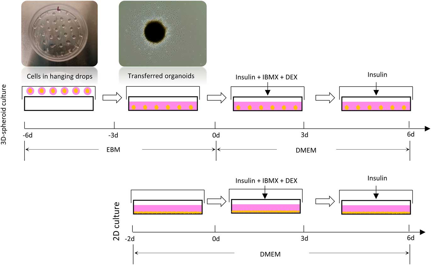
The thawed cells were cultured in DMEM until 80% confluence was reached. The cells were then resuspended in endothelial basal medium-2 (EBM-2, Cat. no. CC-3156; Lonza, Basel, Switzerland) at a concentration of 4×10⁴ cells/ml. The lid of a bacteriological dish was flipped over and about 40 drops of medium containing 103 cells with the volume of 25 µl each were pipetted onto the lid. The bacteriological dish was filled with 5 ml phosphate-buffered saline (PBS). The lid was quickly flipped back and put onto the dish, avoiding any movement of the drops. The hanging drops were maintained on the lid for 3 days, when cells formed spherical structures. After that, the spheroids were transferred one by one using a pipette with 200 µl tips (cut the top to fit the spheroids). The spheroids were cultured in EBM-2 for 3 additional days to settle onto the cell culture plates (1.5 spheroids/cm2). Endothelial basal medium-2 was replaced by DMEM supplemented with adipogenic induction medium (MDI): insulin (1 μg/ml), dexamethasone (0.1 μg/ml) and isobutylmethylxanthine (27.8 μg/ml) for the following 3 days, and then just DMEM supplemented with insulin (1 μg/ml) for the last 3 days of culture. The timing of culturing is showed in Figure 1.
Schematic diagram showing timelines of cell culture and differentiation. 3D=three dimensional; 2D=two dimensional; EBM=endothelial basal medium; IBMX=isobutylmethylxanthine; DEX=dexamethasone; DMEM=Dulbecco’s Modified Eagle’s Medium.

Oil-Red O staining
The adipocytes were fixed with 10% formalin for 10 min then washed with PBS for three times. Adipocytes were stained with Oil-Red O solution for 10 min then rinsed with PBS to remove excessive Oil-Red O dye. Following microscopic observation, Oil-Red O dye retained in cells were solubilized with isopropanol and the light absorbance was measured at 510 nm using a Synergy H1 Multi-Mode Reader (BioTek, Winooski, VT, USA).
Quantitative real-time PCR
Total RNA was isolated using TRIzol (Life Technologies, Grand Island, NY, USA), and complementary DNA was synthesized using the iScript™ cDNA Synthesis Kit (Bio-Rad, Hercules, CA, USA). The quantitative real-time PCR was then performed using the CFX RT-PCR detection system (Bio-Rad) with the SYBR green RT-PCR kit from Bio-Rad. Primer sequences are listed in Table 1. All values were normalized to the level of a housekeeping gene: UXT1.
Quantitative PCR primer sequences used for analyzing adipogenic differentiation

Statistical analyses
All data are presented as means±SEM. An unpaired, two-tailed Student’s t test was used to compare differences between two groups. All analyses were performed using SAS 9.0 (SAS Institute Inc., Cary, NC, USA). Statistical significance was assigned at P<0.05.
Results and discussion
Necessity of an alternative adipogenic culture system
Though adipogenesis in rodents and humans has been extensively studied during the past few decades, the same is not true for ruminant species. This knowledge is only further obscured due to the differences in lipid metabolism between ruminant and non-ruminant species. While rodents and humans synthesize lipids primarily in the liver (Hillgartner et al., Reference Hillgartner, Salati and Goodridge1995), ruminant animals rely on their adipose tissue to generate adipocyte droplets from acetate moieties (Vernon, Reference Vernon1980). Insulin is a critical component for inducing lipid accumulation in the adipogenic protocol for rodent and human cells. However, ruminants are less responsive to insulin compared with rodents and humans because glucose is not the major source of fatty acid synthesis in ruminants (Vernon, Reference Vernon1980). This is further supported by the fact that circulating insulin levels do not influence the in vivo lipogenesis of bovine animals to the degree observed in rodents (Smith et al., Reference Smith, Prior and Mersmann1983). Non-ruminants absorb large amounts of lipids in circulation while ruminants synthesize long-chain fatty acids in adipocytes de novo for triglyceride synthesis (Roh et al., Reference Roh, Hishikawa, Hong and Sasaki2006). Unlike humans and rodents, which use glucose as the precursor for de novo lipogenesis (Bergen and Mersmann, Reference Bergen and Mersmann2005), ruminant animals use acetate as the principal precursor for de novo lipogenesis (Vernon, Reference Vernon1980; Bouchard and Paquin, Reference Bouchard and Paquin2013). Several studies added acetate in the adipogenic medium of bovine cells (Grant et al., Reference Grant, Ortiz-Colon, Doumit and Buskirk2008a; Taniguchi et al., Reference Taniguchi, Guan, Zhang, Dodson, Okine and Moore2008), but failed to show comparative lipid accumulation as seen in murine cells. These differences arise the need for a new method which can effectively induce adipogenesis in ruminant SVCs.
Adipogenic differentiation of spheroid culture of bovine stromal vascular cells
While a two-dimensional culture system allows limited cell–cell interactions, three-dimensional culturing provides an efficient microenvironment for organogenesis in vitro (Huch and Koo, Reference Huch and Koo2015). To compare the efficiency of adipogenic differentiation in a conventional two-dimensional culture system, bovine SVCs derived from intramuscular fat, mouse SVCs derived from inguinal subcutaneous fat, porcine SVCs derived from intramuscular fat, C3H10T1/2 and 3T3-L1 cells were cultured. Upon 100% confluence (C3H10T1/2 cells were pre-treated with 10 ng/ml BMP4 before 100% confluence in order to gain adipogenic capacity (Tang et al., Reference Tang, Otto and Lane2004), and adipogenesis was induced by MDI for 3 days, followed by 3 days of insulin. The SVCs from mouse and porcine origins, and the 3T3-L1 and C3H10T1/2 cell lines, successfully accumulated a large amount of lipid droplets. However, only few, small, lipid droplets were observed in the bovine SVCs (Figure 2). Thus, the generic adipogenic protocol used for 3T3-L1 cells is not entirely applicable for bovine SVCs.
Differential adipogenic potential of cells isolated from different species. Bovine stromal vascular cells (SVCs) derived from intramuscular fat, mouse SVCs derived from inguinal subcutaneous fat, porcine SVCs derived from intramuscular fat, C3H10T1/2 and 3T3-L1 cells differentiated in two-dimensional (2D) cell culture plates; lipid droplets were stained by Oil-Red O. Bars show 100-µm length.

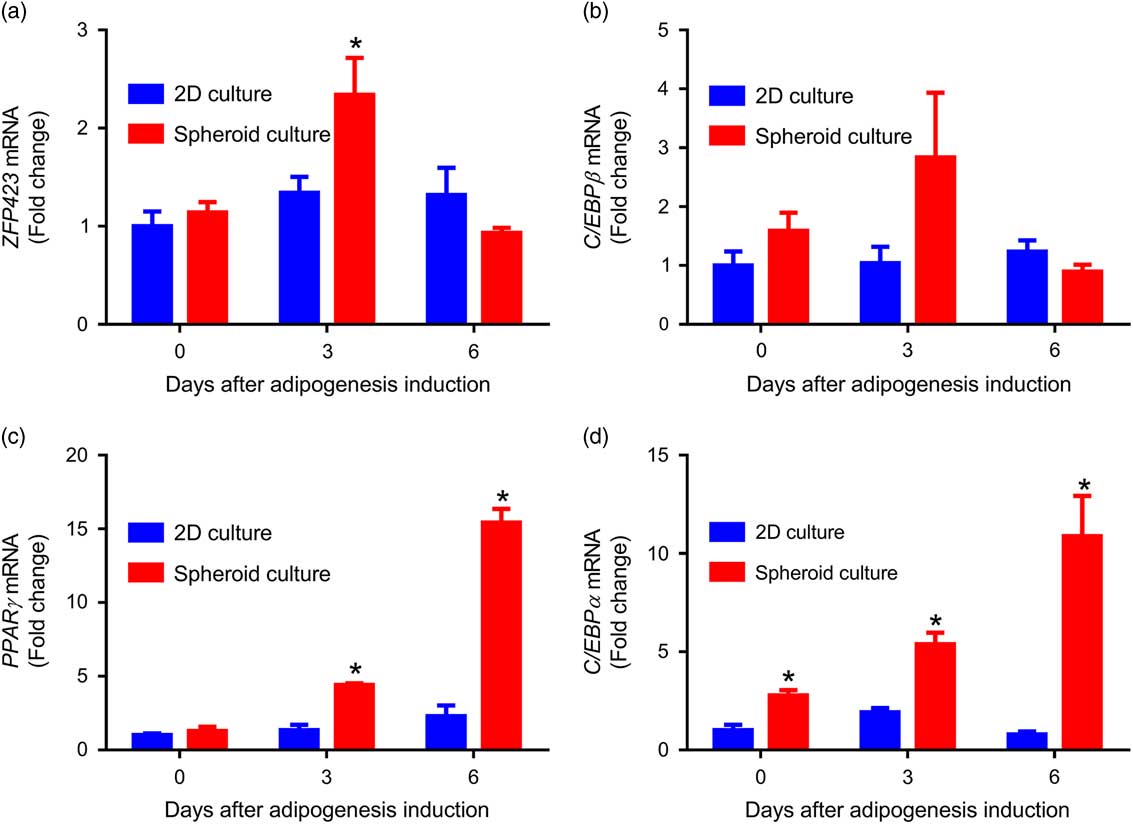
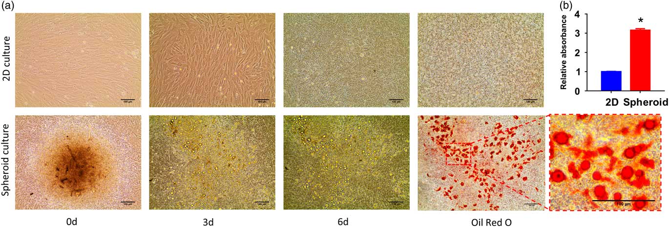
Thus, we tested three-dimensional spheroid culture method. To form a spheroid, SVCs were cultured in the hanging drops for 3 days. Cells then aggregated to form multicellular spheroids after 2 days in hanging drops. The cell spheroids were then transferred to a plate with some cells migrating out of spheroids and spreading (Figure 1). When compared with the two-dimensional culture (Figure 1) at 3 days following adipogenic induction, when its expression is at the highest, cells in a spheroid culture had higher expression of zinc-finger protein 423 (ZFP423, Figure 3a), a transcription factor that induces preadipocyte determination, the expression of PPARγ and adipocyte differentiation (Gupta et al., Reference Gupta, Arany, Seale, Mepani, Ye, Conroe, Roby, Kulaga, Reed and Spiegelman2010; Hudak et al., Reference Hudak, Gulyaeva, Wang, Park, Lee, Kang and Sul2014). In addition, the protein contents of PPARγ (Chawla et al., Reference Chawla, Schwarz, Dimaculangan and Lazar1994) (Figure 3c) and CEBPα (Rosen et al., Reference Rosen, Hsu, Wang, Sakai, Freeman, Gonzalez and Spiegelman2002) (Figure 3d) were also higher in three-dimensional spheroids compared with two-dimensional culture. After 6 days of adipogenesis, bovine SVCs in two-dimensional culture had very few visible lipid droplets, whereas bovine SVCs in the spheroid culture had a higher amount of large lipid droplets (Figure 4a). The increased lipid droplets in the spheroid culture were further confirmed by Oil-Red O staining (Figure 4a and b). In short, this spheroid culture system effectively enhances adipogenesis in bovine SVCs. These results are consistent with our previous observation in mouse SVCs, where three-dimensional spheroid culture effectively promotes adipogenesis (Wang et al., Reference Wang, Fu, Liang, Wang, Yang, Zou, Nie, Zhao, Gao, Zhu, De avila, Maricelli, Rodgers and Du2017a).
Adipogenic gene expression during adipogenic spheroid culture. Gene expression of zinc-finger protein 423 (ZFP423) (a), C/EBPβ (b), peroxisome proliferator-activated receptor γ (PPARγ) (c) and C/EBPα (d) at different time points during adipogenesis. Data are presented as mean±SEM, n=6, unpaired two-tailed t test, *P<0.05. mRNA=messenger RNA.

Comparison of adipogenesis in two-dimensional (2D) conventional culture and 3D spheroid culture. (a) Representative images at different time points showing bovine stromal vascular cells undergoing adipogenic differentiation in 2D and spheroid culture systems. Cells on day 9 were stained with Oil-Red O. (b) Quantification of Oil-Red O absorbance. Bars show 100-µm length. Data are presented as mean±SEM, n=6, unpaired two-tailed t test, *P<0.05.

Endothelial induction at the early stage of spheroid culture is required for adipogenesis
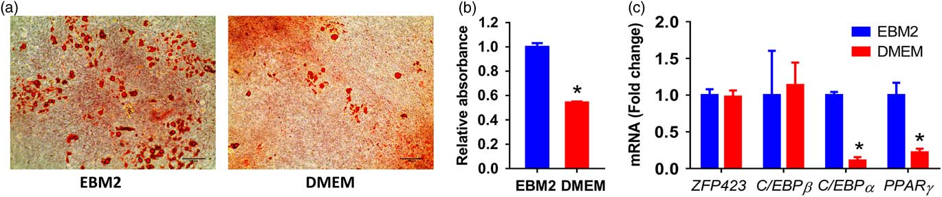
Adipogenesis is spatially and temporally associated with vascular development (Cao, Reference Cao2007). During adipose tissue development in vivo, angiogenesis precedes adipogenesis, which provides a niche environment necessary for adipocyte formation (Cao, Reference Cao2013). Adipose progenitor cells like platelet-derived growth factor receptor (PDGFR)α+ (Lee et al., Reference Lee, Petkova, Mottillo and Granneman2012) and PDGFRβ+ cells (Vishvanath et al., Reference Wang, Fu, Liang, Deavila, Wang, Zhao, Tian, Zhao, Gomez, Trombetta, Zhu and Du2015) can also be located perivascularly (Wang et al., Reference Villena, Kim and Sul2017b), which give rise to both white and brown adipocytes (Tran et al., Reference Tran, Gealekman, Frontini, Zingaretti, Morroni, Giordano, Smorlesi, Perugini, De Matteis, Sbarbati, Corvera and Cinti2012). Thus, we tested whether a medium inducing endothelial development promotes adipogenesis in a spheroid culture. Although DMEM-cultured spheroids generated adipocytes, this occurred significantly less than that of the EBM-2-cultured spheroids, where angiogenesis was promoted (Figure 5a and b). The messenger RNA expression of adipogenic genes at 3 days after adipogenesis, including C/EBPα and PPARγ was significantly higher in EBM-2-cultured spheroids than those of DMEM-cultured spheroids (Figure 5c). Thus, endothelial induction in spheroids prior to adipogenesis substantially increased the adipogenic capacity of bovine SVCs, in line with in vivo adipose tissue development. The endothelial cells generated in the spheroid may provide an appropriate environment for adipogenic differentiation of bovine SVCs, a condition absent in two-dimensional adipogenic cultures. Of note, in a three-dimensional culture model of rodent cells, we previously showed that vascular endothelial growth factor a signalling played a role in differentiation of beige adipose progenitors (Wang et al., Reference Wang, Fu, Liang, Wang, Yang, Zou, Nie, Zhao, Gao, Zhu, De avila, Maricelli, Rodgers and Du2017a). Right now, related spheroid culture has been increasingly used for studying morphogenesis and tissue development (Iles and Bartholomeusz, Reference Iles and Bartholomeusz2016), and in this study the spheroid culture was utilized to induce adipogenesis in bovine SVCs.
Endothelial induction enhances adipogenesis in spheroid culture. (a) Representative images of differentiated bovine stromal vascular cells with or without endothelial growth induction with endothelial basal medium-2 (EBM-2) before adipogenesis. (b) Quantification of Oil-Red O absorbance. (c) Gene expressions. Bars show 100-µm length. Data are presented as mean±SEM, n=6, unpaired two-tailed t test, *P<0.05. mRNA=messenger RNA.

In summary, we developed a new protocol to induce the adipogenic differentiation of SVCs derived from bovine intramuscular fat, in a spheroid culture system. This method dramatically improves adipogenesis of bovine SVCs, which is likely due to the supportive environment generated in the three-dimensional spheroids. Compared with murine or human cells, ruminant cells require more factors other than standard MDI to effectively differentiate into mature adipocytes. In short, the spheroid culture system developed in this study is an efficient model for in vitro studies of adipogenesis concerning ruminant-derived SVCs. In addition, this model can also be used for studying the role of angiogenesis in adipose tissue development in general.
Acknowledgements
This project was supported by the China Scholarship Council’s award to Y.N.M. (File no. 201508625024) and B.W. (File no. 201206350033), and the Agriculture and Food Research Initiative Competitive Grant nos. 2015-67015-23219 and 2016-68006-24634 from the USDA National Institute of Food and Agriculture, and NIH R01HD067449 to M.D.
Declaration of interest
None.
Ethics statement
All procedures of animal handling were approved by IACUC at Washington State University (#04354).
Software and data repository resources
None.