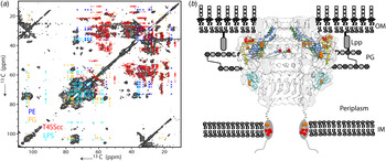
1. Introduction
Compartmentalization via membranes underlies many of the most fundamental biological processes, including bioenergetics, communication, sensing and organization (van Meer et al. Reference van Meer, Voelker and Feigenson2008; von Heijne, Reference von Heijne2007). These processes are assisted by membrane proteins (MPs) that represent about one-third of the proteome of every cell and over half of the current drug targets. A genome-wide approach revealed the membrane–protein interaction landscape of Saccharomyces cerevisiae (Babu et al. Reference Babu, Vlasblom, Pu, Guo, Graham, Bean, Burston, Vizeacoumar, Snider, Phanse, Fong, Tam, Davey, Hnatshak, Bajaj, Chandran, Punna, Christopolous, Wong, Yu, Zhong, Li, Stagljar, Conibear, Wodak, Emili and Greenblatt2012) suggesting that similarly as to what has been observed in the cytosol, most proteins occur and function in complexes rather than as isolated entities in the membrane (Zorman et al. Reference Zorman, Botte, Jiang, Collinson and Schaffitzel2015). Complexation events critical for MP function can range from the binding of small molecules such as ions, water, lipids or substrates to the formation of large protein clusters of MDa size. Indeed, homo-oligomerization is a well-known phenomenon for both prokaryotic (Hazelbauer et al. Reference Hazelbauer, Falke and Parkinson2008) as well eukaryotic (Bessman et al. Reference Bessman, Freed and Lemmon2014) MPs. This process serves to amplify signaling or, as in the case of β-barrel outer MPs in bacteria, helps to establish patterning of the cell membrane into micro-domains and is the basis of β-barrel protein turnover (Kleanthous et al. Reference Kleanthous, Rassam and Baumann2015). In addition, a significant fraction of MPs exists as part of larger (hetero-) molecular assemblies and exert their function via a range of molecular conformations and subunits that are active during different stages of their functional cycle (Chakrapani et al. Reference Chakrapani, Cordero-Morales, Jogini, Pan, Cortes, Roux and Perozo2011; Vardy & Roth, Reference Vardy and Roth2013).
While systems biology approaches help to catalog the membrane content and membrane–protein interactions, significant progress is needed for the determination of their stoichiometry, structure and cellular function. For more than four decades NMR has been used to investigate membranes and MPs (Oldfield et al. Reference Oldfield, Kinsey and Kintanar1982; Seelig & Gally, Reference Seelig and Gally1976). Here, we review recent methodological progress to study membrane–protein complexes (MPCs) by NMR, starting with expression, isotope-labeling and reconstitution protocols. Next we will discuss tailored NMR methods to deal with spectral complexity and limited spectral spectroscopic sensitivity that are usually encountered in NMR-based studies of MPCs. In general, we will put the emphasis on the development and use of solid-state NMR (ssNMR) spectroscopy. However, we also review studies using solution-state NMR methods on protein complexes in membrane mimetics. We will highlight recent progress in using NMR to study MPCs including G-protein-coupled receptors (GPCRs), ion channels and retinal proteins, and extend our discussion to protein–protein complexes that span entire cellular compartments or orchestrate processes such as protein transport across or within membranes.
We also refer the interested reader to related NMR publications on the subject of MP preparations using, for example, micelles or nanodiscs (Dürr et al. Reference Dürr, Soong and Ramamoorthy2013; Oxenoid & Chou, Reference Oxenoid and Chou2013) and their potential influence on MP structure (Zhou & Cross, Reference Zhou and Cross2013). Furthermore, ssNMR reviews have appeared recently on the subject of MP structure determination (Hong et al. Reference Hong, Zhang and Hu2012; Radoicic et al. Reference Radoicic, Lu and Opella2014; Tang et al. Reference Tang, Comellas and Rienstra2013; Wang & Ladizhansky, Reference Wang and Ladizhansky2014), MP–protein interactions (Miao & Cross, Reference Miao and Cross2013) and function (Baker & Baldus, Reference Baker and Baldus2014; Kimata et al. Reference Kimata, Reeves and Smith2015). When discussing ssNMR applications, we will largely concentrate on randomly oriented systems that are subject to Magic Angle Spinning (MAS, Andrew et al. Reference Andrew, Bradbury and Eades1958; Lowe, Reference Lowe1959). The combination of orientational constraints and ssNMR is described in various reviews; see e.g., (Opella, Reference Opella2013).
2. NMR on MPCs
2.1 Sample preparation for NMR
2.1.1 Protein expression and Isotope labeling
Virtually all NMR studies conducted today on MPCs use isotope-labeling to increase spectral sensitivity and resolution. Uniform 13C, 15N labeling is attractive since this strategy potentially generates the maximum amount of spectroscopic information from a single sample. However, with increasing size, the NMR analysis may suffer from spectral overlap. Furthermore, larger biomolecules can be characterized by a high repetitiveness of hydrophobic amino acids (as regularly seen in MPs) and a dominant influence of a single type of secondary structure (such as seen in α-helical MPs or β-sheet-rich amyloids). Such effects lead to additional ambiguities in the interpretation of the spectra, calling for more refined labeling approaches for protein production, such as selective isotope-labeling. Bacteria supplemented with labeled amino acids just after cell induction will produce proteins with the corresponding amino-acid labeling pattern as a result of direct incorporation of these residues during protein synthesis. Selective amino-acid labeling (which has to be selected carefully to decrease scrambling) can thus not only reduce spectral overlap, but may also reveal the specific protein topological regions or domains of special interest in the NMR spectra. Such ‘forward’ labeling has, for example, been used extensively to study MPs and other large biomolecules (see, e.g. (Jelinski et al. Reference Jelinski, Sullivan and Torchia1980; Lewis et al. Reference Lewis, Harbison, Herzfeld and Griffin1985) for early demonstrations). In a next stage, position-specific labeling can be achieved by substitution of uniformly labeled glucose with specifically labeled precursors such as [1,3-13C]-glycerol or [2-13C]-glycerol in the minimal growth medium. This approach leads to a characteristic distribution of 13C and 12C isotopes within each amino acid (Hong & Jakes, Reference Hong and Jakes1999; LeMaster & Kushlan, Reference Lemaster and Kushlan1996). Such labeling patterns can provide in principle highly resolved spectra and facilitate sequential resonance assignment arising from the appearance of characteristic cross-peak patterns for specific types of amino acids (Higman et al. Reference Higman, Flinders, Hiller, Jehle, Markovic, Fiedler, van Rossum and Oschkinat2009). Another example includes characteristic 13C-labeling patterns within methylated and aromatic residues by using [1-13C]-glucose, as the sole carbon source. Further labeling schemes, including labeling isolated 13C methyl sites are for example reviewed in (Renault et al. Reference Renault, Cukkemane and Baldus2010).
While 13C, 15N-detected spectroscopy has so far represented the work horse to conduct MP studies by ssNMR spectroscopy, 1H-detection offers the potential to significantly increase spectral sensitivity and resolution (Ishii & Tycko, Reference Ishii and Tycko2000). The prevailing method to detect protons in solid proteins is perdeuteration, i.e., the complete deuteration and subsequent reintroduction of exchangeable protons in protonated buffers. The later process has proven difficult in MPs that retain a lipid-detergent belt or, in the case of cellular studies (vide infra) that are largely protected from the exterior by their cellular membrane environment. In principle, fully protonated MPs can be studied using fast MAS frequencies (Weingarth et al. Reference Weingarth, van der Cruijsen, Ostmeyer, Lievestro, Roux and Baldus2014). Moreover, approaches such as ILV (Goto & Kay, Reference Goto and Kay2000; Huber et al. Reference Huber, Hiller, Schanda, Ernst, Böckmann, Verel and Meier2011), RAP (Asami & Reif, Reference Asami and Reif2013), proton cloud (Sinnige et al. Reference Sinnige, Daniëls, Baldus and Weingarth2014a) or SAIL labeling (Kainosho et al. Reference Kainosho, Torizawa, Iwashita, Terauchi, Mei Ono and Güntert2006) can be used to probe contacts between side-chain protons of specific types of amino acids. For example, Weingarth and co-workers have described an approach using specific sets of (1H,13C,15N) labeled amino acids (Sinnige et al. Reference Sinnige, Daniëls, Baldus and Weingarth2014a) in an otherwise deuterated NMR-silent background that does not require molecular unfolding, keeping molecular complexes intact. Such preparations generate local proton ‘clouds’ that facilitate assignment and structure elucidation in complex systems. Another option is the use of ‘fractional deuteration’ in 1H-detected ssNMR spectroscopy. This labeling scheme, based on protonated 13C-glucose and D2O in the growth medium, was previously proposed in solution NMR spectroscopy as an alternative to ILV labeling (Rosen et al. Reference Rosen, Gardner, Willis, Parris, Pawson and Kay1996) and in 13C-detected ssNMR spectroscopy (Nand et al. Reference Nand, Cukkemane, Becker and Baldus2012) for spectral editing. As recently shown, this approach retains sizable 1H levels and provides an avenue to high-resolution 1H ssNMR (Mance et al. Reference Mance, Sinnige, Kaplan, Narasimhan, Daniëls, Houben, Baldus and Weingarth2015b).
An intrinsic aspect of studying protein complexes, including those associated with membranes, refers to the use of mixed-labeling strategies to identify intermolecular interactions. For example, such approaches use samples in which one protein partner is 15N labeled, while the second is produced separately in a 13C-enriched medium (Etzkorn et al. Reference Etzkorn, Bockmann, Lange and Baldus2004). The protein interface is then analyzed by ssNMR experiments that encode magnetization transfer between 15N and 13C. Segmental labeling can also be an option as the molecular size increases. In this approach, only a fraction of the protein is studied and data are compared with larger constructs. Such ‘divide-and-conquer’ strategies were, for example, employed with reassembled multidomain MPs (Etzkorn et al. Reference Etzkorn, Kneuper, Dünnwald, Vijayan, Krämer, Griesinger, Becker, Unden and Baldus2008, Reference Etzkorn, Seidel, Li, Martell, Geyer, Engelhard and Baldus2010). Along the same lines, spectral crowding can be reduced by studying biomolecules after protein segments have been removed by enzymatic cleavage (Schneider et al. Reference Schneider, Ader, Lange, Giller, Hornig, Pongs, Becker and Baldus2008).
While producing 13C, 15N labeled proteins is a well-established method in bacteria, expression and isolation of 13C, 15N labeled proteins for NMR experiments in eukaryotic cells is not straightforward. One difficulty relates to the fact that not all eukaryotic cells can grow in suspension and have to be attached to plates in order to grow. Consequently, large amounts of cells have to be cultured to obtain one NMR sample. However, significant progress has been made in the last years to express and purify proteins from eukaryotic cells. Fully and specifically 13C, 15N labeled proteins suitable for NMR studies were produced from different types of eukaryotic cells, namely, yeast, insect and mammalian cells. For example, Pichia pastoris was successfully used to produce 15N labeled Ubiquitin for in-cell NMR studies (Bertrand et al. Reference Bertrand, Reverdatto, Burz, Zitomer and Shekhtman2012) and isotope-labeled MPs (Emami et al. Reference Emami, Fan, Munro, Ladishansky and Brown2013; Liu et al. Reference Liu, Liu, Fan, Munro, Ladishansky, Brown and Wang2016). Moreover, Sf9 insect cells were used to produce fully labeled proteins for in-cell NMR experiments (GB1 (Hamatsu et al. Reference Hamatsu, O'Donovan, Tanaka, Shirai, Hourai, Mikawa, Ikeya, Mishima, Boucher, Smith, Laue, Shirakawa and Ito2013)) and specifically labeled GPCR proteins (Nygaard et al. Reference Nygaard, Zou, Dror, Mildorf, Arlow, Manglik, Pan, Liu, Fung, Bokoch, Thian, Kobilka, Shaw, Mueller, Prosser and Kobilka2013). Human cells, namely, HEK293T cells were also used to produce 13C, 15N-labeled GPCRs (Opefi et al. Reference Opefi, Tranter, Smith and Reeves2015) and SOD1 (Barbieri et al. Reference Barbieri, Bertini, Luchinat, Secci, Zhao, Banci and Aricescu2013). In our laboratory, we (Kaplan et al. 2016, under revision) could produce fully and specifically labeled membrane vesicles derived from A431 cells (human cancer cells), which are known to express high levels of epidermal growth factor receptor (EGFR), and we performed NMR studies on EGFR in situ. Finally, we note that cell free protein expression has also emerged as a potentially viable route for MP expression for structural studies; however, due to the complexities of the systems we refer the reader to (Schwarz et al. Reference Schwarz, Dötsch and Bernhard2008) for a review.
2.1.2 Membrane mimetics
NMR studies have been conducted on MPs in detergents and other membrane mimetics for solution and ssNMR. While such approaches can stabilize protein plasticity, it is well known that the choice of membrane mimetic can change the structural and functional properties of MPs (Zhou & Cross, Reference Zhou and Cross2013). Indeed, more specific and stronger binding of cytochrome P450 to its reductase was observed in phospholipid bicelles as compared with detergents (Zhang et al. Reference Zhang, Huang, Im, Waskell and Ramamoorthy2015). Significant changes in the protein dynamics were also detected by Shimada and co-workers (Kofuku et al. Reference Kofuku, Ueda, Okude, Shiraishi, Kondo, Mizumura, Suzuki and Shimada2014) when comparing detergent and nanodisc reconstitutions of the deuterated β2-adrenergic receptor (β2AR). Related to the use of detergents or maltose–neopentyl glycol (MNG) amphiphiles (Chae et al. Reference Chae, Rasmussen, Rana, Gotfryd, Chandra, Goren, Kruse, Nurva, Loland, Pierre, Drew, Popot, Picot, Fox, Guan, Gether, Byrne, Kobilka and Gellman2010) by solution-state NMR (see, e.g. (O'Connor et al. Reference O'Connor, White, Doncescu, Didenko, Roth, Czaplicki, Stevens, Wüthrich and Milon2015; Sounier et al. Reference Sounier, Mas, Steyaert, Laeremans, Manglik, Huang, Kobilka, Demene and Granier2015; Zhou et al. Reference Zhou, Cierpicki, Jimenez, Lukasik, Ellena, Cafiso, Kadokura, Beckwith and Bushweller2008)), several groups have used microcrystallization approaches to study homo- (Shahid et al. Reference Shahid, Markovic, Linke and van Rossum2012b) as well as heteromeric MPCs (Sperling et al. Reference Sperling, Tang, Berthold, Nesbitt, Gennis and Rienstra2013) by ssNMR. Also, nanodiscs based on proteins such as membrane scaffold protein (Ritchie et al. Reference Ritchie, Grinkova, Bayburt, Denisov, Zolnerciks, Atkins and Sligar2009) or polymers, including Amphipols (Tribet et al. Reference Tribet, Audebert and Popot1996) or SMALPS (Dörr et al. Reference Dörr, Koorengevel, Schäfer, Prokofyev, Scheidelaar, van der Cruijsen, Dafforn, Baldus and Killian2014) have been considered.
2.1.3 Reconstitution versus coexpression
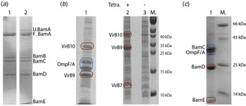
Reconstitution of MPCs into lipid bilayers is one of the preferred methods for studying these proteins by ssNMR. The incorporation of detergent-solubilized MPs into such lipid bilayers occurs spontaneously in the presence of lipids when the concentration of detergents is reduced. Parameters such as lipid composition, nature of the salts, pH value and temperature, or the presence of an endogenous ligand as well as the method for detergent removal (dialysis, biobeads) can be varied to maximize a functional reconstitution at a high protein to lipid ratio. Reasons for unsuccessful reconstitution of MPCs from the purified subunits or subcomplexes can be the absence of interaction partners that are needed for stable folding and function or because the protein–protein interactions are formed by interfaces within the bilayer, which are masked by the detergent used for solubilization. Coexpression of the complex subunits can in part overcome this problem. Co-transformation of several plasmids with single or multiple expression cassettes and with different origins of replication and antibiotic resistance can be expressed simultaneously and subsequently purified as a functional unit, providing the interactions between the partners is strong enough. For example, for the BAM complex (see Subsection 3.6), Bernstein and co-workers (Roman-Hernandez et al. Reference Roman-Hernandez, Peterson and Bernstein2014) showed that the entire complex can be reconstituted via expression of the full construct on one plasmid or via reconstitution of the two subcomplexes, BamAB and BamCDE, in detergents. In spite of seemingly identical compositions and molecular weight (Fig. 1a ), the activity of the complex expressed as a single complex was superior to that of the reconstituted complex. In addition, we could co-express three proteins that form the so-called core complex of bacterial Type IV secretion system (T4SS), these being VirB7, VirB9 and VirB10 (Kaplan et al. Reference Kaplan, Cukkemane, van Zundert, Narasimhan, Daniëls, Mance, Waksman, Bonvin, Fronzes, Folkers and Baldus2015). SDS–PAGE analysis of the isolated 13C, 15N-labeled cell envelopes from induced cells showed high expression of these three components in bacterial membranes (Fig. 1b ).
Expression and sample preparation strategies for BAM and T4SS core complex proteins. (a) Purification of proteins from membranes containing overexpressed material and reconstitution into liposomes. Lanes 1 and 2 show reconstituted Bam(AB)(CDE) subcomplexes and the co-expressed BamABCDE complex, respectively. (b) Overexpression of T4SS core complex in wild-type E. coli cell envelope (lane 1). Lanes 2 and 3 highlight the reduction in endogenous expression of OmpF/A in a double-mutant strain and overexpression of the proteins of interest upon induction with tetracyclin. (c) Outer membrane fraction of wild-type E. coli cells overexpressed with BamCDE using rifampicin during expression.

2.1.4 Cellular ssNMR studies
While synthetic lipid bilayers can mimic the physical environment of cellular membranes, they often lack their chemical heterogeneity. In addition to more closely resembling the cellular environment, the use of native membranes can remove the need to purify the MP of interest, eliminating any potential for structural disruptions during the solubilization process and speeding up the process of sample preparation which can be very cumbersome. Several groups have demonstrated that ssNMR can be used to study the structure and dynamics of MPs embedded in native lipid membranes (Etzkorn et al. Reference Etzkorn, Martell, Andronesi, Seidel, Engelhard and Baldus2007; Fu et al. Reference Fu, Wang, Li, Santiago-Miranda, Pielak and Tian2011; Jacso et al. Reference Jacso, Franks, Rose, Fink, Broecker, Keller, Oschkinat and Reif2012; Kulminskaya et al. Reference Kulminskaya, Pedersen, Bjerring, Underhaug, Miller, Frigaard, Nielsen and Nielsen2012; Ward et al. Reference Ward, Wang, Munro, Ritz, Hung, Gor'kov, Jiang, Liang, Brown and Ladizhansky2015). The previous work has also demonstrated that it is possible to study MPs in cellular envelopes and whole cells of Escherichia coli (E. coli) by conventional (Kaplan et al. Reference Kaplan, Cukkemane, van Zundert, Narasimhan, Daniëls, Mance, Waksman, Bonvin, Fronzes, Folkers and Baldus2015; Renault et al. Reference Renault, Pawsey, Bos, Koers, Nand, Tommassen-van Boxtel, Rosay, Tommassen, Maas and Baldus2012a) and dynamic nuclear polarization (DNP)-enhanced ssNMR (Kaplan et al. Reference Kaplan, Cukkemane, van Zundert, Narasimhan, Daniëls, Mance, Waksman, Bonvin, Fronzes, Folkers and Baldus2015; Renault et al. Reference Renault, Tommassen-Van Boxtel, Bos, Post, Tommassen and Baldus2012b; Yamamoto et al. Reference Yamamoto, Caporini, Im, Waskell and Ramamoorthy2015). In particular, we could show that studies using cellular envelopes or whole cells of Gram-negative bacteria are facilitated by the use of an E. coli deletion strain, to remove signals from naturally highly abundant outer MPs, i.e., OmpA and OmpF (Fig. 1b lanes 1 and 2 show the same protein expression in WT and deletion E. coli strains, respectively). In these preparations, signals from other cellular components, such as lipids, nucleotides, peptidoglycan and lipopolysaccharides, remain visible at intensities similar to that of the (overexpressed) protein of interest (Baldus, Reference Baldus2015). Such non-proteinaceous correlations can help refine the supramolecular structure of a protein in a membrane setting (see, e.g. (Weingarth & Baldus, Reference Weingarth and Baldus2013)). Furthermore, these signals can be used to study the structure of other molecular components such as RNA (Renault et al. Reference Renault, Pawsey, Bos, Koers, Nand, Tommassen-van Boxtel, Rosay, Tommassen, Maas and Baldus2012a) or components of the cell walls of bacteria and plants (see, e.g. (Wang et al., Reference Wang, Salazar, Zabotina and Hong2014)), as well as helping to clarify the cellular distribution of added reagents, such as radicals for DNP experiments (Takahashi et al. Reference Takahashi, Ayala, Bardet, de Paëpe, Simorre and Hediger2013).
On the other hand, these cellular signals can complicate the analysis of spectra of MPs that have not been previously characterized in synthetic bilayers. To reduce the challenges posed by the high background levels in cellular membranes we have devised ways of minimizing background labeling by removing highly abundant endogenous MPs as mentioned above, or by adding Rifampicin to the growth medium in order to specifically label the target protein of interest by inhibiting expression of endogenous proteins during expression (Baker et al. Reference Baker, Daniëls, van der Cruijsen, Folkers and Baldus2015) (Fig. 1c ). Hitherto, such cellular studies have been limited to MPs in prokaryotic environment; recent work in our laboratory illustrates the feasibility of extending such studies to include eukaryotic MPs in cellular environments (M. Kaplan et al. 2016, under revision).
2.2 From assignment to three-dimensional (3D) structure and supramolecular information
As molecular tumbling is significantly suppressed in such MPC samples, their ssNMR spectra are broadened by strong anisotropic interactions. Most commonly, MAS (Andrew et al. Reference Andrew, Bradbury and Eades1958; Lowe, Reference Lowe1959) in combination with high-power decoupling is utilized to average out these interactions and to re-establish high resolution for low gamma nuclei such as 13C or 15N. In contrast, the line-widths of protons remain prohibitively broad at moderate MAS frequencies (10–20 kHz) due to the strong inter-proton dipolar couplings. In the last years, protocols have been established to determine entire 3D protein structures from MAS ssNMR data obtained on randomly oriented biomolecules. Although progress has been made to incorporate 1H ssNMR spectroscopy, virtually all of these methods rely on 13C, 15N spectroscopy for: (1) spectral assignment and (2) the subsequent collection of distance restraints (see, e.g. (Baldus, Reference Baldus2002; Hong et al. Reference Hong, Zhang and Hu2012; Lange et al. Reference Lange, Becker, Seidel, Giller, Pongs and Baldus2005; Radoicic et al. Reference Radoicic, Lu and Opella2014; Tang et al. Reference Tang, Comellas and Rienstra2013; Wang & Ladizhansky, Reference Wang and Ladizhansky2014)). 3D structural information has also been obtained for MPs in lipid bilayers (Wang et al. Reference Wang, Munro, Shi, Kawamura, Okitsu, Wada, Kim, Jung, Brown and Ladizhansky2013), using microcrystals for orientational restraints (Radoicic et al. Reference Radoicic, Lu and Opella2014). Extending ssNMR experiments to additional spectral dimensions and the use of non-uniform sampling schemes (NUS) provides further opportunities to reduce complexity without prohibitively increasing NMR acquisition times (Heise et al. Reference Heise, Seidel, Etzkorn, Becker and Baldus2005b; Paramasivam et al. Reference Paramasivam, Suiter, Hou, Sun, Palmer, Hoch, Rovnyak and Polenova2012). In parallel, multiple acquisition schemes that make efficient use of spectrometer times by dual acquisition have been successfully demonstrated in ssNMR of MPs (Gopinath & Veglia, Reference Gopinath and Veglia2015).
Comparing chemical shifts before and after complex formation is a convenient means to probe protein-binding interfaces. While chemical shift perturbation studies provide useful insights into residues affected by complex formation (see Section 3), specific ssNMR pulse schemes in combination with isotope labeling strategies have been developed to obtain distance restraints across molecular interfaces. The direct detection of intermolecular contacts at the protein–protein interface in uniformly labeled samples is usually prohibited by spectral crowding and dipolar truncation. As discussed above, mixed labeling, for example the combination of 13C- and 15N-labeled proteins, is then employed. Polarization transfer across the molecular interface can then be established via a variety of pulse schemes, including REDOR/-TEDOR-based or NHHC and PAIN transfer schemes (see, e.g. (Weingarth & Baldus, Reference Weingarth and Baldus2013) for further details).
Spectral resolution and sensitivity are critical factors when structural studies of MPCs are attempted. Hence, an interesting addition to the toolbox of structure-elucidation methods is the use of paramagnetic relaxation enhancement (PRE's) that extends the distance range available to NMR to 10–20 Å and are thus ideal tools to probe the structure of membrane complexes by NMR (Nadaud et al. Reference Nadaud, Helmus, Höfer and Jaroniec2007; Otting, Reference Otting2010). PRE effects have already been successfully used in the context of MP oligomers (van der Cruijsen et al. Reference van der Cruijsen, Koers, Sauvee, Hulse, Weingarth, Ouari, Perozo, Tordo and Baldus2015; Wang et al. Reference Wang, Munro, Kim, Jung, Brown and Ladizhansky2012). Lipids containing paramagnetic head groups (see, e.g. (Zhuang & Tamm, Reference Zhuang and Tamm2014)) may further enhance the use of PRE's for the study of MPs and MPCs in the future.
2.3 Signal enhancement by DNP
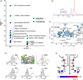
A critical parameter for studying complex molecular systems such as MPCs refers to spectroscopic sensitivity. This challenge has been addressed in the field of ssNMR by the advent of commercial DNP (Ni et al. Reference Ni, Daviso, Can, Markhasin, Jawla, Swager, Temkin, Herzfeld and Griffin2013)–NMR setups that can operate at 400, 600 and up to 800 MHz NMR frequency. High-field conditions (Koers et al. Reference Koers, van der Cruijsen, Rosay, Weingarth, Prokofyev, Sauvee, Ouari, van der Zwan, Pongs, Tordo, Maas and Baldus2014) are particularly attractive for complex biomolecules such as MPCs and we have observed enhancement factors of almost two orders of magnitude when studying MPCs in their natural cell membrane environment at 400 MHz. In Fig. 2a , data obtained in our group at both 400 and 800 MHz DNP conditions are shown for the case of EGFR-rich plasma membrane vesicles of A431 cells (see also Subsection 3.5) as well as MPs (PagL (Renault et al. Reference Renault, Pawsey, Bos, Koers, Nand, Tommassen-van Boxtel, Rosay, Tommassen, Maas and Baldus2012a)) and MPCs, i.e., the type 4 secretion system core complex (T4SScc, Subsection 3.6 and (Kaplan et al., Reference Kaplan, Cukkemane, van Zundert, Narasimhan, Daniëls, Mance, Waksman, Bonvin, Fronzes, Folkers and Baldus2015)) embedded in bacterial cell envelopes. In addition, Fig. 2a contains DNP enhancement factors obtained on proteoliposomal preparations of KcsA (Koers et al. Reference Koers, van der Cruijsen, Rosay, Weingarth, Prokofyev, Sauvee, Ouari, van der Zwan, Pongs, Tordo, Maas and Baldus2014; van der Cruijsen et al. Reference van der Cruijsen, Koers, Sauvee, Hulse, Weingarth, Ouari, Perozo, Tordo and Baldus2015) as well as YidC (Baker et al. Reference Baker, Daniëls, van der Cruijsen, Folkers and Baldus2015) and DNP data obtained on reference systems such as liposomal Aβ peptides (Koers et al. Reference Koers, López-Deber, Weingarth, Nand, Hickman, Mlaki Ndao, Reis, Granet, Pfeifer, Muhs and Baldus2013) and 13C,15N labeled biosilica from S. turris (Jantschke et al. Reference Jantschke, Koers, Mance, Weingarth, Brunner and Baldus2015). For comparison, we also included a limited set of published DNP values, namely for a toxin bound to the acetycholine receptor (Linden et al. Reference Linden, Lange, Franks, Akbey, Specker, van Rossum and Oschkinat2011), Mistic in natural membranes (Jacso et al. Reference Jacso, Franks, Rose, Fink, Broecker, Keller, Oschkinat and Reif2012) as well as ligands of the M2 channel (Andreas et al. Reference Andreas, Barnes, Corzilius, Chou, Miller, Caporini, Rosay and Griffin2013) and SecYEG (Reggie et al. Reference Reggie, Lopez, Collinson, Glaubitz and Lorch2011).
DNP on MPs and MPCs. (a) Comparison between the DNP enhancement factors of published and in-house measured MPs and peptides using 400 MHz/263 GHz and 800 MHz/527 GHz DNP setups. (b) 1H–13C cross-polarization experiments of GSLV labeled bacterial cell envelope expressing T4SScc performed at 400 MHz DNP conditions (red: microwaves on, blue: microwaves off, from (Kaplan et al. Reference Kaplan, Cukkemane, van Zundert, Narasimhan, Daniëls, Mance, Waksman, Bonvin, Fronzes, Folkers and Baldus2015). (c) Comparison of an NCA 2D experiment on U–13C,15N labeled KcsA at ambient temperatures (black) to data on a AMUpol-tagged G116C KcsA variant at LT-DNP conditions (blue) (van der Cruijsen et al. Reference van der Cruijsen, Koers, Sauvee, Hulse, Weingarth, Ouari, Perozo, Tordo and Baldus2015). (d) Comparison of the experimentally determined 13C line widths of residues located in the KcsA filter and turret region (solid lines) to predictions based on MD runs (adapted from (Koers et al. Reference Koers, van der Cruijsen, Rosay, Weingarth, Prokofyev, Sauvee, Ouari, van der Zwan, Pongs, Tordo, Maas and Baldus2014)). (e) Results of T1–15N relaxation measurements on membrane-embedded KcsA–Kv1.3 as reported by Ader et al. (Ader et al. Reference Ader, Pongs, Becker and Baldus2010). For reference, the binding modes of Kaliotoxin and porphyrin (see Section 3.3) are included.

Interestingly, considerable variations are seen in Fig. 2a , with the largest DNP enhancements, and observed for MPCs in natural cell membrane preparations. In addition, it is well known that the efficiency of the DNP transfer mechanisms strongly depends on the biradicals used and those molecules such as TOTAPOL (Song et al. Reference Song, Hu, Joo, Swager and Griffin2006) or AMUPol (Sauvee et al. Reference Sauvee, Rosay, Casano, Aussenac, Weber, Ouari and Tordo2013) designed to exploit cross-effect DNP suffer from a reduced efficiency with increasing magnetic fields. In fact, studies in our laboratory have shown an approximately fourfold decrease in DNP enhancements when moving from 400 to 800 MHz conditions. In recent theoretical work (Mance et al. Reference Mance, Gast, Huber, Baldus and Ivanov2015a), we could show that this effect can be well explained by an effective hyperfine coupling that describes the initial electron–nuclear polarization transfer step (occurring over a distance of approximately 10 Å). Nevertheless, sizable signal enhancements have been observed including applications on cellular preparations such as the type 4 secretion system (Kaplan et al. Reference Kaplan, Cukkemane, van Zundert, Narasimhan, Daniëls, Mance, Waksman, Bonvin, Fronzes, Folkers and Baldus2015) as shown in Fig. 2b . Moreover, recent studies suggest that trityl-based biradicals may be better suited for ultrahigh field DNP studies in the future (Mathies et al. Reference Mathies, Caporini, Michaelis, Liu, Hu, Mance, Zweier, Rosay, Baldus and Griffin2015).
Next to sensitivity, spectroscopic resolution is another critical factor for DNP applications on complex (bio)molecules. We recently compared the spectral resolution of CC and NC two-dimensional (2D) data sets on fully 13C, 15N labeled variants of the KcsA potassium channel (see Subsection 3.3) at 800 MHz DNP conditions using soluble (Koers et al. Reference Koers, van der Cruijsen, Rosay, Weingarth, Prokofyev, Sauvee, Ouari, van der Zwan, Pongs, Tordo, Maas and Baldus2014) and tagged (van der Cruijsen et al. Reference van der Cruijsen, Koers, Sauvee, Hulse, Weingarth, Ouari, Perozo, Tordo and Baldus2015) AMUPol variants to data obtained at ambient temperatures.
In Fig. 2c , such NCA data are compared using a KcsA sample carrying an AMUPol biradical covalently attached to protein position G116 that was mutated to Cys. This position is rather distant from the turret and selectivity filter region of the channel (van der Cruijsen et al. Reference van der Cruijsen, Koers, Sauvee, Hulse, Weingarth, Ouari, Perozo, Tordo and Baldus2015) shown in Figs 2d and 2e . In Fig. 2c , several correlations are visible that compare favorably to data obtained at higher temperatures (black). On the other hand, peaks such as G79 or I60 are absent from the spectrum. Comparing experimental line width (Fig. 2d , solid lines) with predictions on the basis of MD calculations (histograms, Fig. 2d ) for specific residues (Koers et al. Reference Koers, van der Cruijsen, Rosay, Weingarth, Prokofyev, Sauvee, Ouari, van der Zwan, Pongs, Tordo, Maas and Baldus2014) suggests that local backbone fluctuations are most likely the prominent source of line broadening that leads to disappearance of signals such as G79 or I60 in Fig. 2c . In detail, Fig. 2d compares experimental data and MD predictions for residues of the KcsA selectivity filter (T74–G79). In each case, a good correlation between experimental line-widths and MD amplitudes (bar graphs) is seen. In fact, the large broadening of Gly79 and Thr74 is in line with our previous T1 15N relaxation measurements (Ader et al. Reference Ader, Pongs, Becker and Baldus2010) that predicted enhanced dynamics for the filter residues of the KcsA–Kv1.3 channel (Fig. 2e ).
These findings along with previous ssNMR low temperature (LT) work on unfolded (Heise et al. Reference Heise, Luca, de Groot, Grubmüller and Baldus2005a), globular (Havlin & Tycko, Reference Havlin and Tycko2005) and, most recently, needle-forming proteins (Fricke et al. Reference Fricke, Mance, Chevelkov, Giller, Becker, Baldus and Lange2016) suggest that low-temperature DNP experiments are sensitive to structural disorder (see, e.g. (Koers et al. Reference Koers, van der Cruijsen, Rosay, Weingarth, Prokofyev, Sauvee, Ouari, van der Zwan, Pongs, Tordo, Maas and Baldus2014)) and thus provide a means to probe motional amplitudes by comparing the line broadening under different sample conditions, for example in the presence and absence of a specific ligand that binds the MPC. Finally as demonstrated by several groups for the case of retinal MPCs (see Subsection 3.1), LT-DNP will continue to offer a powerful means to trap functional intermediates, for example in the context of MPC substrates.
2.4 Dynamics and computational methods
A powerful aspect of NMR studies on MPCs relates to the study of molecular dynamics ranging from the level of single atom or protein groups to the observations of large-scale domain motion. Related to such studies is the identification of MPC regions that exhibit dynamics, for example prior to or during interaction with the substrate or due to spatial proximity of the lipid bilayer or the lipid–water interface. Moreover, for many channels, receptors and transporters conformational dynamics are key to an often allosteric functional mechanism. Several NMR methods are well suited to probe exchange between conformers in MPCs. For solution NMR, methyl-TROSY-based CPMG or ZZ-exchange experiments are suitable for the generally large MPCs (Rosenzweig & Kay, Reference Rosenzweig and Kay2014; Sprangers et al. Reference Sprangers, Velyvis and Kay2007). In the case of ssNMR, measurements of dipolar couplings and relaxation rates provide a valuable source for detailed analysis of MPC dynamics (Hong et al. Reference Hong, Zhang and Hu2012). Tailored ssNMR methods that are sensitive to fast (nanosecond), medium (microsecond to millisecond) or slow motion are available today. Already in the case of microcrystalline proteins, a variety of motional degrees of freedom can be probed that may at least in part be related to the molecular environment. Dynamics on specific MPCs are described in more detail in Section 3.
As discussed in the previous section, the comparison to MD simulations suggests that there is strong relationship between motional amplitudes and the degree of local disorder seen in LT DNP ssNMR. Hence, combining NMR experiments at ambient and low temperatures provides a route to probe both motional rates and amplitudes by ssNMR data. In addition, MD simulations can assist in both the structural and topological refinement of protein structure and dynamics and in the case of MPCs in defining lipid–protein interactions (van der Cruijsen et al. Reference van der Cruijsen, Nand, Weingarth, Prokofyev, Hornig, Cukkemane, Bonvin, Becker, Hulse, Perozo, Pongs and Baldus2013; Weingarth et al. Reference Weingarth, Ader, Melquiond, Nand, Pongs, Becker, Bonvin and Baldus2012) as well as in probing protein dynamics relevant for activation (Mertz et al. Reference Mertz, Struts, Feller and Brown2012).
Computational methods are in fact becoming an integral aspect of applying NMR methods to large molecular complexes. For example, several programs such as FANDAS (Gradmann et al. Reference Gradmann, Ader, Heinrich, Nand, Dittmann, Cukkemane, Dijk, Bonvin, Engelhard and Baldus2012) or VirtualSpectrum (Nielsen & Nielsen, Reference Nielsen and Nielsen2014) can support the investigation of MPCs in various phases, starting with tailored protein labeling, assignment and evaluation of protein structure and complex formation.
Computational techniques such as molecular docking can also be of tremendous help in ‘divide-and-conquer’ approaches in which information of MP subunits from X-ray or NMR (solution and ssNMR) is available and used in the study MPCs by NMR. Docking programs such as HADDOCK (Dominguez et al. Reference Dominguez, Boelens and Bonvin2003) or the ROSETTA based ‘dock-and fold’ (Das et al. Reference Das, Andre, Shen, Wu, Lemak, Bansal, Arrowsmith, Szyperski and Baker2009) allow for protein flexibility during the docking and can incorporate experimental data, including NMR chemical shifts or chemical shift perturbations. More recently, NMR has been successfully used in combination with other techniques, including cryo-EM, super-resolution light microscopy or mass spectrometry to investigate molecular structures (Kaplan et al. 2016, under revision). The combination of these different and complementary techniques extends the range of molecular complexity that can be investigated by NMR.
3. Examples
3.1 Retinal proteins and light-harvesting complexes
Light-sensitive MPCs, in particular, retinal proteins containing covalently bound retinal have been the subject of ssNMR spectroscopic investigations for more than three decades with pioneering studies on bacteriorhodpsin (bR) (see, e.g. (Herzfeld & Lansing, Reference Herzfeld and Lansing2002) and references therein) and rhodopsin (see, e.g. (Kimata et al. Reference Kimata, Reeves and Smith2015; Mertz et al. Reference Mertz, Struts, Feller and Brown2012) and references therein).
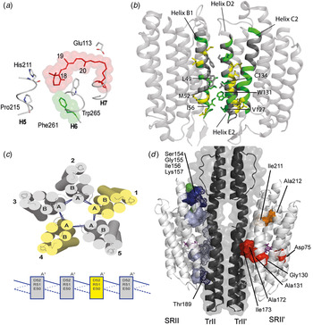
In the case of rhodopsin, it has been possible to dissect the topology and dynamics of the retinal ligand as well as the activation profile of the receptor itself (Kimata et al. Reference Kimata, Reeves and Smith2015). In Fig. 3a , a view of the 11-cis retinal chromophore in rhodopsin from the extracellular surface is shown revealing the hydrogen-bonding network between Tyr191, Tyr268 and Glu181, and between Glu113 and the Schiff base proton. Smith and co-workers have proposed that retinal isomerization drives the Schiff base proton away from Glu113, which facilitates deprotonation, ultimately allowing the extracellular end of helix H6 to rearrange (Kimata et al. Reference Kimata, Reeves and Smith2015).
NMR-based studies of retinal MPCs. (a) Amino acids associated with rhodopsin activation using a view of the 11-cis retinal chromophore in rhodopsin from the extracellular surface. The hydrogen-bonding network is shown between Tyr191, Tyr268 and Glu181, and between Glu113 and the Schiff base proton (Figure reproduced from Kimata et al. Reference Kimata, Reeves and Smith2015 with permission from publisher). (b) Comparison of ssNMR studies on ASR in synthetic and E. coli membranes with unperturbed (green) residues and residues experiencing significant changes (yellow) in chemical shifts between both settings. Side view from the extracellular side of the interaction interface (helices B1, E2 and D2). The side chains of perturbed residues and residues involved in the interaction interface are shown (Figure reproduced from Ward et al. Reference Ward, Wang, Munro, Ritz, Hung, Gor'kov, Jiang, Liang, Brown and Ladizhansky2015 with permission from publisher). (c) Cross-protomer interactions in the homo-oligomeric MPC Proteorhodopsin by DNP-enhanced ssNMR (Reprinted with permission from (Maciejko et al. Reference Maciejko, Mehler, Kaur, Lieblein, Morgner, Ouari, Tordo, Becker-Baldus and Glaubitz2015)). Copyright 2015 American Chemical Society. (d) SRII receptor topology, complex formation with its cognate transducer and activation in a natural membrane environment. Residues that are affected by transducer binding are highlighted on the left on the SRII structure (dark/light blue stand for identified/potential chemical shift perturbations, respectively). Green residues denote protein residues that exhibit a reduction in molecular mobility in the complex. Residues highlighted on the right hand of the SRII structure illustrate areas that underwent chemical shift changes upon light activation (adapted from (Etzkorn et al. Reference Etzkorn, Seidel, Li, Martell, Geyer, Engelhard and Baldus2010)).

SsNMR spectroscopy has also been used to characterize different functional states of bR by using DNP-enhanced ssNMR and irradiation with laser light (Bajaj et al. Reference Bajaj, Mak-Jurkauskas, Belenky, Herzfeld and Griffin2009; Mak-Jurkauskas et al. Reference Mak-Jurkauskas, Bajaj, Hornstein, Belenky, Griffin and Herzfeld2008). More recently, such experiments have been conducted on 15N-labeled channelrhodopsin-2 carrying 14,15-13C2 retinal reconstituted into lipid bilayers, which allowed characterization of three distinct intermediates (Becker-Baldus et al. Reference Becker-Baldus, Bamann, Saxena, Gustmann, Brown, Brown, Reiter, Bamberg, Wachtveitl, Schwalbe and Glaubitz2015) providing novel insight into the photoactive site of channelrhodopsin-2 during the photocycle.
In addition to the details of the photocycle, an important aspect for activation refers to the question of how retinal proteins assemble into homo- as well as hetero-protein complexes to establish maximum functionality. Ladishansky and co-workers not only succeeded in determining a 3D structure of Anabaena Sensory Rhodopsin (ASR) in lipid bilayers (Wang et al. Reference Wang, Munro, Shi, Kawamura, Okitsu, Wada, Kim, Jung, Brown and Ladizhansky2013) but they also used cellular ssNMR to obtain information on which side chains and residues are involved in stabilizing the in situ structure of ASR (Ward et al. Reference Ward, Wang, Munro, Ritz, Hung, Gor'kov, Jiang, Liang, Brown and Ladizhansky2015). In Fig. 3b , a view from the extracellular side of the interaction interface (helices B1, E2 and D2) between two ASR monomers and helix C2, which is tightly packed against many of the perturbed residues in helix D2, is shown. The side chains of perturbed residues and residues involved in the interaction interface are shown in yellow, unperturbed residues (compared with their NMR shifts seen in reconstituted bilayers) are given in green. In addition, Glaubitz and co-workers recently visualized specific cross-protomer interactions in the homo-oligomeric MP Proteorhodopsin by DNP-enhanced ssNMR (Maciejko et al. Reference Maciejko, Mehler, Kaur, Lieblein, Morgner, Ouari, Tordo, Becker-Baldus and Glaubitz2015). In the WT protein, the salt bridge R51−D52 stabilizes the pentamer and acts as an ‘oligomerization switch’ between pentamer and hexamer formation (Fig. 3c ).
Next to information about the (supramolecular) structure of retinal proteins, ssNMR methods provide an increasingly accurate description of MP dynamics in a membrane environment. For example, conformational dynamics in ASR in liposomes (Good et al. Reference Good, Wang, Ward, Struppe, Brown, Lewandowski and Ladizhansky2014) could be characterized by determination of order parameters from MAS dipolar coupling measurements and motional time-scales from rotating frame spin lattice (R1rho) relaxation times. Using the 3D GAF approach, these data could be explained by a model of collective motions by entire helices or loops.
We have previously shown how ssNMR can be used in the case of sensory rhodopsin II (SRII), to examine receptor topology, complex formation with its cognate transducer, and activation in a natural membrane environment (Etzkorn et al. Reference Etzkorn, Martell, Andronesi, Seidel, Engelhard and Baldus2007, Reference Etzkorn, Seidel, Li, Martell, Geyer, Engelhard and Baldus2010). To infer residue-specific information about receptor dynamics before and after complex formation we combined scalar-based correlation experiments that probe fast motion (Andronesi et al. Reference Andronesi, Becker, Seidel, Heise, Young and Baldus2005) with 2D double-quantum (13C,13C) correlation experiments which can provide information about local C–C bond motions without the need of an extra frequency dimension (Etzkorn et al. Reference Etzkorn, Seidel, Li, Martell, Geyer, Engelhard and Baldus2010; Schneider et al. Reference Schneider, Seidel, Etzkorn, Lange, Becker and Baldus2010). Together with the examination of chemical-shift changes due to complex formation or light activation, we could identify SRII residues that are critical for forming the complex and the early events of light activation. Figure 3d shows residues that are affected by transducer binding and are highlighted on the left on the SRII structure (dark/light blue stand for identified/potential chemical shift perturbations, respectively). Green residues denote protein residues that exhibit a reduction in molecular mobility in the complex. Residues highlighted on the right hand of the SRII structure illustrate areas that underwent chemical shift changes upon light activation (adapted from (Etzkorn et al. Reference Etzkorn, Seidel, Li, Martell, Geyer, Engelhard and Baldus2010)).
Finally, we also note that another family of light-sensing MPCs extensively studied by ssNMR is light-harvesting complexes. Here, NMR was used to study the electronic as well as spatial structures (see, e.g. (Alia et al. Reference Alia, Ganapathy and de Groot2009; Egorova-Zachernyuk et al. Reference Egorova-Zachernyuk, Hollander, Fraser, Gast, Hoff, Cogdell, de Groot and Baldus2001; McDermott et al. Reference Mcdermott, Zysmilich and Polenova1998; Pandit et al. Reference Pandit, Reus, Morosinotto, Bassi, Holzwarth and de Groot2013)) of the MPCs and their ligands.
3.2 GPCRs
GPCRs are a class of integral MPs composed of seven transmembrane helices that are involved in signaling for a number of essential biological processes and functioning as drug receptors. They convert a large variety of extracellular stimuli into intracellular responses through the activation of heterotrimeric G-proteins making them key regulatory elements and important targets for pharmaceutical drug discovery. In the last years, X-ray crystallography has obtained structural snapshots of a variety of GPCRs by combining advanced biochemical procedures (Maeda & Schertler, Reference Maeda and Schertler2013). Yet, essential features of the structure and dynamics, the ligand binding and the entire activation landscape of GPCRs in the presence of the native membrane environment are still to be determined.
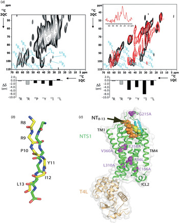
In addition to work mentioned earlier on rhodopsin (Kimata et al. Reference Kimata, Reeves and Smith2015), several studies have been conducted to study ligand binding to GPCRs. In collaboration with the laboratory of R. Grisshammer, we previously studied the conformation of Neurotensin (residues 8–13, NT(8–13)) bound to its GPCR (Luca et al. Reference Luca, White, Sohal, Filippov, van Boom, Grisshammer and Baldus2003) by 2D ssNMR in detergents (Fig. 4a , left) as well as in lipid bilayers (right, indicated in red). In the latter case, Fig. 4a (right) also includes ssNMR data of free frozen [13C,15N]–NT(8–13) in black. Using secondary chemical-shift information (indicated by the secondary chemical shifts Δδ as defined by Luca et al. (Reference Luca, Filippov, van Boom, Oschkinat, de Groot and Baldus2001)), we proposed that the ligand largely adopts an extended conformation in the bound state (Fig. 4b , yellow). About ten years later, Grisshammer and co-workers (White et al. Reference White, Noinaj, Shibata, Love, Kloss, Xu, Gvozdenovic-Jeremic, Shah, Shiloach, Tate and Grisshammer2012) succeeded in capturing crystals of a variant of the NT receptor that was thermostabilized by mutagenesis and C-terminally fused with T4 lysozyme (T4L, Fig. 4c ) that revealed a NT conformation (Fig. 4b , green) that largely agreed (Fig. 4b , green) with the ssNMR predictions (Fig. 4 b, yellow). Notably, we also conducted ssNMR studies of the free ligand at low temperatures to sample the conformational space of the free peptide. These studies suggested that in the free state, neurotensin is largely unstructured (Heise et al. Reference Heise, Luca, de Groot, Grubmüller and Baldus2005a). Further studies on isotope-labeled ligands bound to GPCRs investigated human histamine in complex with the H1 receptor (Ratnala et al. Reference Ratnala, Kiihne, Buda, Leurs, de Groot and Degrip2007), for which two different ligand protonation states were found. Furthermore, 2D ssNMR studies of the bradykinin peptide bound to the human bradykinin β2 receptor in DDM (dodecyl β-D-maltopyranoside) postulated a double-S-shape structure of the ligand (Lopez et al. Reference Lopez, Shukla, Reinhart, Schwalbe, Michel and Glaubitz2008).
Binding of Neurotensin to its GPCR as seen by ssNMR and X-ray. (a) 2D 2Q (13C,13C) data on U[13C,15N] Neurotensin (NT8–13) in complex with its receptor in detergents (left) as well as in lipid bilayers (right). On the right, data on free frozen NT(8–13) are given in black as reference. From the resonance positions, secondary chemical shifts were computed for the peptide leading to a model of the backbone fold given in yellow in (b) (Luca et al. Reference Luca, White, Sohal, Filippov, van Boom, Grisshammer and Baldus2003) superimposed on the X-ray structure (White et al. Reference White, Noinaj, Shibata, Love, Kloss, Xu, Gvozdenovic-Jeremic, Shah, Shiloach, Tate and Grisshammer2012). (c) A 3D X-ray structure of a thermostabilized receptor version containing a C-terminally fused T4 lysozyme (White et al. Reference White, Noinaj, Shibata, Love, Kloss, Xu, Gvozdenovic-Jeremic, Shah, Shiloach, Tate and Grisshammer2012) observed the Neurotensin fragment as indicated in green in (B).

More recently, several NMR groups have investigated the dynamics of the DDM solubilized β2AR. Using reductive 13C-dimethylation of lysine residues, Bokoch et al. (Reference Bokoch, Zou, Rasmussen, Liu, Nygaard, Rosenbaum, Fung, Choi, Thian, Kobilka, Puglisi, Weis, Pardo, Prosser, Mueller and Kobilka2010) could apply STD-filtered 13C-HMQC experiments to identify the ligand-specific conformational changes in the extracellular loops. This work provided the first insights into the conformational coupling between the orthosteric binding site and the extracellular surface, whose exact conformation remains elusive from crystal structures. Labeling of three cytosolic cysteine residues with trifluoroethanthiol (TET) allowed using 19F-NMR on β2AR to study structural changes occurring upon binding of several ligands to the extracellular side of the receptor (Liu et al. Reference Liu, Horst, Katritch, Stevens and Wuthrich2012). In addition, the temperature dependence of the 19F line-width was used to probe dynamics of the receptor. Employing the fast dynamics of the methionine methyl group, Met-13Cε labeled β2AR was employed to study the receptor's conformational dynamics in the absence or presence of different ligands, using a combined solution NMR and MD approach (Nygaard et al. Reference Nygaard, Zou, Dror, Mildorf, Arlow, Manglik, Pan, Liu, Fung, Bokoch, Thian, Kobilka, Shaw, Mueller, Prosser and Kobilka2013). Recently, signal propagation upon opioid agonist binding was studied using solution NMR on 13C-dimethylated μ-Opioid receptor (μOR) in MNG amphiphiles (Sounier et al. Reference Sounier, Mas, Steyaert, Laeremans, Manglik, Huang, Kobilka, Demene and Granier2015). This work revealed a possible allosteric pathway of activation regulating μOR subdomain conformations. Finally, a complete 3D structural model of the GPCR CXCR1 undergoing rapid rotational diffusion in lipid bilayers has been reported using ssNMR spectroscopy (Park et al. Reference Park, Das, Casagrande, Tian, Nothnagel, Chu, Kiefer, Maier, De Angelis, Marassi and Opella2012).
3.3 Ion channels
Ion channels are pore-forming MPs whose functions include establishing a resting membrane potential, shaping action potentials and other electrical signals by gating the flow of ions across the cell membrane, thereby controlling the flow of ions across secretory and epithelial cells, and regulating cell volume.
The oligomeric influenza M2 proton channel has been studied extensively by NMR in a variety of different membrane mimetics and lipid bilayers. Using solution-state NMR, the tetrameric structure, comprising the membrane helix and the juxtamembrane domain, was solved in DHPC micelles (Schnell & Chou, Reference Schnell and Chou2008). In this structure, the drug rimantadine was bound in fourfold to the exterior of the channel, stabilizing the closed tetrameric structure, in contrast to the X-ray structure where one amantadine was identified in the core of the pore (Stouffer et al. Reference Stouffer, Acharya, Salom, Levine, Di Costanzo, Soto, Tereshko, Nanda, Stayrook and DeGrado2008). ssNMR data of the membrane domain of the channel in phospholipid bilayers however confirmed the high-affinity site, as found in the X-ray structure, for a single amantadine in the N-terminal channel lumen (Cady et al. Reference Cady, Schmidt-Rohr, Wang, Soto, Degrado and Hong2010). The second low-affinity site on the C-terminal protein surface was only observed at high concentrations of the drug in the lipid bilayer. Protein–drug distances determined from 13C(2H) REDOR experiments allowed determining a structural model of the drug-bound channel in phospholipid bilayers. 13C(15N) REDOR and 1H15N and 1H13C DIPSHIFT experiments allowed to describe in detail the structural changes and dynamics of His37 at different stages of the channel (Hu et al. Reference Hu, Luo and Hong2010). In parallel, ssNMR data on M2 in oriented lipid bilayers, including the juxtamembrane helices, provided further details on the mechanism of the proton channel (Sharma et al. Reference Sharma, Yi, Dong, Qin, Peterson, Busath, Zhou and Cross2010). Recently, the structure of the drug-resistant S31N mutant was solved with ssNMR, showing a dimer of dimers, rather than a tetrameric structure (Andreas et al. Reference Andreas, Reese, Eddy, Gelev, Ni, Miller, Emsley, Pintacuda, Chou and Griffin2015). For this study, several isotope-labeling strategies were applied, including (partial) deuteration for 1H detection and mixed samples for inter-monomer contacts.
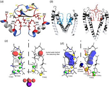
Bacterial potassium channels such as KcsA and chimera's with eukaryotic K+ channels (including KcsA–Kv1.3) have served as valuable model systems to understand the structural and dynamical aspects of potassium channel function by a variety of structural and biophysical methods. For example, our group has examined the effects of ligand binding and pH-induced activation upon channel structure in lipid bilayers. A particular interesting class of ion channel ligands is toxins from various spiders and scorpions, representing an important source of natural drugs. We obtained 3D structural information on Kaliotoxin bound to a chimeric KcsA–Kv1.3 potassium channel (Fig. 5a ) by forming mixed complexes in which either the channel or the ligand was labeled (Lange et al. Reference Lange, Giller, Hornig, Martin-Eauclaire, Pongs, Becker and Baldus2006). Subsequently, we also probed the inactivated state of the channel (Fig. 5b , left) and ligand-binding states for natural and synthetic blockers such as porphyrin using ssNMR spectroscopy (Ader et al. Reference Ader, Schneider, Hornig, Velisetty, Wilson, Lange, Giller, Ohmert, Martin-Eauclaire, Trauner, Becker, Pongs and Baldus2008, Reference Ader, Schneider, Hornig, Velisetty, Vardanyan, Giller, Ohmert, Becker, Pongs and Baldus2009) (Fig. 5b , right). These studies not only provided detailed insight into which channel residues are critical for complex formation and inactivation (Fig. 5b ), but also revealed a remarkable conformational plasticity of the selectivity filter (the core unit for ion channel function) in membranes. More recently, we also examined how the lipid bilayer contributes to channel structure as well as to the functional cycle (van der Cruijsen et al. Reference van der Cruijsen, Nand, Weingarth, Prokofyev, Hornig, Cukkemane, Bonvin, Becker, Hulse, Perozo, Pongs and Baldus2013; Weingarth et al. Reference Weingarth, Prokofyev, van der Cruijsen, Nand, Bonvin, Pongs and Baldus2013). 1H-detected ssNMR allowed us to infer the location of water molecules in two different channel states (Figs 5c and 5d , taken from (Weingarth et al. Reference Weingarth, van der Cruijsen, Ostmeyer, Lievestro, Roux and Baldus2014)). In addition, McDermott and co-workers examined the effect of protonation state of E71 for inactivation (Bhate & McDermott, Reference Bhate and Mcdermott2012). Methyl-TROSY experiments, including 13C ZZ-exchange experiments, on truncated KcsA tetramers revealed a correlation between tetramer stability and the equilibrium between conductive and inactivated channels at low pH (Imai et al. Reference Imai, Osawa, Mita, Toyonaga, Machiyama, Ueda, Takeuchi, Oiki and Shimada2012). In addition, the population of the conductive state was lower in lipid-nanodiscs as compared with DDM micelles, stressing the effect of the membrane environment on MPC conformational dynamics. Similar studies should be possible in larger ion channels. For example, our group already reported ssNMR studies on the ligand-gated ion channel mlCNG (Cukkemane & Baldus, Reference Cukkemane and Baldus2013; Cukkemane et al. Reference Cukkemane, Nand, Gradmann, Weingarth, Kaupp and Baldus2012).
Potassium channels studied by NMR. (a). Model of the KTX–KcsA-Kv1.3 complex. Residues (yellow labels, toxin; black labels, channel) affected or unperturbed by complex formation (according to ssNMR chemical-shift mapping) are indicated in red and blue, respectively (adapted from Lange et al. Reference Lange, Giller, Hornig, Martin-Eauclaire, Pongs, Becker and Baldus2006). (b) Structural models for the KcsA–Kv1.3 channel at pH 4·0 (after inactivation, left, and with bound porphyrin at pH 7·5, right). Residues for which Cα chemical shift changes were observed are blue (left, pH 4·0) and red (right, porphyrin-bound). Residues for which no Cα chemical shift changes larger than 0·6 ppm have been found are black; unassigned residues are in gray (adapted from (Ader et al. Reference Ader, Schneider, Hornig, Velisetty, Wilson, Lange, Giller, Ohmert, Martin-Eauclaire, Trauner, Becker, Pongs and Baldus2008)) (c and d) represent snapshots of MD simulations of the closed-conductive and open-inactivated channel state, respectively. Protons for which 1H-detected ssNMR experiments observe the proximity and the absence of buried water are color-coded in yellow and green, respectively (adapted from (Weingarth et al. Reference Weingarth, van der Cruijsen, Ostmeyer, Lievestro, Roux and Baldus2014)).

3.4 ATPases and transporters
Cellular calcium concentrations are modulated by sarco/endoplasmic reticulum calcium ATPases (SERCA), members of the P-type ATPase family important for muscle contraction and cellular transport. The activity of SERCA in muscle cells is inhibited by the binding of phospholamban (PLN), a transmembrane protein whose phosphorylation state changes its interaction with SERCA. We have used ssNMR, in particular the combination of scalar and dipolar ssNMR methods, to characterize conformational substates of free (Andronesi et al. Reference Andronesi, Becker, Seidel, Heise, Young and Baldus2005) and SERCA-bound (Seidel et al. Reference Seidel, Andronesi, Krebs, Griesinger, Young, Becker and Baldus2008) conformations of PLN variants. In the latter case, we observed a C-terminal α-helix that more recently was confirmed by X-ray crystallography (Akin et al. Reference Akin, Hurley, Chen and Jones2013). On the other hand, the N-terminal region exhibits both in free state as well as in the presence of SERCA considerable structural disorder (Andronesi et al. Reference Andronesi, Becker, Seidel, Heise, Young and Baldus2005) which may be critical for the functional regulation of SERCA (Gustavsson et al. Reference Gustavsson, Verardi and Mullen2013).
Furthermore, ssNMR studies were recently conducted to elucidate the active-site structure of the thermophilic Foc-subunit ring in membranes (Kang et al. Reference Kang, Todokoro, Yumen, Shen, Iwasaki, Suzuki, Miyagi, Yoshida, Fujiwara and Akutsu2014). FoF1–ATP synthase uses the electrochemical potential across membranes or ATP hydrolysis to rotate the Foc-subunit ring. To elucidate the underlying mechanism, 2D 13C–13C correlation spectra of TFoc rings labeled with SAIL (Kainosho et al. Reference Kainosho, Torizawa, Iwashita, Terauchi, Mei Ono and Güntert2006)-Glu and -Asn were recorded. The resulting chemical-shift assignments provided insights into the chemical mechanism underlying the proton locking in Foc rings and H+ transfer at the interface of the c and a subunits.
In addition, ssNMR was used to study ATP-binding cassette (ABC) transport systems, which facilitate the translocation of substances, such as amino acids, across cell membranes energized by ATP hydrolysis. Glaubitz and co-workers (Hellmich et al. Reference Hellmich, Mönkemeyer, Velamakanni, van Veen and Glaubitz2015) and references therein) have examined dynamics and, more recently, nucleotide binding on LmrA, which belongs to the family of homodimeric multidrug ABC transporters from Lactococcus lactis that respond to a large number of chemically unrelated substrates. Oschkinat and co-workers utilized microcrystals of the ABC transporter ArtMP from Geobacillus stearothermophilus in different nucleotide-bound or -unbound states (Lange et al. Reference Lange, Becker-Baldus, Kunert, van Rossum, Casagrande, Engel, Roske, Scheffel, Schneider and Oschkinat2010). From selectively 13C, 15N-labeled ArtP, several sequence-specific assignments were obtained, most of which could be transferred to spectra of ArtMP. Distinct sets of NMR shifts were obtained for ArtP with different phosphorylation states of the ligand. Indications were found for an asymmetric or inhomogeneous state of the ArtP dimer bound with triphosphorylated nucleotides.
E. coli EmrE, a homodimeric multidrug antiporter, has been suggested to offer a convenient paradigm for secondary transporters due to its small size. It contains four transmembrane helices and forms a functional dimer. Ong et al. (Reference Ong, Lakatos, Becker-Baldus, Pos and Glaubitz2013) have probed the specific binding of substrates TPP+ and MTP+ to EmrE reconstituted into 1,2-dimyristoyl-sn-glycero-3-phosphocholine (DMPC) liposomes by 31P MAS NMR, showing that both substrates occupy the same binding pocket but also indicate some degree of heterogeneity of the bound ligand population, reflecting the promiscuous nature of ligand binding by multidrug efflux pumps. Direct interaction between 13C-labeled TPP+ and key residues within the EmrE dimer has been probed by through-space 13C–13C correlation spectroscopy. This was made possible by the use of ssNMR enhanced by DNP through which a nineteenfold signal enhancement was achieved. Ligand binding was also studied using oriented ssNMR methods (Gayen et al. Reference Gayen, Banigan and Traaseth2013). Dynamics of EmrE in isotropic bicelles and in the presence of the drug TPP+ could be characterized using solution NMR 1H-15N TROSY based experiments, providing deuteration of the non-labile protons (Morrison et al. Reference Morrison, DeKoster, Dutta, Vafabakhsh, Clarkson, Bahl, Kern, Ha and Henzler-Wildman2011). These data showed the protein exchanging between two asymmetric conformations with an exchange rate of about 4 to 5 s−1, as determined by ZZ-exchange experiments. Using derivatives of TPP+ however showed the interconversion rate to be dependent on the substrate identity (Morrison & Henzler-Wildman, Reference Morrison and Henzler-Wildman2014). These conformational dynamics were confirmed by ssNMR on magnetically aligned bicelles as well as MAS experiments in DMPC liposomes (Cho et al. Reference Cho, Gayen, Banigan, Leninger and Traaseth2014). A combined solution and ssNMR study on the pH dependency of the conformational dynamics in EmrE showed that the protonation state of a membrane-embedded glutamate residue is key in the allosteric modulation of the global conformational motions (Gayen et al. Reference Gayen, Leninger and Traaseth2016).
Finally, ssNMR was used to study the Yersinia adhesin A (YadA), which is a prototypical member of the TAA family and, like other family members, it forms a stable trimeric pore inside the outer membrane (OM) and functions as the translocation pore for its extracellular adhesive domain. The original ssNMR structure of YadA-M was solved from a microcrystalline sample (Shahid et al. Reference Shahid, Bardiaux, Franks, Krabben, Habeck, van Rossum and Linke2012a) and a stretch of four residues (Ala, Ser, Ser and Ala) in the translocation domain was found to be highly flexible based on its non-helical NMR chemical shifts, reduced dipolar couplings, higher random-coil index and predicted low-order parameter values. Comparison of the dynamics between the microcrystalline and membrane-embedded samples indicates greater flexibility of the ASSA region in the outer-membrane preparation at physiological temperatures (Shahid et al. Reference Shahid, Nagaraj, Chauhan, Franks, Bardiaux, Habeck, Orwick-Rydmark, Linke and van Rossum2015).
3.5 Kinases
Kinase-mediated signaling is initiated by stimulation of cell surface receptors, which undergo conformational changes to activate their cellular kinase in eukaryotes (Lemmon & Schlessinger, Reference Lemmon and Schlessinger2010) or bacteria, with the latter containing two-component systems consisting of a sensor kinase and a response regulator (Mascher et al. Reference Mascher, Helmann and Unden2006). Compared to the structural biology of GPCRs or ion channels, high-resolution structures of such cell surface receptors are still elusive (Kovacs et al. Reference Kovacs, Zorn, Huang, Barros and Kuriyan2015). The difficulty in using crystallography to determine such 3D structures may be related to the fact that kinase receptors typically contain only one or two transmembrane helices, which allows for considerable mobility for the extra-membrane regulatory units. Indeed, a prominent role of protein dynamics as well as of the surrounding cell membrane for receptor function has been discussed in the literature (Bessman et al. Reference Bessman, Freed and Lemmon2014).
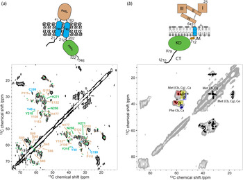
An example of such systems is DcuS, the C4-dicarboxylate sensor of E. coli, that is, a member of the periplasmic sensing histidine kinases. To allow transmembrane sensing, the multidomain protein DcuS possesses functional domains in the periplasm, within the membrane and in the cytoplasm. Periplasmic signal perception is achieved by a Per-Arnt-Sim domain (PASP). A membrane-integral domain consisting of two transmembrane helices transmits the signal to a cytoplasmic region. This region comprises a second PAS domain (PASC) and the C-terminal transmitter or kinase region consisting of the conserved DHp (dimerization and HisP-transfer) and catalytic (HATPase) domains (Fig. 6a , top).
NMR studies on receptor kinases. (a) 2D 13C–13C spin diffusion ssNMR data on a reverse-labeled DcuS in lipid bilayers with tentative resonance assignments of different protein domains colored as in the cartoon (top). (see also main text and (Etzkorn et al. Reference Etzkorn, Kneuper, Dünnwald, Vijayan, Krämer, Griesinger, Becker, Unden and Baldus2008). (b) 13C–13C experiment of 13C methionine and phenylalanine labeled EGFR-rich A431 membrane vesicles. Black crosses represent EGFR chemical-shift predictions for residues M and F based on the available crystal/NMR structures of the different domains of EGFR. Red, yellow and blue boxes represent where (Cα, Cβ) correlations of helical, random coil and β-strand M and F are expected. A schematic representation of EGFR different domains is given on the top.

Previously, we (Etzkorn et al. Reference Etzkorn, Kneuper, Dünnwald, Vijayan, Krämer, Griesinger, Becker, Unden and Baldus2008) have used a combination of ssNMR, structural modeling and mutagenesis to establish a structure–function relationship for membrane-embedded DcuS. We compared 2D ssNMR data of an individual cytoplasmic PAS domain to structural models generated in silico using ROSETTA (Yarov-Yarovoy, Reference Yarov-Yarovoy, Schonbrun and Baker2006). These studies, together with previous NMR work on the periplasmic PAS domain, enabled structural investigations of a membrane-embedded 40 kDa construct, comprising both PAS segments and the membrane domain. In Fig. 6a , 2D ssNMR data of a reverse-labeled DcuS–[PASP/TM1,2/PASC], with tentative assignments obtained from solution-state assignments in PASp and ShiftX (see (Han et al. Reference Han, Liu, Ginzinger and Wishart2011) and references therein) chemical-shift predictions. According to our studies, structural alterations were largely limited to protein regions close to the transmembrane segment. Data from isolated and multidomain constructs favor a disordered N-terminal helix in the cytoplasmic domain. Mutations of residues in this region strongly influence function, suggesting that a change in protein flexibility is related to signal transduction toward the kinase domain and regulation of kinase activity (Etzkorn et al. Reference Etzkorn, Kneuper, Dünnwald, Vijayan, Krämer, Griesinger, Becker, Unden and Baldus2008).
Receptor activation can be influenced by stimulus strength (such as ligand type and concentration) and receptor concentration, as for members of the receptor tyrosine kinase (RTK) family (Kovacs et al. Reference Kovacs, Zorn, Huang, Barros and Kuriyan2015). Using ssNMR, Matsushita and co-workers (Matsushita et al. Reference Matsushita, Tamagaki, Miyazawa, Aimoto, Smith and Sato2013) have examined the activation of the ErbB2 RTK by focusing on the membrane-spanning (MS) and juxta-membrane domains. They exploited an oncogenic mutation in the MS region that results in constitutively active signaling, as well as the ability to induce dimerization through fusion to a Put3 sequence. After reconstitution in synthetic lipid bilayers with phosphatidylinositol-4,5-bisphosphate (PIP2), an integral player in the downstream events following the activation of RTKs, ssNMR studies were conducted. Measurements of deuterated leucine residues in the MS region confirmed dimerization of the mutant. Conformations exhibiting different levels of molecular mobility were distinguished by dipolar and direct excitation experiments that are largely insensitive to motion. Taken together, the results suggest a specific mechanism by which extracellular ligand binding can be converted into intracellular signaling in which the lipid environment plays an important role (Matsushita et al. Reference Matsushita, Tamagaki, Miyazawa, Aimoto, Smith and Sato2013).
On the other hand, Her1 or the EGFR establishes cellular signaling as many other tyrosine kinase receptors via a single transmembrane helix (Fig. 6b ). Solution-state NMR has been used to infer the structure of the transmembrane region in membrane mimetics (see. e.g. (Endres et al. Reference Endres, Das, Smith, Arkhipov, Kovacs, Huang, Pelton, Shan, Shaw, Wemmer, Groves and Kuriyan2013) and references therein). EGFR is a 1286 amino-acid receptor and is implicated in the development of many types of cancer. While crystal and NMR structures are available for the different domains of the receptor, the structure and dynamics of the full-length EGFR are still elusive, let alone in its native environment. In our lab, we successfully developed protocols (Kaplan et al. 2016 under revision) to study EGFR-containing native membrane vesicles isolated from human cancer cells (A431) grown on fully or specifically 13C, 15N media. In Fig. 6b , results of a 2D 13C–13C spin diffusion experiment of specifically 13C Met and Phe labeled A431 membrane vesicles are shown. Comparison of the experimental data to FANDAS (Gradmann et al. Reference Gradmann, Ader, Heinrich, Nand, Dittmann, Cukkemane, Dijk, Bonvin, Engelhard and Baldus2012)-based predictions on the basis of the structural information available on EGFR segments confirms the presence of Met and Phe resonances in different secondary structures suggesting a folded EGFR in the isolated vesicles. These results provided the basis to investigate the effect of EGFR activation due to its natural ligand EGF at different temperatures in a natural membrane environment by ssNMR methods (Kaplan et al. 2016 under revision).
3.6 Translocation and insertion machines
3.6.1 β-barrel assembly machinery (β-BAM)
The process of protein folding and insertion into bacterial membranes is essential for physiological, pathogenic, and drug resistance functions. In Gram-negative bacteria, and E. coli in particular, the β-BAM complex (BAM complex) inserts β-barrel proteins into the OM of this organism. This system is conserved across diverse bacteria and homologues have been identified and studied in mitochondria (SAM) and chloroplasts (TOC) (Walther et al. Reference Walther, Papic and Bos2009).
Unfolded OM proteins upon entering into the periplasm are shuttled by periplasmic chaperones, to the 200 kDa heteropentameric BAM complex. Although the exact mechanism is still to be elucidated, it is postulated that β-sheets from the β-barrel of the core component, BamA, and its associated POTRA domains (P1–P5) serve as templates for β-sheet formation in a process termed β-augmentation (Hagan et al. Reference Hagan, Silhavy and Kahne2011).
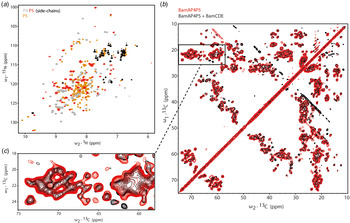
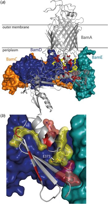
While a P4P5 tandem construct of BamA is well folded (Gatzeva-Topalova et al. Reference Gatzeva-Topalova, Warner, Pardi and Sousa2010; Sinnige et al. Reference Sinnige, Houben, Pritisanac, Renault, Boelens and Baldus2015a), studies on BamAP5 constructs (Morgado et al. Reference Morgado, Zeth, Burmann, Maier and Hiller2015; Sinnige et al. Reference Sinnige, Houben, Pritisanac, Renault, Boelens and Baldus2015a) highlight the requirement of a stabilizing interface with POTRA 4 for correct folding of POTRA 5 (Fig. 7a ). Solution NMR 15N-CPMG experiments performed on the P4P5 construct then showed that several residues of the POTRA 5 domain of BamA exhibit conformational dynamics (Sinnige et al. Reference Sinnige, Weingarth, Daniëls, Boelens, Bonvin, Houben and Baldus2015b) (Fig. 8b residues in red) and form a surface with residues whose amide resonances were, as a result of this conformational dynamics, broadened beyond detection (Fig. 8b residues in yellow). Interestingly, many of these residues exchanging between one or more conformations in absence of the binding lipoproteins map remarkably well onto the interaction surface with BamD in the recently published crystal structures of the BamACDE (Bakelar et al. Reference Bakelar, Buchanan and Noinaj2016) (Fig. 8a ) and BamABCDE complexes (Gu et al. Reference Gu, Li, Dong, Zeng, Zhang, Paterson, Stansfeld, Wang, Zhang, Wang and Dong2016; Han et al. Reference Han, Zheng, Wang, Yang, Liu, Sun, Cao, Zhou, Ni, Lou, Zhao and Huang2016) (Fig. 8b , residues in surface and stick representation). Residue E373, which has been shown previously to be central in the BamAP5/BamD interaction (Ricci & Silhavy, Reference Ricci and Silhavy2012) is located within this region. Remarkably, the remaining dynamic residues map to the opposite side of POTRA 5 and cluster to the β 2-sheet and α 2-helix (Fig. 8b red residues). This region has been previously shown as a site for substrate binding/β-augmentation in POTRA 2 (Knowles et al. Reference Knowles, Jeeves, Bobat, Dancea, McClelland, Palmer, Overduin and Henderson2008) and POTRA 3 (Gatzeva-Topalova et al. Reference Gatzeva-Topalova, Walton and Sousa2008; Kim et al. Reference Kim, Malinverni, Sliz, Silhavy, Harrison and Kahne2007), respectively.
NMR studies on the β-BAM complex. (a) Solution NMR 1H–15N HSQC spectra of P4P5 tandem BamA POTRA domains (Gray – P4, red – P5, black – side-chains P4P5) compared with P5 alone (orange) (see also (Sinnige et al. Reference Sinnige, Houben, Pritisanac, Renault, Boelens and Baldus2015a, Reference Sinnige, Weingarth, Daniëls, Boelens, Bonvin, Houben and Baldusb)). (b) Solid-state PARIS-CC spin-diffusion NMR spectra of specifically labeled BamAP4P5 comprising the beta-barrel and domains P4 and P5 reconstituted alone (red) or in the presence of unlabeled BamCDE (black) in DLPC liposomes. (c) Zoom-in on the Valine–Threonine region of the spectrum in (b).

P5 dynamics represented on the crystal structure of BamACDE sub-complex (Bakelar et al. Reference Bakelar, Buchanan and Noinaj2016). (a) Residues in P5 that undergo conformational exchange in the tandem P4P5 construct are indicated in balls, with residues broadened beyond detection in yellow and residues with clear 15N-relaxation profiles in red. (b) Zoom-in of the crystal structure with the dynamic BamA P5 residues from (b), which are at the interface of BamD/E, displayed in surface representation and sticks.

The effect BamA has on the lipid bilayer is quite significant as evidenced in a study that combined ssNMR and EM to study the protein in various bilayers (Sinnige et al. Reference Sinnige, Weingarth, Renault, Baker, Tommassen and Baldus2014b). Electron micrographs of BamA proteoliposomes show notches at the surface, which was suggested to be from the protein destabilizing the bilayer in its proximity. This study also showed that the β-barrel is itself extremely robust, largely preserving its structure and dynamics in bilayers of varying membrane thicknesses (21–27 Å in the liquid crystalline phase) (Sinnige et al. Reference Sinnige, Houben, Pritisanac, Renault, Boelens and Baldus2015a, Reference Sinnige, Weingarth, Renault, Baker, Tommassen and Baldusb). These observations were corroborated upon determination of the structure of the β-barrel, which showed that the transmembrane β-barrel itself could destabilize the membrane due to its asymmetric hydrophobic thickness, induced by the asymmetry of the β-barrel, presumably to allow for more efficient protein insertion (Noinaj et al. Reference Noinaj, Rollauer and Buchanan2015).
In such a system where the lipid bilayer plays such a crucial role in function, it is imperative to study the effects which occur on the complex in environments, which mimic their native environment. Such influences were highlighted in a study on BamA precipitates which showed that in the absence of a membrane the POTRA domains exhibit fast-motion on the NMR time-scale (sub-μs or faster), as they were not present in spectra that probe rigid protein segments. However, these domains show much reduced mobility once the protein is reconstituted into lipid bilayers (Renault et al. Reference Renault, Bos, Tommassen and Baldus2011).
Currently we have data that show formation of the complex in liposome preparations upon reconstitution of the unlabeled BamCDE sub-complex to a specifically labeled BamAP4P5 construct in liposomes (Fig. 7b ), as evidenced by chemical shift perturbations, such as those shown, for example, in the zoom-in of the valine-threonine region of the CC spin-diffusion experiment (Fig. 7c ) (Pinto et al., Manuscript in preparation).
3.6.2 Secretion systems in Gram-negative bacteria
Bacteria have evolved a wide variety of highly specialized macromolecular nanomachines that secrete a wide range of substrates, including small molecules, proteins and DNA. These substrates have key roles in the response of a bacterium to its environment and also in several physiological processes such as adhesion, pathogenicity, adaptation and survival. Depending on the secretion system, the secreted substrates have three possible fates: they remain associated with the bacterial OM, they are released into the extracellular space, or they are injected into a target cell (either a eukaryotic or bacterial cell). In Gram-negative bacteria, these machineries can be divided into two categories: those spanning both the inner membrane and the OM, and those that span the OM only. Five double-MS secretion systems have been identified to date and are classified as type I secretion system (T1SS), T2SS, T3SS, T4SS and T6SS (Costa et al. Reference Costa, Felisberto-Rodrigues, Meir, Prevost, Redzej, Trokter and Waksman2015).
For both the T3SS as well as the T4SS, ssNMR studies have in the last years been conducted to elucidate the structural arrangement of individual components or to obtain insight into their cell-embedded fold. For the T3SS, needle filament structural arrangements compared to the monomer unit could be elucidated (Poyraz et al. Reference Poyraz, Schmidt, Seidel, Delissen, Ader, Tenenboim, Goosmann, Laube, Thünemann, Zychlinsky, Baldus, Lange, Griesinger and Kolbe2010) as well as the 3D structure could be obtained (Loquet et al. Reference Loquet, Sgourakis, Gupta, Giller, Riedel, Goosmann, Griesinger, Kolbe, Baker, Becker and Lange2012). More recently, we have examined the structural arrangement of the entire T4SS core complex (T4SScc) in the bacterial cell envelope. T4SS is responsible for the transfer of a wide variety of effector proteins and nucleic acids between bacteria and to host cells and has been implicated in many diseases like gastrointestinal diseases through Helicobacter pylori. Moreover, it plays a vital role in bacterial conjugation thereby helping in the spread of antibiotic resistance. T4SS consists of 12 proteins VirB1 to VirB11 and VirD4 and three of these proteins (VirB7, VirB9 and VirB10) form the so-called core complex (T4SScc), which is a 1·1 MDa complex with tetradecamer symmetry spanning the whole bacterial cell envelope. Thus far, high-resolution structural data are available for only half of the complex, including a low-resolution EM map (Rivera-Calzada et al. Reference Rivera-Calzada, Fronzes, Savva, Chandran, Lian, Laeremans, Pardon, Steyaert, Remaut, Waksman and Orlova2013). However, these results obtained on purified T4SScc cannot explain how this complex is located in its physiological environment and how substrates interact with this machine in a native setting (Low et al. Reference Low, Gubellini, Rivera-Calzada, Braun, Connery, Dujeancourt, Lu, Redzej, Fronzes, Orlova and Waksman2014). For this reason, we resorted to investigate T4SScc in a native bacterial cell envelope by DNP enhanced ssNMR. We could successfully produce fully and specifically [13C, 15N]-labeled T4SScc in E. coli lacking OmpA/F (thereby decreasing the NMR background signal) and isolate the cell envelope for in situ DNP-enhanced ssNMR studies (Kaplan et al. Reference Kaplan, Cukkemane, van Zundert, Narasimhan, Daniëls, Mance, Waksman, Bonvin, Fronzes, Folkers and Baldus2015). In addition to T4SScc, cellular endogenous non-proteinaceous compounds were also visible in ssNMR spectra, for example in conventional 2D 13C,13C spin diffusion experiments at ambient temperatures (Fig. 9a ). In our DNP studies using selective labeling, we could for the first time confirm that segments of the T4SScc seen in protein crystals retain their fold in the cellular envelope and we obtained new insight regarding the structure and dynamics of the hitherto elusive part of T4SScc, in particular its embedding in the inner bacterial membrane (Fig. 9b ). Further NMR studies to refine structure and dynamics of the T4SScc in a native setting and in response to substrates are ongoing.
NMR studies on Type-4 Secretion System core-complex. (a) 13C–13C correlation experiment of a fully 13C, 15N labeled bacterial cell envelope expressing T4SScc. Red crosses represent T4SScc chemical-shift predictions based on the available crystal structure and modeling of the structurally elusive parts. Chemical-shift predictions of non-proteinaceous endogenous molecules like lipids (PE), peptidoglycans (PG) and lipopolysaccharide (LPS) are shown in dark blue, orange and light blue, respectively. (b) Figure representing the T4SScc electron-microscopy map with the crystal structure of the outer membrane complex (PDB = 3JQO) docked in, all embedded in the bacterial cell envelope. Red and orange spheres represent the identified probes in different specifically 13C, 15N labeled cell envelopes expressing T4SScc. For further information see (Kaplan et al. Reference Kaplan, Cukkemane, van Zundert, Narasimhan, Daniëls, Mance, Waksman, Bonvin, Fronzes, Folkers and Baldus2015).

4. Conclusions and future perspectives
In this work, we have described how NMR has been used to study membrane-protein complexes. Advancements in NMR instrumentation in particular in the field of ssNMR, as well as in the area of sample preparation and labeling in pro- and eukaryotes have strongly increased the utility of NMR to infer structural and/or dynamic information at the most detailed, i.e., atomic level in virtually all applications/areas discussed in the review. Indeed, NMR methods today can be applied to study receptors and MPCs encompassing several hundreds of amino-acids and more by using tailored labeling schemes in combination with high-sensitivity methods such as DNP and high-field NMR.
Not surprisingly, NMR has contributed significantly to understanding the structural and dynamic aspects of ligand binding or other (in)activation events in the case of ion channels and GPCRs. In addition, NMR provides unique opportunities to elucidate the influence of the lipid bilayer, small molecules or, as in the case of cellular ssNMR, other cellular components including glycans or other proteins for the formation and functional pathway of MPCs.
The information to be obtained by NMR not only delivers structural information, but also helps deciphering protein dynamics before and after complex formation or related to activation at atomic resolution. Both aspects are increasingly realized as critical parameters for understanding protein function. Already, NMR on soluble proteins has become a leading technology to understand allostery within conformational ensembles (Motlagh et al. Reference Motlagh, Wrabl, Li and Hilser2014) and the examples given here underline the growing potential of NMR to obtain such information in the context of MPCs. An important focus area for future NMR-studies of MPCs will hence likely be the examination of protein structure, dynamics and protein–protein interactions in natural prokaryotic or eukaryotic membranes, cell compartments and whole cells. Reference information obtained in different membrane mimetics, starting from detergents or nanodiscs and leading to synthetic lipid bilayers will help to establish a general framework of how lipid–protein interactions and the surrounding bilayer and other molecular players contribute to functioning under physiological conditions.
Ultimately, NMR-based studies of MPCs may, in combination with other structural biology tools including X-ray and cryo-EM, provide unprecedented insight into the energetics of protein–ligand and protein–protein interactions that underlie the basis for essential biological functions in cellular compartments within and between cells.
Acknowledgements
We gratefully acknowledge our collaborators and colleagues for their invaluable contributions to cited publications from our own research group. These studies were supported through grants from NWO, the EU and NIH as well as the DFG, the Max-Planck-Society and the Volkswagen foundation. We also are indebted to our colleagues S.O. Smith, V. Ladishansky, C. Glaubitz and R. Grisshammer for providing electronic figure versions.