


Fe, Zn and Ca are essential minerals that play a crucial role in human health, supporting functions such as oxygen transport, immune response and bone health, respectively(Reference Singh and Prasad1,Reference Chasapis, Loutsidou and Spiliopoulou2) . However, widespread deficiencies of micronutrients exist globally with recent estimates indicating that 69% of women of reproductive age and 56% of preschool-aged children have at least 1 micronutrient deficiency with Fe and Zn among the top three most deficient micronutrients(Reference Stevens, Beal and Mbuya3). In many cases, diets may not necessarily be low in Fe, Zn or Ca, but plant-based diets dominant in regions and populations with high prevalence of deficiencies are characterised by poor mineral bioavailability(Reference Chasapis, Loutsidou and Spiliopoulou2,Reference Muleya, Bailey and Bailey4) . Depending on the type of diet, Fe and Zn bioavailability can range from 5 to 15% and 15–50%, respectively, with the low end of the range typical for unrefined, high-fibre plant-based diets while the high end of the range corresponds to refined diets with a high proportion of animal source foods(5). Ca bioavailability, on the other hand, is highly variable amongst plant sources and can be as low as 9% for high-oxalate vegetables such as spinach and over 50% for low-oxalate vegetables like kale(Reference Weaver, Wastney and Fletcher6).
The poor bioavailability of minerals in some matrices is due to several factors; presence of digestion and absorption inhibitors such as phytic acid, tannins, oxalates and fibre(Reference Singh and Prasad1,Reference Gabaza, Muchuweti and Vandamme7) ; structural and cellular integrity(Reference Duijsens, Gwala and Pallares8,Reference Glahn, Tako and Cichy9) ; physicochemical state and matrix interactions such as the solubility of tri-calcium phosphate used in fortification, or the interaction of Ca with fats forming Ca soaps(Reference Fardet, Dupont and Rioux10). Although these issues affecting mineral bioavailability are dominant in plant-based matrices, they are also relevant to emerging novel food and feed sources being developed for sustainable food security, such as edible insects, algal and fungal based products. It is now increasingly recommended to reduce consumption of animal source foods and increase consumption of plant and alternative sources of food for both health and environmental sustainability(Reference Willett, Rockström and Loken11). As a result, future diets are likely to include fewer animal source foods and greater proportions of plant and emerging food sources whose mineral bioavailability is likely to be poor, highly variable or unknown. Understanding the mineral bioavailability of these foods is now more critical than ever. Bioavailability data is urgently needed to update dietary recommendations and inform strategies to enhance bioavailable mineral supplies in food systems, particularly as shortfalls of Fe, Zn and Ca are predicted in populations consuming diets with minimal animal source foods(Reference Beal, Ortenzi and Fanzo12). Bioavailability should now be considered as an integral part of food composition data, recognising that gross composition alone indicates the potential of food, not its exact nutritional value.
To date, there is only a limited amount of Fe, Zn and Ca bioavailability data for foods and even more limited data for emerging food and feed ingredients. A major challenge is the reliance on in vivo gold standard methods used to determine mineral bioavailability. These are costly, time consuming, invasive and in some cases ethically problematic, factors that have limited research progress in this area despite the rapid pace of ingredient discovery and product innovation. There is an urgent need for robust in vitro methods for routine evaluation of in vitro mineral bioavailability (bioaccessibility), particularly for screening purposes and gaining mechanistic insights that are not feasible in vivo. Ultimately these methods can reduce the need for in vivo experiments. Going forwards, it may also be valuable to explore how in vitro methods could be integrated with in vivo data, perhaps through robust algorithms or in silico models built on specifically generated data sets. In vitro methods are used to evaluate mineral bioaccessibility across a wide range of food matrices. However, there remains a lack of unified direction and common objectives among researchers to validate and standardise these methods for broader application in policy and dietary guideline development. The diversity of approaches, including static, semi-dynamic and dynamic models, and considerable variation in experimental parameters, such as enzyme concentrations, digestion times, pH control and sampling procedures, make cross-study comparisons difficult.
To address these inconsistencies, a standardised static in vitro digestion method was proposed by the COST Action INFOGEST(Reference Minekus, Alminger and Alvito13) and later updated by Brodkorb et al. (Reference Brodkorb, Egger and Alminger14). This protocol presents a valuable opportunity for the research community to align efforts and collaboratively generate harmonised, comparable data on mineral bioaccessibility. The purpose of this review is to examine the current use of the INFOGEST in vitro digestion model to assess Fe, Zn and Ca bioaccessibility and to highlight key challenges and limitations. The goal is to suggest strategies to enhance its utility for generating results that are comparable, actionable and informative for product development and dietary intake assessment.
Overview of digestion and absorption of Fe, Zn and Ca
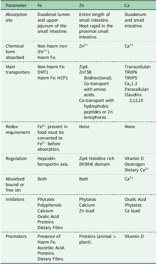
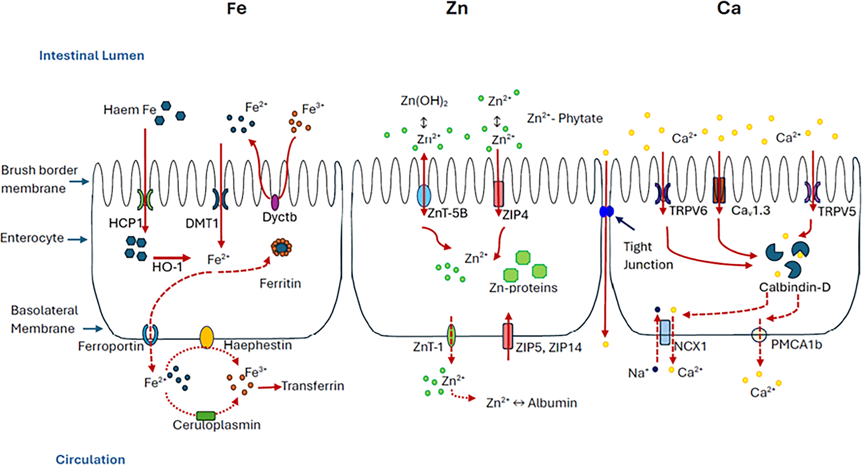
Understanding the digestion and absorption mechanisms of Fe, Zn and Ca is essential for interpreting mineral bioavailability data in the context of both in vivo and in vitro studies. These minerals differ in their chemical behaviour, interactions with dietary digestion and absorption inhibitors and enhancers, sites of absorption and absorption and transport pathways. These differences not only influence how much of each mineral is ultimately absorbed but also determine the extent to which current in vitro digestion methods can effectively replicate physiological reality. This section provides a brief overview of the key absorption processes for Fe, Zn and Ca, laying the groundwork for subsequent discussion on how these processes are modelled, or not modelled, in in vitro assessments of mineral bioaccessibility. Table 1 and Figure 1 show the summary of mechanisms involved in the digestion and absorption of Fe, Zn and Ca.
Summary table of absorption mechanisms for Fe, Zn and Ca

Table 1. Long description
A table comparing the absorption mechanisms of iron, zinc, and calcium. The table has six rows and four columns. The columns are labeled Parameter, Fe, Zn, and Ca. The rows are labeled Absorption site, Chemical form absorbed, Main transporters, Redox requirement, Regulation, Inhibitors, Promoters. Row 1: Absorption site, Fe: Duodenal lumen and upper jejunum of the small intestine, Zn: Entire length of small intestine. Most rapid in the proximal small intestine, Ca: Duodenum and small intestine. Row 2: Chemical form absorbed, Fe: Non-haem iron (Fe2+). Haem Fe, Zn: Zn2+, Ca: Ca2+. Row 3: Main transporters, Fe: Non-haem Fe: DMT1. Haem Fe: HCP1, Zn: Zip4. ZnT5B (bidirectional). Co-transport with amino acids. Co-transport with hydrophobic peptides or Zn ionophores, Ca: Transcellular: TRVP6. TRVP5. Ca1.3. Paracellular: Claudins 2,12,15. Row 4: Redox requirement, Fe: Fe3+ present in food must be converted to Fe2+ before absorption, Zn: None, Ca: None. Row 5: Regulation, Fe: Hepcidin-ferroportin axis, Zn: Zip4 histidine rich (M3M4) domain, Ca: Vitamin D. Oestrogen. Dietary Ca2+. Row 6: Inhibitors, Fe: Phytates. Polyphenols. Calcium. Oxalic Acid. Proteins. Dietary Fibre, Zn: Phytates. Calcium. Zn load, Ca: Oxalic Acid. Phytates. Ca load. Row 7: Promoters, Fe: Presence of Haem Fe. Ascorbic Acid. Proteins. Dietary Fibre, Zn: Proteins (animal > plant), Ca: Vitamin D.
Summary of the absorption mechanisms of Fe, Zn and Ca.

Before absorption can occur, dietary minerals must first be released from the complex food matrices in which they are embedded. In most foods, minerals are not present in free, soluble form, but are bound to or entrapped within macronutrient structures such as proteins, digestible and non-digestible polysaccharides, phytate complexes and polyphenolic structures. Mechanical breakdown during mastication and peristalsis, followed by chemical and enzymatic digestion in the stomach and small intestine, plays a critical role in liberating Fe, Zn and Ca into the intestinal lumen(Reference Duijsens, Gwala and Pallares8,Reference Glahn, Tako and Cichy9,Reference Sandberg15,Reference Kyomugasho, Gwala and Christiaens16) . Proteolytic digestion is particularly important for Fe and Zn, which are often tightly associated with proteins in both animal and plant tissues. For example, in cereals and legumes, Fe may be complexed with storage proteins or trapped within cell walls(Reference Glahn, Tako and Cichy9), while Zn is strongly associated with proteins in plant tissues(Reference Ebrahim, Alnajjar and Mohammed17). The hydrolysis of these proteins is important for the release of minerals into the intestinal lumen. Starch digestion, although not targeting mineral binding structure directly, can contribute to matrix loosening and with the combination of the mechanical effects of mastication and peristalsis, helps to disrupt plant cell integrity, facilitating the release of minerals that are otherwise trapped within intact cells or complexed with indigestible components(Reference Glahn, Tako and Cichy9,Reference Kyomugasho, Gwala and Christiaens16) . Therefore, accurate simulation of macronutrient digestion is paramount when using in vitro models to estimate the bioaccessible fraction of minerals. This point will be revisited in the next section.
Fe absorption occurs mainly via enterocyte cells in the duodenum and upper jejunum(Reference Piskin, Cianciosi and Gulec18). Apical uptake and transport of Fe across the brush border differs for haem and non-haem Fe which typically represent 10 and 90% of dietary Fe respectively(Reference Piskin, Cianciosi and Gulec18). Non-haem Fe (usually Fe3+) is first reduced to Fe2+ by ferric reductases, that is, duodenal cytochrome b (Dcytb) before being transferred into the enterocytes by divalent metal transporter 1 (DMT1)(Reference Conrad and Umbreit19,Reference Correnti, Gammella and Cairo20) . Haem Fe is transferred into enterocyte cells via haem carrier protein 1 (HCP1) and is then converted to Fe2+ by haem oxygenase 1 (HO-1)(Reference Conrad and Umbreit19). From the enterocyte cells the majority of Fe2+ is released across the basolateral membrane and into the bloodstream through conformational changes in the ferroportin protein with the remaining Fe2+ used by the enterocyte cells or incorporated into ferritin(Reference Correnti, Gammella and Cairo20). In the bloodstream Fe2+ is oxidised to Fe3+ by haephestin on the basolateral membrane or circulating ceruloplasmin to enable binding to transferrin for distribution(Reference Correnti, Gammella and Cairo20).
Zn homeostasis is regulated by transporters on both the apical and basolateral membranes of enterocyte cells and the Zn-binding protein metallothionein(Reference Maares and Haase21). Absorption from the small intestine across the intestinal brush border membrane into enterocyte cells is mainly via the ZIP4 transporter and subsequent release into the bloodstream is via the ZnT-1 basolateral membrane protein(Reference Maares and Haase21,Reference Devarshi, Mao and Grant22) . The transporters ZIP5 and ZIP14 on the basolateral surface import Zn from the bloodstream to the enterocytes and ZnT-5B on the apical surface allows bidirectional transfer of Zn(Reference Maares and Haase21,Reference Hall and King23) . Within the enterocyte Zn may be present as cytoplasmic-free (complexed by low molecular weight ligands), protein-bound (e.g. bonded to metallothionein) and free Zn(Reference Maares and Haase21).
Absorption of Ca can be through an active transcellular or passive paracellular pathway(Reference Bronner24,Reference Shkembi and Huppertz25) . The transcellular pathway dominates in the duodenum and jejunum and is regulated by vitamin D(Reference Bronner24,Reference de Barboza, Guizzardi and de Talamoni26) . Active transport of Ca2+ into enterocyte cells is through transient receptor potential vanilloid 6 (TRPV6), 5 (TRPV5) and Cav1.3. Transporter TRPV6 is the dominant transporter present in the duodenum with Ca v1.3 present mainly from the proximal jejunum to the mid-ileum, a distribution that supports the hypothesis that TRPV6 is mainly active between meals with Ca v1.3 active when Ca2+ concentrations are elevated during digestion of food(Reference de Barboza, Guizzardi and de Talamoni26). Ca is transferred to the basolateral surface by calbindin proteins with a high Ca2+ affinity, that is, calbindin-DK9, before release to the bloodstream via Ca-ATPase (PMCA1b) and sodium-calcium exchanger (NCX1)(Reference Shkembi and Huppertz25,Reference de Barboza, Guizzardi and de Talamoni26) . Paracellular transport, a linear function of lumen Ca2+ concentration, occurs throughout the intestine via transmembrane proteins of tight junctions, that is, claudins 2, 12 and 15(Reference de Barboza, Guizzardi and de Talamoni26).
Digestion and absorption mechanisms of minerals in vivo are critical to consider when assessing mineral bioavailability using in vitro digestion models, which may simulate chemical and mechanical processes of digestion but may lack the cellular and hormonal components that regulate absorption.
Translating in vivo to in vitro
In vitro digestion methods aim to simulate the chemical and mechanical breakdown of food that occurs in the gastrointestinal tract (GIT)(Reference Etcheverry, Grusak and Fleige27). These models typically simulate three major phases of digestion: the oral, gastric and intestinal stages, where most nutrient release and absorption occurs. Among current approaches, static models are the most widely used due to their practicality, cost-effectiveness and reproducibility and are the focus of this review. More complex models include semi-dynamic systems (with dynamic gastric phases) and fully dynamic systems like the dynamic gastrointestinal model (TIM), which better reflect temporal and physiological complexity(Reference Xavier and Mariutti28,Reference Hur, Lim and Decker29) .
The digestion and absorption of minerals involve four general steps: (1) release of minerals from the food matrix, (2) diffusion to the brush border membrane (3) cellular uptake into enterocytes and (4) basolateral transport into circulation(Reference Sandberg30). Most in vitro methods simulate the first step reasonably well, particularly solubilisation of minerals through enzymatic and pH driven matrix breakdown. Some studies also simulate the second step by separating the digesta into low and high molecular weight fractions(Reference Muleya, Bailey and Bailey4,Reference Miller, Schricker and Rasmussen31,Reference Muleya, Young and Bailey32) but steps 3 and 4, cellular uptake and systemic transport, are often not captured unless the digestion model is coupled with cell culture systems such as Caco-2 cell monolayers commonly used to estimate Fe bioavailability(Reference Glahn, Wien and van Campen33). This review focuses on static in vitro digestion systems without cell culture components, as these are commonly used, more practical and offer greater reproducibility in assessing Fe, Zn and Ca bioaccessibility.
Standard static methods show great variation in the parameters simulated: pH, oral 6.8–7.2; gastric 1.5–4.0; intestinal 6.5–7.5, temperature (typically 37°C), mixing/agitation (using e.g. shaking water baths or rotators), enzyme combinations (amylase, pepsin, pancreatin, trypsin, chymotrypsin, proteases normally from porcine) and bile salts. Digestion times commonly range from 2 minutes in the oral phase and 1–2 hours in the gastric and intestinal phase(Reference Hur, Lim and Decker29). However, these parameters are fixed, whereas in vivo digestion is dynamic and responsive to food properties via feedback mechanisms. Importantly, in vitro methods do not account for site-specific absorption differences among minerals. For example, Fe is primarily absorbed in the duodenum, Zn in the jejunum and Ca throughout the small intestine(Reference Piskin, Cianciosi and Gulec18,Reference Maares and Haase21,Reference Bronner24) . They also cannot model individual-level variation (e.g. age, sex, mineral status), or the influence of the intestinal microbiota on mineral metabolism(Reference Sandberg30). Additional limitations include the use of fixed transit times that ignore food-specific gastric emptying rates. In general, longer transit times are associated with a greater level of mineral release(Reference Sandberg30). In vitro, however, transit time is often fixed regardless of food composition. Likewise, enzyme concentrations and composition of digestive fluids in vivo are modulated by meal composition and hormonal feedback, features absent in static models(Reference Sandberg30,Reference Lynch34) .
Despite these constraints, in vitro models have generated a substantial body of evidence that underpins much of our current understanding of mineral bioavailability. They have elucidated the effects of digestion and absorption inhibitors like phytate, polyphenols, tannins and fibre, enhancing effects of ascorbic acid on Fe bioavailability and impact of food processing on mineral bioaccessibility(Reference Gabaza, Muchuweti and Vandamme7,Reference Gabaza, Shumoy and Muchuweti35–Reference Theodoropoulos, Turatti and Greiner37) . Although the magnitude of effects sometimes differs, these models have proven indispensable as screening tools and in mechanistic research(Reference Lynch34).
A key area of divergence across studies is the definition of the bioaccessible fraction. In some cases, bioaccessibility is equated with solubility: the amount of mineral released and found in the supernatant after centrifugation of digested samples. However, others further distinguish low molecular weight soluble fractions (normally < 6–14 kDa) as truly bioaccessible, based on their potential to diffuse through the mucus layer and reach transport sites(Reference Sandberg30). Methods such as dialysis, membrane filtration and less frequently size exclusion chromatography are used to separate such fractions(Reference Muleya, Bailey and Bailey4,Reference Miller, Schricker and Rasmussen31) . These distinctions matter, as dialysis and filtration can yield different outcomes and solubility alone may overestimate bioaccessibility.
Recent commentary by Grundy et al. (Reference Grundy, Moughan and Wilde38) highlights persistent confusion around the use of the term bioaccessibility, with differing definitions applied across studies and nutrient types. Grundy et al. (Reference Grundy, Moughan and Wilde38) therefore advocate for clearer and more harmonised terminology. In our view, bioaccessibility in the context of minerals should be defined as the fraction released from the food matrix and present in absorbable, low molecular weight forms, as this best reflects the fraction that is physiologically available for uptake at the intestinal epithelium. This definition is broadly in line with the definition later proposed by Grundy et al. (Reference Grundy, Deglaire and Le Feunteun39), where bioaccessibility should be defined as ‘the fraction of an ingested compound that is biochemically and/or physically released from the food matrix and solubilised within the gastrointestinal fluids in an absorbable form’. This definition accounts for the processes occurring in the GIT, leading to the formation of the absorbable fractions, that is, physical release from food matrix, solubilisation, biochemical reactions and interactions which can happen in any order(Reference Grundy, Deglaire and Le Feunteun39).
To address the variability and improve inter-laboratory comparability, the INFOGEST protocol was introduced as a standardised static digestion model. It harmonises pH values, enzyme activities, electrolyte concentrations and digestion times. In the next sections, we explore the application of the INFOGEST method in assessing Fe, Zn and Ca bioaccessibility and highlight the challenges associated and opportunities for broader application.
INFOGEST digestion model to determine Fe, Zn and Ca bioaccessibility
The INFOGEST static in vitro digestion method was first developed under the COST Action FA1005 initiative to harmonise in vitro digestion experimental conditions across laboratories studying nutrient digestion in humans(Reference Minekus, Alminger and Alvito13). Its primary objective was to offer a reproducible, physiologically relevant framework that standardises key experimental conditions simulated during gastrointestinal digestion such as types of enzymes and their activity, pH, ionic strength, digestion times and mixing, thereby enabling better comparability of data across studies. The protocol was updated in 2019(Reference Brodkorb, Egger and Alminger14) to improve physiological relevance, notably refining enzyme activity analysis and incorporating gastric lipase, which had been omitted from INFOGEST 1.0. Today, it is widely used to assess the digestibility and bioaccessibility of food components including starch, proteins, fats, vitamins, minerals and phytochemicals. The protocol simulates three major phases of gastrointestinal digestion, that is, oral, gastric and intestinal, using well-defined parameters; oral (masticated food mixed 1:1 with simulated salivary fluid + salivary amylase (75 U/mL), pH 7, 37°C, 2 min), gastric (oral bolus mixed 1:1 with simulated gastric fluid + porcine pepsin (2000 U/mL), rabbit gastric lipase (60 U/mL), pH 3, 37°C, 2 h) intestinal (mix gastric chyme 1:1 with simulated intestinal fluid + pancreatin (based on trypsin activity 100 U/mL), bovine bile (10mM), pH 7, 37°C, 2 h).
While the method provides a strong foundation for simulating food breakdown, there is no consensus on how to handle samples post-digestion for evaluating different physiological endpoints. Brodkorb et al. (Reference Brodkorb, Egger and Alminger14) offer general recommendations, for example, using dialysis membranes or centrifugal filters with a 3–10 kDa cut-off to assess bioaccessibility, but these are not standardised. As a result, there is no single agreed method for separating the bioaccessible fraction of minerals. This flexibility has led to varied interpretations of what constitutes bioaccessible Fe, Zn or Ca, as noted in the previous section. These differences significantly impact reported outcomes and hinder direct comparisons across studies, even when the same digestion protocol is used.
Another notable challenge when applying the INFOGEST method to mineral bioaccessibility is the background contamination of minerals, from digestion reagents. Enzyme preparations such as pancreatin, pepsin and bile salts may contain significant trace levels of these minerals, potentially confounding analytical results, especially when assessing foods such as cereals and legumes, where endogenous mineral concentrations are relatively low. Some studies have attempted to address this by purifying reagents using Chelex-100 resin, a metal-chelating resin that removes divalent cations from solutions(Reference Glahn, Lee and Yeung40). However, there is no harmonised or standardised guidance currently provided in the INFOGEST protocol regarding background correction or reagent treatment, making this a persistent source of variability across studies. While the INFOGEST model is physiologically informed, it was primarily developed for evaluating macronutrient digestibility. Its application to mineral bioaccessibility presents distinct challenges due to the physicochemical complexity of mineral - matrix interactions and the modifying effects of digestion and absorption inhibitors and enhancers. Despite these limitations, the INFOGEST method remains the most widely adopted static digestion protocol, and its application in assessing mineral bioaccessibility continues to expand. The following sections explore how key challenges, that is, high background mineral concentrations and inconsistencies in separating the bioaccessible fraction, can be addressed to improve its reliability and utility.
Resolving the high mineral contribution of enzymes used in the INFOGEST model
A defining feature of the INFOGEST protocol is the standardisation of enzyme sources and activities, which distinguishes it from earlier in vitro digestion methods. While previous models, such as the widely used method for evaluating in vitro Fe bioavailability proposed by Miller et al. (Reference Miller, Schricker and Rasmussen31), adapted by Glahn et al. (Reference Glahn, Lee and Yeung40) and used by many others(Reference Kose, de Bie and Wang41), quantified enzyme additions by weight (e.g. grams of pancreatin, bile etc), INFOGEST standardises enzyme activities (e.g. U/mL), acknowledging that different enzyme batches vary substantially in potency. This shift improves physiological relevance, but introduces new analytical challenges, particularly when assessing mineral bioaccessibility. INFOGEST enzyme concentrations are substantially higher than those used in earlier protocols. For instance, Kose et al. (Reference Kose, de Bie and Wang41) used pancreatin and bile preparations at approximately 1.4 and 8.5 mg/mL, respectively. In contrast, INFOGEST requires pancreatin and bile concentrations of approximately 133 and 200 mg/mL, respectively, based on typical measured trypsin activity of 6 U/mg and bile salt concentration of 0.667 mmol/g(Reference Brodkorb, Egger and Alminger14). These are orders of magnitude higher than in previous methods. The implications are significant; trace levels of Fe contamination from enzymes can meaningfully affect bioaccessibility measurements, as evidenced by Glahn et al. (Reference Glahn, Lee and Yeung40) who used Chelex-100 resin to reduce residual metals despite working with much lower enzyme concentrations.
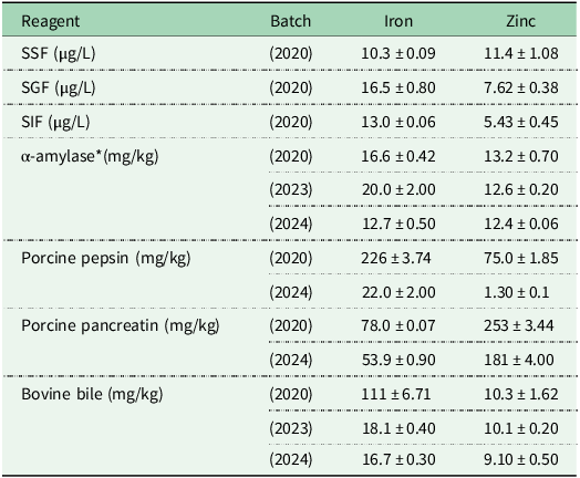
We assessed the mineral content of simulated digestive solutions, enzymes and bile across multiple batches. Table 2 shows Fe and Zn concentrations for α-amylase, porcine pepsin, porcine pancreatin and bovine bile, revealing marked batch-to-batch variability. The highest Fe and Zn concentration were observed in pepsin, pancreatin and bile. Pepsin is typically available in pure, high-activity forms, allowing its use in small quantities. In contrast, pancreatin and bile are less refined and require higher concentrations to meet INFOGEST-specified activity levels. Consequently, the elevated enzyme dosages recommended by INFOGEST amplify the contribution of reagent-derived minerals, which becomes especially problematic when analysing foods with low intrinsic Fe and Zn levels, such as cereals and legumes.
Mineral concentrations of digestive enzymes and bile recommended in the INFOGEST method

Table 2. Long description
A table comparing mineral content of digestive solutions, enzymes, and bile across different batches. The table has 10 rows and 4 columns. The columns are labeled Reagent, Batch, Iron, and Zinc. The row labels include different reagents and batches. Row 1: Reagent, SSF, Batch, 2020, Iron, 10.3 ± 0.09, Zinc, 11.4 ± 1.08. Row 2: Reagent, SGF, Batch, 2020, Iron, 16.5 ± 0.80, Zinc, 7.62 ± 0.38. Row 3: Reagent, SIF, Batch, 2020, Iron, 13.0 ± 0.06, Zinc, 5.43 ± 0.45. Row 4: Reagent, α-amylase, Batch, 2020, Iron, 16.6 ± 0.42, Zinc, 13.2 ± 0.70. Row 5: Reagent, α-amylase, Batch, 2023, Iron, 20.0 ± 2.00, Zinc, 12.6 ± 0.20. Row 6: Reagent, α-amylase, Batch, 2024, Iron, 12.7 ± 0.50, Zinc, 12.4 ± 0.06. Row 7: Reagent, Porcine pepsin, Batch, 2020, Iron, 226 ± 3.74, Zinc, 75.0 ± 1.85. Row 8: Reagent, Porcine pepsin, Batch, 2024, Iron, 22.0 ± 2.00, Zinc, 1.30 ± 0.1. Row 9: Reagent, Porcine pancreatin, Batch, 2020, Iron, 78.0 ± 0.07, Zinc, 253 ± 3.44. Row 10: Reagent, Porcine pancreatin, Batch, 2024, Iron, 53.9 ± 0.90, Zinc, 181 ± 4.00. Row 11: Reagent, Bovine bile, Batch, 2020, Iron, 111 ± 6.71, Zinc, 10.3 ± 1.62. Row 12: Reagent, Bovine bile, Batch, 2023, Iron, 18.1 ± 0.40, Zinc, 10.1 ± 0.20. Row 13: Reagent, Bovine bile, Batch, 2024, Iron, 16.7 ± 0.30, Zinc, 9.10 ± 0.50.
* α-amylase is derived from Bacillus sp.
SSF = Simulated Salivary Fluid, SGF = Simulated Gastric Fluid, SIF = Simulated Intestinal Fluid.
Data is based on Muleya et al. (Reference Muleya, Young and Bailey32) for 2020 batches and Fioroto et al. (Reference Fioroto, Muleya and Wilson42) for subsequent batches.
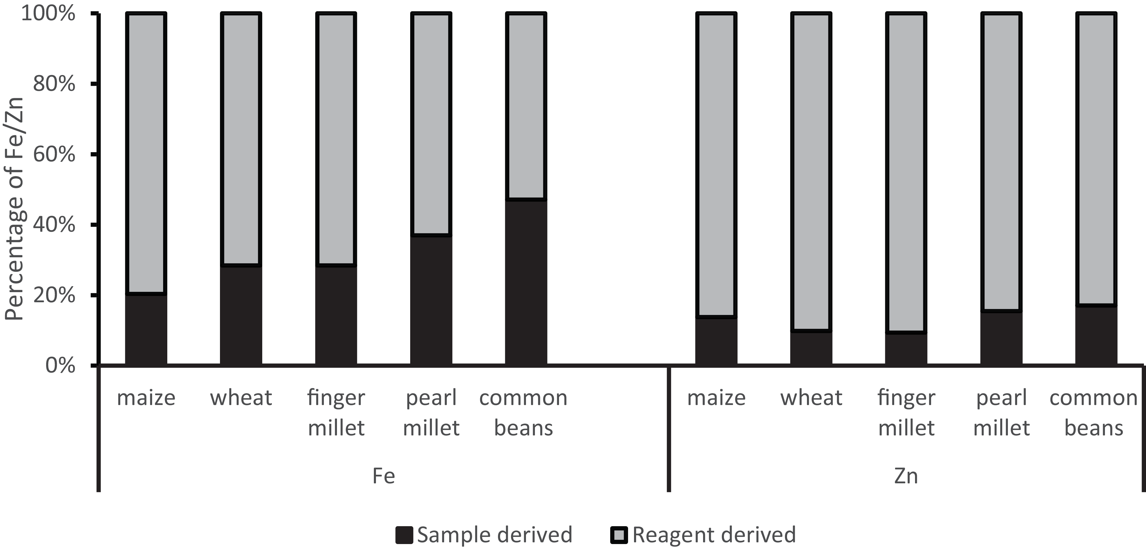
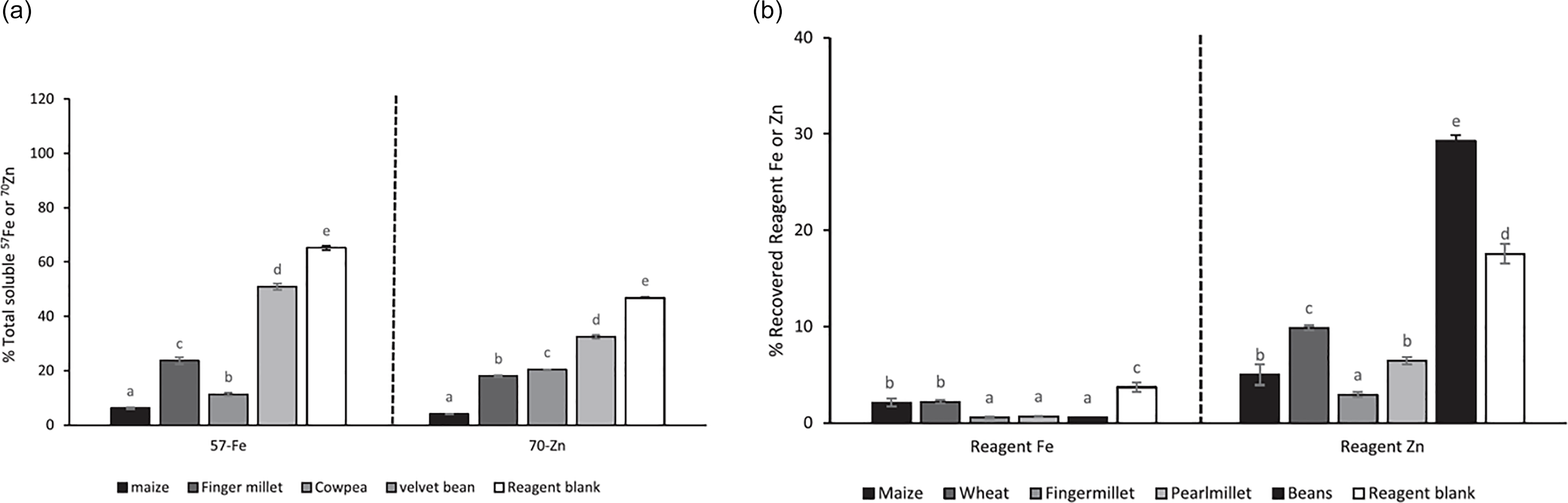
To understand the relevance of these background concentrations, we compared the expected Fe and Zn contributions from simulated digestive reagents to typical endogenous levels in cereal and legume matrices under standard INFOGEST conditions (Figure 2). In all cases, reagent-derived Fe and Zn exceeded the sample mineral content, presenting major analytical challenges. These minerals are not inert, they contribute to the total soluble mineral pool and may alter the ligand environment, influencing how minerals partition into absorbable or non-absorbable forms. To explore this further, we spiked stable isotopes of 57Fe and 70Zn in digests of maize, finger millet, cowpea and velvet bean, alongside a reagent blank (enzymes only without food). We expected 100% recovery of the spike in the blank. However, we recovered ∼60% of Fe and <50% of Zn (Figure 3(a)), indicating that even in the absence of food, the digestive reagents modified mineral solubility. Recovery in the presence of food matrices varied further, demonstrating how each matrix interacted uniquely with the applied stable isotope; likewise, reagent Fe and Zn will be expected to behave in a similar way with the spiked isotopes (Figure 3(b)).
Distribution of Fe and Zn in digests containing typical cereals and legumes under INFOGEST in vitro digestion conditions.
Data is derived from Muleya et al. (Reference Muleya, Young and Bailey32).

Recovery of 57Fe and 70Zn (a) and of reagent Fe and Zn (b) from cereal and legume digests.
Data is derived from Muleya et al. (Reference Muleya, Young and Bailey32).

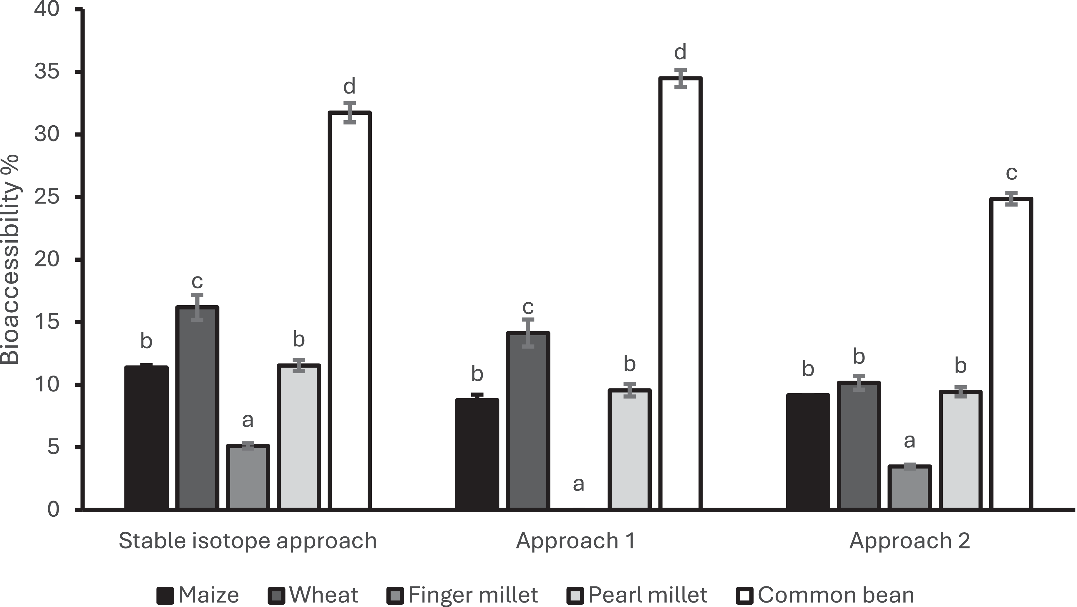
To correct for this analytical interference, we propose reducing the enzyme concentrations as well as applying stable isotopes as reagent mineral tracers. Simulated digestive fluids across all digestion phases are pre-equilibrated with 57Fe, 70Zn or 43Ca prior to digestion(Reference Muleya, Bailey and Bailey4,Reference Muleya, Young and Bailey32) . The recovery of these isotopes in the bioaccessible fractions post-digestion provides a robust means of estimating and correcting for reagent-derived mineral contributions. Figure 3(b) presents recovery data for reagent Fe and Zn with notable variability observed across different cereal and legume matrices. This further highlights the role of matrix-specific interactions. Once corrected, bioaccessibility values calculated using stable isotope tracers differ significantly from those obtained via conventional blanket blank corrections (Figure 4).
Zinc bioaccessibility based on three ways of calculation: stable isotope approach, approach 1 and 2.
Data is derived from Muleya et al. (Reference Muleya, Young and Bailey32). The stable isotope approach involves applying a specific blank correction based on each matrix interaction with reagent Zn, approach 2 is based on a blanket blank correction on the bioaccessible fraction, Approach 3 does not utilise a blank correction, calculating bioaccessibility based on total recovered Zn from bioaccessible and non bioaccessible fractions.

These findings underscore the importance of accounting for enzyme-derived minerals in INFOGEST-based protocols, particularly when analysing foods with intrinsically low mineral concentrations and/or high levels of mineral chelating compounds. Without appropriate correction, background minerals from reagents can significantly distort bioaccessibility estimates. A combined strategy, reducing enzyme and bile concentrations where analytically feasible, and applying stable isotopes as tracers to monitor and correct for reagent-derived contributions, provides a more accurate and physiologically relevant approach for assessing Fe, Zn and Ca bioaccessibility.
Iron and zinc binding under low and high pancreatin/bile conditions
In the previous section, we demonstrated that applying stable isotopes under reduced pancreatin and bile concentrations improved the accuracy of mineral bioaccessibility measurements. These reduced conditions were necessary because standard INFOGEST enzyme concentrations introduce background levels of Fe and Zn that exceed those found in many test foods. However, a key limitation of this approach is the uncertainty surrounding how reduced enzymatic activity may impact macronutrient hydrolysis and subsequent mineral release and solubility. As highlighted previously, macronutrient hydrolysis plays a role in releasing associated bound minerals and in matrix loosening.
To test this, foods with differing macronutrient compositions; starch-rich, protein-rich and lipid-rich matrices were digested under low (optimised stable isotope approach described in previous section) and high pancreatin/bile conditions (standard INFOGEST including stable isotopes)(Reference Fioroto, Muleya and Wilson42). Indeed, under the standard INFOGEST conditions, macronutrient hydrolysis was consistently increased cross all food matrices(Reference Fioroto, Muleya and Wilson42). However, Fe and Zn solubility was not increased consistently across matrices under low and high pancreatin/bile conditions. In fact, in some foods, solubility was decreased or did not change under the standard INFOGEST conditions. It should be noted however, that lower solubility does not necessarily mean the minerals were not released from the food matrix but based on the balance of ligand interactions in the system, released minerals could have been bound into insoluble complexes leading to lower solubility under the standard INFOGEST conditions. The balance of ligand interactions in the in vitro digestion system could be influenced by 1. breakdown products from digestion 2. background minerals and their associated ligands which will be higher under the standard INFOGEST with higher concentrations of pancreatin/bile. This therefore means that the combined influence of enzymatic action and background minerals makes it difficult to isolate the effects of macronutrient digestion alone on mineral solubility.
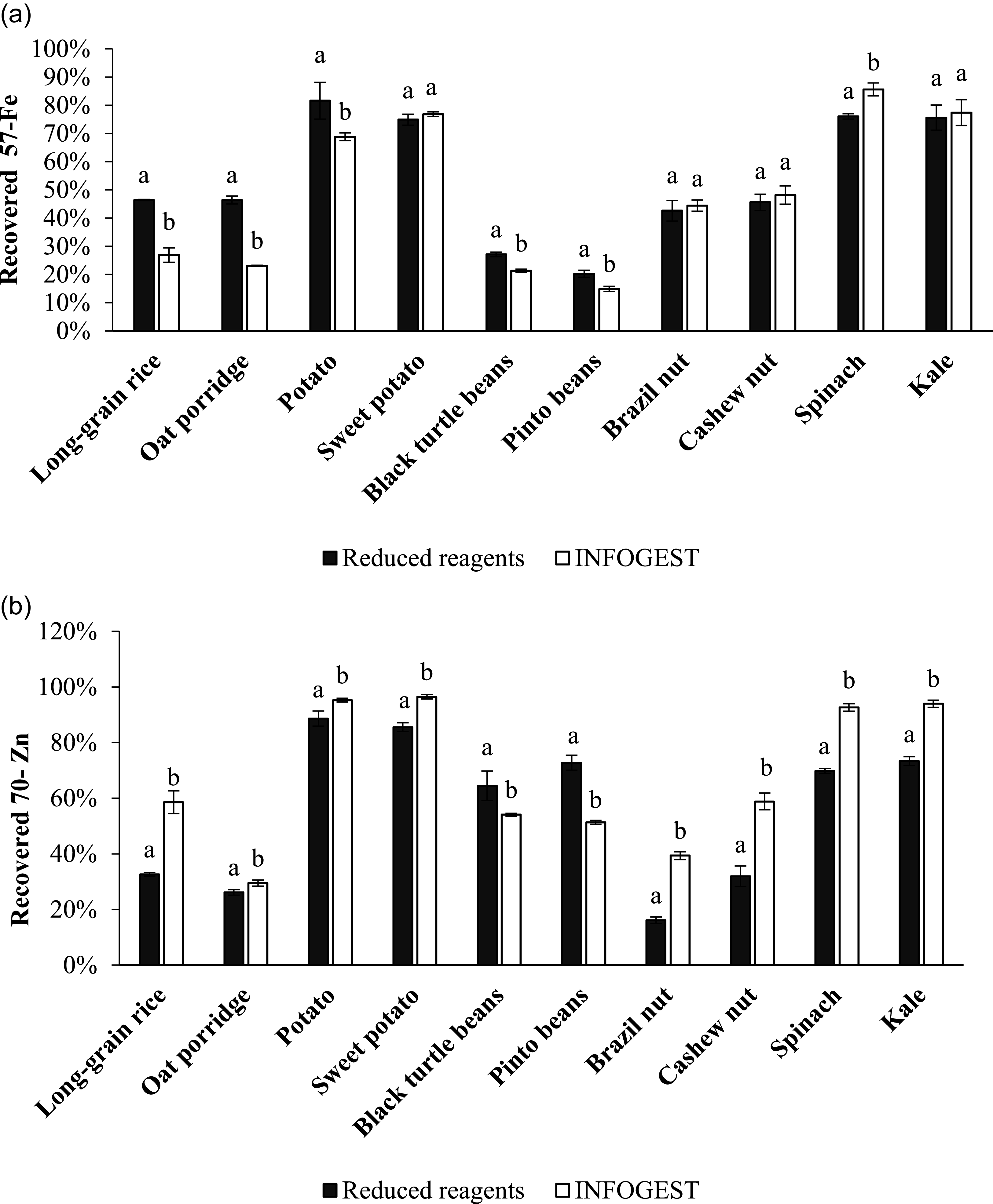
To prove that the ligand environment changes under the two conditions of pancreatin/bile across the different food matrices, we can trace the recovery of the applied stable isotopes of 57Fe and 70Zn. The applied isotopes are already soluble and do not need to be released from a food matrix, such that the direction of their recovery demonstrates the change in the ligand environment, that is, whether it becomes inhibitory, promoting or unchanged under higher pancreatin/bile conditions. Recovery of Fe and Zn varied by matrix and pancreatin/bile condition(Reference Fioroto, Muleya and Wilson42) (Figure 5). For Fe, no significant change in recovery was observed in sweet potato, Brazil nut, cashew nut or kale. However, under standard INFOGEST conditions (high enzyme), Fe recovery decreased in long grain rice, oat porridge, potato and black turtle beans, while it increased in spinach. Zn recovery showed a similar trend. Recovery increased under high enzyme conditions for long grain rice, oat porridge, potato, sweet potato, Brazil nut, cashew nut, spinach and kale but decreased for black turtle beans and pinto beans. This wide disparity in Fe and Zn recovery were despite macronutrient digestion, that is, starch and protein being significantly increased under the standard INFOGEST conditions(Reference Fioroto, Muleya and Wilson42).
Recovered 57Fe (a) and 70Zn (b) in different types of food matrices after in vitro digestion under low and high pancreatin/bile conditions.
Data is based on Fioroto et al. (Reference Fioroto, Muleya and Wilson42), supplementary pages.

These findings suggest that while macronutrient digestion may support mineral release, the elevated concentrations of reagent-derived minerals under high enzyme conditions along with breakdown products from digestion alter the ligand environment in unpredictable ways. Notably, the direction of stable isotope recovery comparing the reduced pancreatin/bile and standard INFOGEST conditions was not consistent across food matrices. This matrix-dependent variability underscores the challenge of comparing mineral bioaccessibility across digestion methods utilising differing enzyme concentrations and highlights the risk of confounding due to background mineral contamination and variation in break down products produced. Overall, the data reinforce the need to standardise both enzymatic conditions and background correction methods in in vitro mineral bioaccessibility studies. Variability in enzyme concentrations, reagent mineral concentrations and food matrix composition all influence the ligand environment and thus affect the solubility and bioaccessibility of Fe and Zn. Standardisation is essential to improving the reliability and comparability of findings in this field.
Comparing different separation techniques used in the determination of calcium bioaccessibility
A critical step in assessing mineral bioaccessibility is separating the absorbable fraction from non-absorbable components after digestion. While the INFOGEST protocol recommends a molecular weight cut-off (MWCO) of 3–10 kDa, it does not provide a clear physiological rationale for this range. In contrast, recent work on the determination of in vitro amino acid digestibility has established a 1 kDa MWCO cut-off as more representative of peptide molecules capable of diffusing through the mucus layer and reaching the brush border before being further hydrolysed by peptidases into free amino acids and oligopeptides(Reference Sousa, Recio and Heimo43). Moreover, 80% methanol precipitation has been proposed as a more effective technique for isolating this fraction compared to other common separation methods such as filtration(Reference Sousa, Recio and Heimo43). For minerals like Fe, Zn and Ca, a similar refinement in methodology is urgently needed. For example, two approaches are widely used: dialysis and ultrafiltration. While both aim to separate soluble, low molecular weight Ca species from the insoluble digesta fraction, they differ fundamentally in their driving forces. Dialysis is a diffusion-controlled process, in which Ca transport across the membrane is governed by the concentration gradient between the digesta and the dialysate, thereby mimicking passive diffusion processes in the intestine(Reference Sandberg30). In contrast, ultrafiltration is a pressure-driven technique, typically using centrifugation to force solvent and permeable solutes through a membrane of defined MWCO(Reference Mulder44). As a result, transport during ultrafiltration is influenced not only by pore size but also by centrifugal force.
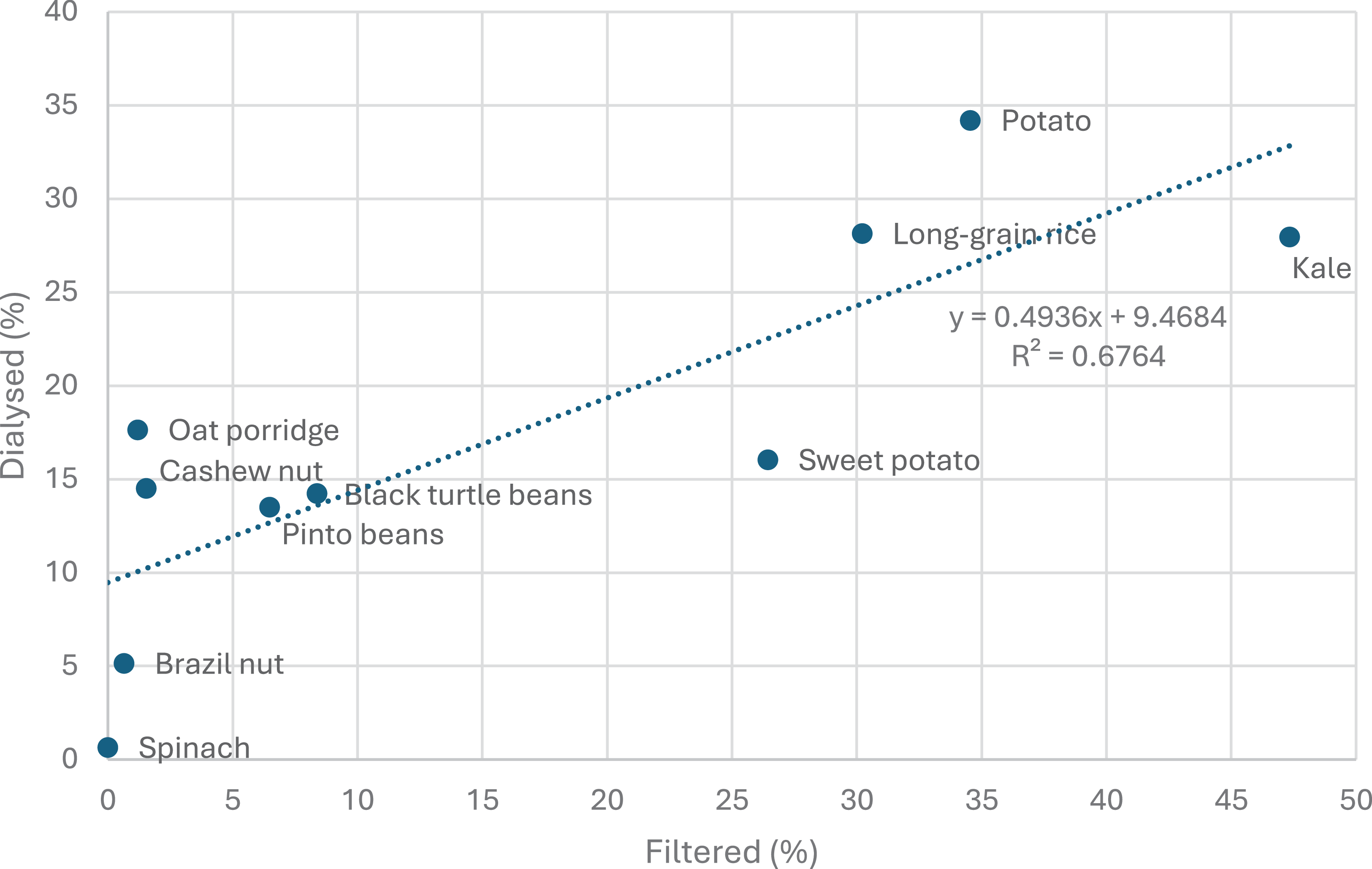
We evaluated the agreement between these two commonly used methods of separation(Reference Fioroto, Muleya and Wilson45). Filtration was carried out after digestion using 3 kDa ultracentrifugal filters(Reference Muleya, Bailey and Bailey4,Reference Lorieau, Le Roux and Gaucheron46) while dialysis (12–14 kDa MWCO) was integrated within the last phase of the gastric digestion and the entirety of the intestinal phase of digestion(Reference Muleya, Young and Bailey32). Our initial hypothesis was that dialysis would consistently yield higher Ca bioaccessibility than ultrafiltration, but differences might not be large as the lower MWCO from ultra filtration might be compensated with the use of centrifugal force. For most of the samples, Ca bioaccessibility was higher by dialysis than by ultrafiltration, as demonstrated by the positive correlation between the two methods (R 2 = 0.68) (Figure 6). However, there were notable exceptions; 1. the magnitude of differences in Ca bioaccessibility for some samples was unexpectedly large. For instance, Ca bioaccessibility for oat porridge and cashew nut respectively was just 1.2% and 1.5% by filtration, compared to 18% and 15% respectively by dialysis, an order of magnitude difference that significantly affects interpretation, despite the direction of difference aligning with expectations. 2. Kale and sweet potato showed higher Ca bioaccessibility when measured by ultrafiltration than by dialysis.
Correlation plot of Ca bioaccessibility measured by dialysis (MWCO of 12–14 kDa) vs. ultrafiltration (MWCO of 3 kDA).
Based on data from Fioroto et al. (Reference Fioroto, Muleya and Wilson45).

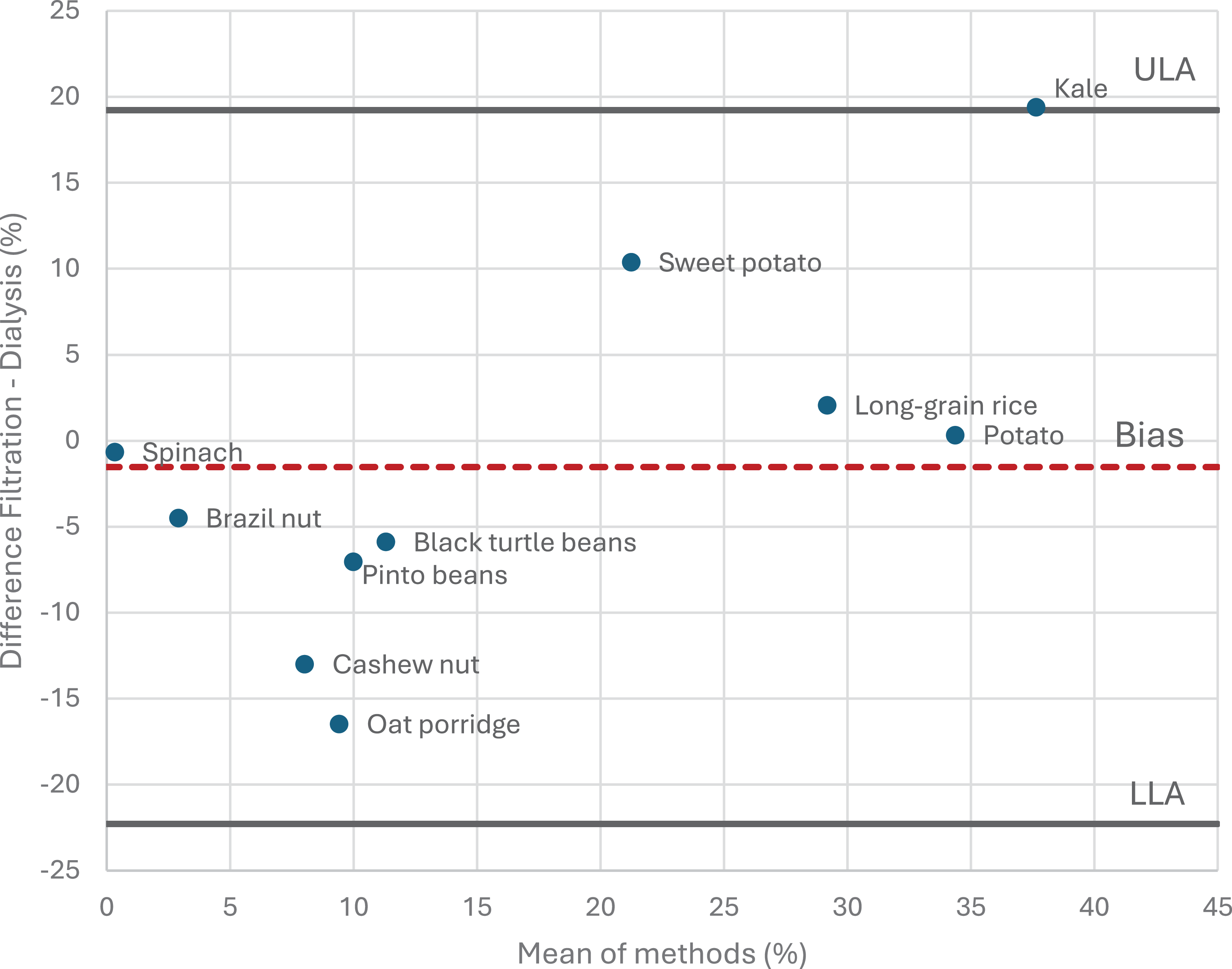
To assess whether the two methods could be used interchangeably, a Bland-Altman analysis was conducted (Figure 7). While the bias was relatively low at –1.5%, the limits of agreement ranged from –22% to +19%, which is unacceptably wide. This variability was largely driven by divergent results in oat porridge, cashew nut, kale and sweet potato. In the case of oat porridge and cashew nut, both matrices contain starch and fibre, which increases digesta viscosity and promote formation of a gel layer at the membrane surface(Reference Belfort, Davis and Zydney47,Reference Dikeman and Fahey48) . This can effectively reduce membrane permeability during pressure-driven ultrafiltration, leading to underestimation of the bioaccessible Ca fraction. In the case of the kale and sweet potato, as dialysis relies on passive diffusion towards equilibrium, Ca transport may be limited by slow diffusion kinetics or re-equilibration reactions during prolonged incubation. On the other hand, short-duration, pressure-driven ultrafiltration may be able to capture a greater proportion of immediately bioaccessible Ca in these matrices(Reference Fioroto, Muleya and Wilson45).
Bland-Altman plot indicating the agreement between in vitro Ca bioaccessibility of various food matrices derived after using filtration (MWCO of 3 kDa) or dialysis (MWCO of 12–14 kDa) as separation techniques.
ULA = Upper limit of agreement, LLA = Lower limit of agreement.
Based on data from Fioroto et al. (Reference Fioroto, Muleya and Wilson45).

These findings highlight that dialysis and ultrafiltration should not be considered interchangeable methods for assessing Ca bioaccessibility. Differences in driving force, transport kinetics and susceptibility to matrix effects can lead to substantial and food-specific discrepancies. These findings highlight the need for standardisation of separation procedures to improve comparability of Ca bioaccessibility across studies.
Future perspectives and conclusion
This review highlights that while the INFOGEST static digestion method provides a robust and harmonised framework for simulating gastrointestinal digestion, it was not originally designed to address the specific analytical and physiological challenges associated with mineral bioaccessibility. To fully realise its potential for Fe, Zn and Ca research, targeted optimisation and further standardisation are required.
An immediate priority is the systematic management of background minerals introduced through digestive reagents, particularly pancreatin and bile. Approaches such as reagent purification using Chelex-100 treatments, reduction of enzyme concentrations where analytically feasible and the use of stable isotopes as reagent tracers offer promising solutions(Reference Muleya, Young and Bailey32,Reference Fioroto, Muleya and Wilson42) . However, harmonised guidance is needed to define acceptable thresholds for reagent-derived minerals relative to food-derived minerals, ensuring values remain physiologically meaningful.
Second, attention must be given to the interplay between macronutrient digestion and mineral release. As demonstrated in this review, enhanced macronutrient hydrolysis does not necessarily translate into increased mineral bioaccessibility, due to matrix-dependent ligand interactions and background mineral effects. Optimising digestion conditions to balance physiological relevance remains a critical challenge.
Third, there is an urgent need to define and standardise the bioaccessible mineral fraction. Current membrane-based separation techniques are highly sensitive to food matrix effects, and MWCO alone is insufficient to ensure comparability across matrices. Alternative separation strategies, including chemical-based approaches successfully applied in protein digestibility research(Reference Sousa, Recio and Heimo43), warrant investigation for minerals.
Finally, progress in this field depends on direct validation against in vivo data and renewed inter-laboratory trials. While limited comparisons exist for Fe(Reference Aragón, Ortiz and Pachón49,Reference Luten, Crews and Flynn50) , robust, coordinated efforts are required across minerals to establish confidence in in vitro data. Such initiatives will be essential for translating bioaccessibility data into food composition databases, dietary modelling and product development. In conclusion, optimised and standardised in vitro methods offer a powerful, ethical and scalable means to integrate bioaccessibility into assessments of mineral nutritional value. Advancing this agenda will be critical as food systems shift towards more plant-based and novel ingredients, where mineral bioavailability is highly variable and often poorly characterised.
Acknowledgements
M.M is grateful to the Organizing Committee for the invitation to speak at the Nutrition Society Summer Conference 2025.
Author contributions
M.M, A.M.F and E.H.B were all responsible for conceptualisation, writing and editing of the manuscript.
Financial support
The authors are grateful to FAPESP (The São Paulo Research Foundation) for supporting A.M.F to conduct a one year research visit at the University of Nottingham (grant number 2023/09766-8 and 2013/07914-8). FAPESP had no role in the design, analysis or writing of this article.
Competing interests
The authors have no conflicts of interests to declare.
Declaration of generative AI use
The authors used generative artificial intelligence (ChatGPT) to assist with grammar correction and improve readability of the text.