Introduction
It is well known that controlling the shape of gold (Au) nanostructures is an efficient strategy to attribute their physical and chemical properties for promising applications in fields such as catalysis, sensing and imaging, and surface-enhanced Raman scattering (SERS) [Reference Sau and Rogach1, Reference Sau, Rogach, Jäckel, Klar and Feldmann2, Reference Daniel and Astruc3, Reference Eustis and El-Sayed4, Reference Sepulveda, Angelome, Lechuga and Liz-Marzan5]. For instance, the localized surface plasmon resonances (SPR) of Au nanoparticles exhibited a much more sensitive shape-dependent response compared with size-dependent response between the visible and near-infrared regions [Reference Nehl and Hafner6]. Furthermore, the remarkably large local electromagnetic field enhancements were induced by these resonances at the nanoparticle surfaces, especially around the sharp tips and edges of the nanostructures, which made the dominant contribution to the SERS enhancement [Reference Mayer and Hafner7, Reference Li, Kang, Dasari and Barman8, Reference Hao, Nehl, Hafner and Nordlander9]. The experiments and the simulations revealed that Au nanostars showed significantly higher SERS enhancement activity than that of Au nanorods and nanospheres [Reference Li, Cushing, Zhang, Lankford, Aguilar, Ma and Wu10]. For this reason, synthesis of Au nanoparticles with morphological features, such as sharp tips and edges, has been significantly developed recently [Reference Jana and Pal11, Reference Grzelczak, Perez-Juste, Mulvaney and Liz-Marzan12, Reference Guerrero-Martinez, Barbosa, Pastoriza-Santos and Liz-Marzan13]. Au nanoparticles with geometrical features, such as nanostars, dendritic, flowerlike, and multibranched nanostructures, have been investigated as one of the most attractive anisotropic nanomaterials [Reference Senthil, Pastoriza-Santos, Rodriguez-González, Javier and Liz-Marzan14, Reference Xia, Luo, Wang, Liu, Yu and Wang15, Reference Jena and Raj16, Reference Wang, Imura and Yamauchi17].
As a mild reducing agent with green and environmentally friendly advantages, hydrogen peroxide (H2O2) is used to synthesize anisotropic Au nanoparticles in the presence of surfactants [Reference Milligan and Morriss18, Reference Paclawski and Fitzner19, Reference Panda and Chattopadhyay20]. For example, with H2O2 used as reducing agent, branched Au nanoparticles are prepared in the presence of sodium citrate using bis(p-sulfonatophenyl) phenylphosphine dihydrate dipotassium (BSPP) or thiol-terminated molecules as ligand molecules [Reference Hao, Bailey, Schatz, Hupp and Li21, Reference Bakr, Wunsch and Stellacci22]. However, the result indicates that the role of BSPP is not essential for formation of branched structures [Reference Hao, Bailey, Schatz, Hupp and Li21]. It means that sodium citrate plays a key role in the formation of branched Au nanoparticles. Sodium citrate is widely employed as a capping agent and a reducing agent to synthesize Au nanoparticles [Reference Turkevich, Stevenson and Hillier23, Reference Frens24]. It could provide enough protection for the stabilization of nanoparticles in the solution and also affect the morphology of nanoparticles [Reference Pei, Mori and Adachi25]. Herein, we report a facile one-pot and environmentally friendly route to synthesize multibranched Au nanostructure by reduction of chlorate gold with hydrogen peroxide in the presence of sodium citrate. The effects of the molar ratio of sodium citrate to HAuCl4 and the concentration of reactants on the morphology of Au nanoparticles were investigated. We also investigated SERS properties of 4-aminothiophenol (PATP) on the multibranched Au nanostructures. The experimental and simulated results both clearly show that the photoinduced catalytic coupling reaction of PATP transformation to 4,4′-dimercaptoazobenzene (DMAB) occurs on the surface of multibranched Au nanostructures at a high power during the SERS measurement.
Results and discussion
Characterization of multibranched flowerlike Au nanostructures
Hydrogen dioxide (H2O2) can be used as an oxidizing agent and also as a reducing agent in a general acknowledgment. Taking into account its reducing ability, it has been used to synthesize Au nanoparticles. The proposed synthesis method is the reduction of the precursor HAuCl4 by H2O2 through the following reaction [Reference Panda and Chattopadhyay20]:
 $$2{\rm{AuCl}}_4^ - + 3{{\rm{H}}_{\rm{2}}}{{\rm{O}}_2} \to 2{\rm{Au}} + 8{\rm{C}}{{\rm{l}}^ - } + 3{{\rm{O}}_2} + 6{{\rm{H}}^ + }\quad .$$
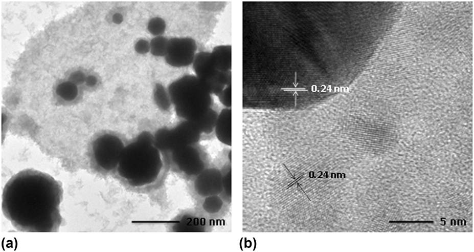
Figure 1 shows the transmission electron microscopy (TEM) and high-resolution transmission electron microscopy (HRTEM) images of the product obtained by reducing HAuCl4 using H2O2 in the absence of sodium citrate. It clearly shows that the product consisted of irregular nanoparticles surrounded with small clusters. The observed fringes with a spacing of 0.24 nm in the nanoparticle and the cluster were in agreement with a (111) plane spacing of Au face-centered cubic (fcc) crystal.
(a) TEM and (b) HRTEM images of the product obtained by reducing HAuCl4 using H2O2 in the absence of sodium citrate at room temperature.

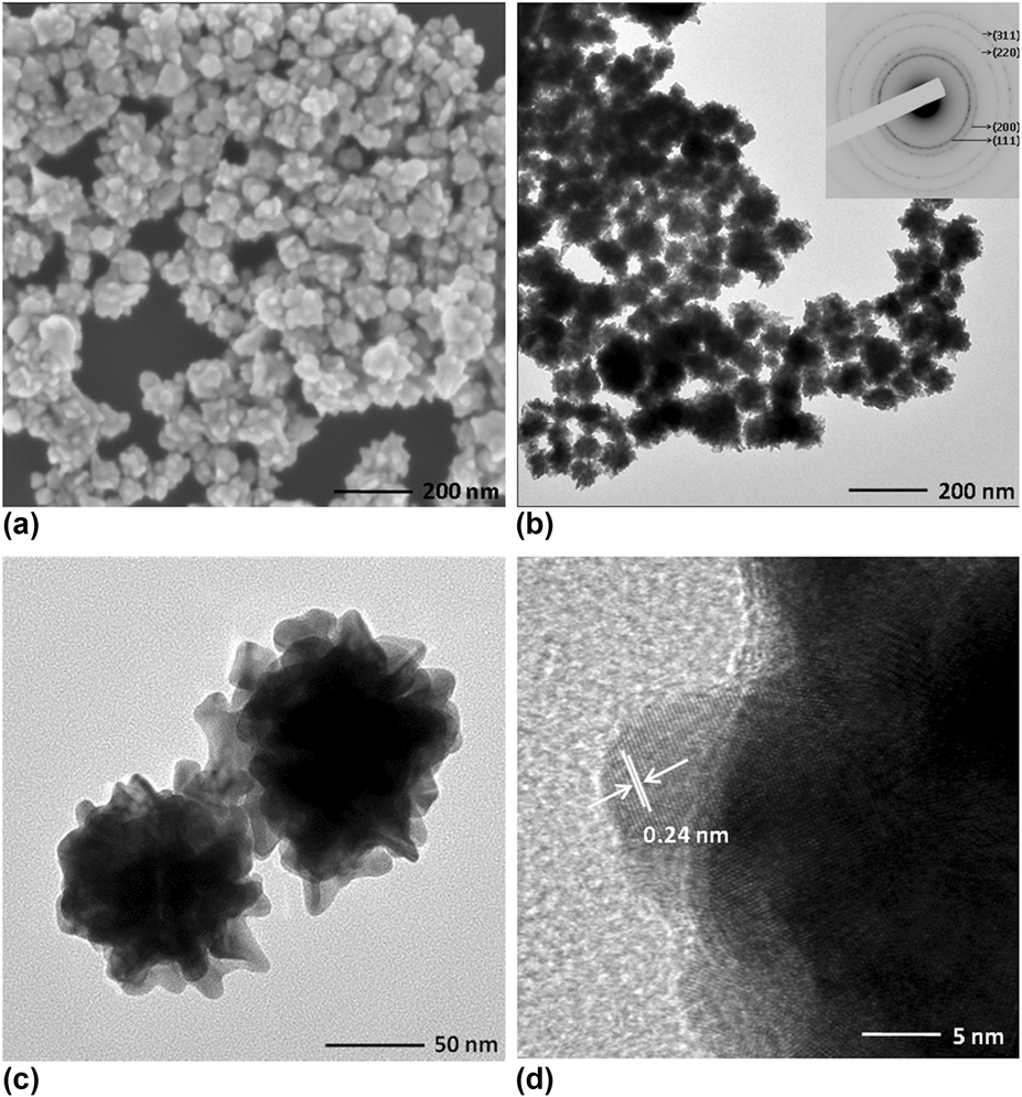
It had been reported that sodium citrate could promote the growth of gold particles at elevated temperature. Sodium citrate could also play a role as a capping agent for the stabilization of the nanoparticles. It acts only as a capping agent in the formation of Au nanoparticles due to its weak reduction capacity at room temperature [Reference Turkevich, Stevenson and Hillier23, Reference Frens24]. Multibranched gold nanostructures could be obtained by reduction of HAuCl4 by H2O2 in the presence of sodium citrate. Figures 2(a)–2(c) show SEM image and TEM images of the sample synthesized by the typical synthesis in the Experimental section, indicating that the product possesses a multibranched shape around hundred nanometers. The chemical composition of the product was determined by energy-dispersive X-ray spectroscopy (EDS). A typical EDS pattern (Fig. S1) recorded from the sample revealed that the multibranched structure is composed of metallic Au. Selected area electron diffraction (SAED) pattern [inset in Fig. 2(b)] of the product shows that all reflections can be indexed as a pure fcc phase of Au with space group Fm3m (JCPDS card No. 04-0784). HRTEM image [Fig. 2(d)] elucidates the single crystalline structure of the individual tip with (111) plane.
(a) SEM image and (b and c) TEM images with different magnifications. (d) HRTEM image of the sample obtained by the typical synthesis. The inset in (b) is the corresponding SAED pattern.

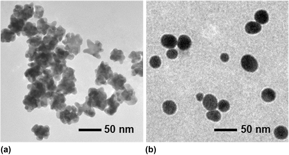
The sodium citrate plays a critical role in the formation of multibranched Au nanostructures. If there is no capping agent, irregular nanoparticles surrounded with small clusters are formed (Fig. 1). When a small quantity of sodium citrate is introduced to the reaction system of HAuCl4 and H2O2, some multibranched Au nanostructures are present with irregular Au nanoparticles (Fig. S2). When the molar ratio of sodium citrate to HAuCl4 is increased to 10:1, multibranched Au nanostructures with diameters of hundred nanometers are obtained (Fig. 2). Due to the weak bonding ability and the repulsion force of the negative charged citrate ions, the citrate ions preferentially adsorb and loosely distributed on the surfaces of Au particles. It has been well demonstrated that the adsorption of chemical species on the surface has a dramatic effect on the surface energies associated with the crystal planes of Au particles [Reference Zou, Ying and Dong26]. The citrate ions on the sites of Au surfaces will decrease the growth rates compared with that of unbounded sites with citrate ions. As a result, the different growth rates lead to the anisotropic growth and the formation of multibranched Au nanostructures [Reference Park and Shumaker-Parry27, Reference Zou, Ren, Shang and Wang28]. When the molar ratio of sodium citrate to HAuCl4 is further increased to 20:1, the size of the multibranched Au nanostructures decreased to several tens of nanometers [Fig. 3(a)]. The growth of multibranched Au nanostructures is hindered by the overabundant presence of sodium citrate. Besides, new nuclei are supposed to form for further growth to produce smaller size multibranched Au nanostructures [Reference Zou, Ren, Shang and Wang28]. The concentration of the reaction reagents (sodium citrate, HAuCl4, and H2O2) has also an important role in the formation of multibranched Au nanostructures. The quasi-spherical Au nanoparticles could be produced at a high concentration of reagents [Fig. 3(b)], probably because the high concentration of reagents leads to the increase in the number of nucleation sites in the reaction solution. Each Au nucleus is completely bound by citrate ions, which results in the isotropic growth and leads to the formation of quasi-spherical Au nanoparticles.
TEM images of Au nanoparticles synthesized at different conditions: (a) the molar ratio of sodium citrate to HAuCl4 is 20:1 and (b) the concentration of the reacted reagents is increased 4 times. The other parameters are same with the typical synthesis.

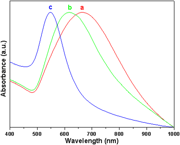
It is well known that Au nanoparticles exhibit strong SPR absorption [Reference Link and El-Sayed29]. There is wide tunable capability of SPR from the Ultraviolet-visible (UV-vis) to near-infrared regions through reliably controlling the size and geometry of nanoparticles [Reference Jain, Lee, El-Sayed and El-Sayed30]. Figure 4 shows UV-vis absorption spectra taken from the aqueous solution of Au nanoparticles synthesized at different experimental conditions. The SPR of multibranched Au nanostructure (as shown in Fig. 2) was located at 665 nm [Fig. 4(a)]. For smaller multibranched Au nanostructure, as shown in Fig. 3(a), the SPR was blue-shifted to 615 nm [Fig. 4(b)]. The peak widths at half maximum of multibranched Au nanostructures are 230 nm [Fig. 4(a)] and 180 nm [Fig. 4(b)]. As observed in Figs. 2 and 3(a), the sizes of multibranched Au nanostructure decreased and the branches also shorten, which result in the blue-shift and narrow peak of SPR [Reference Zou, Ying and Dong26]. The SPR of quasi-spherical Au nanoparticles, as shown in Fig. 3(b), exhibits typical absorption of Au colloid, which is located at 548 nm [Fig. 4(c)], with narrower peak width at half maximum (75 nm).
(a–c) UV-vis absorption spectra of Au nanoparticle solutions for the samples as shown in Fig. 2, Figs. 3(a) and 3(b), respectively.

SERS of PATP on the multibranched Au nanostructures
The SPR induces strong spatially confined electromagnetic field surrounding the sharp tips and edges of the nanoparticles, which could serve as nanoantennas to enhance Raman scattering of the molecules in the region. The experiments and theoretical simulations have shown that branched nanostructures with sharp tips as “hot spots” can provide significantly large near-field enhancements and are thus excellent candidates for SERS substrates. We evaluated the performance of the multibranched Au nanostructures for SERS application using PATP as a target molecule with a 785 nm excitation.
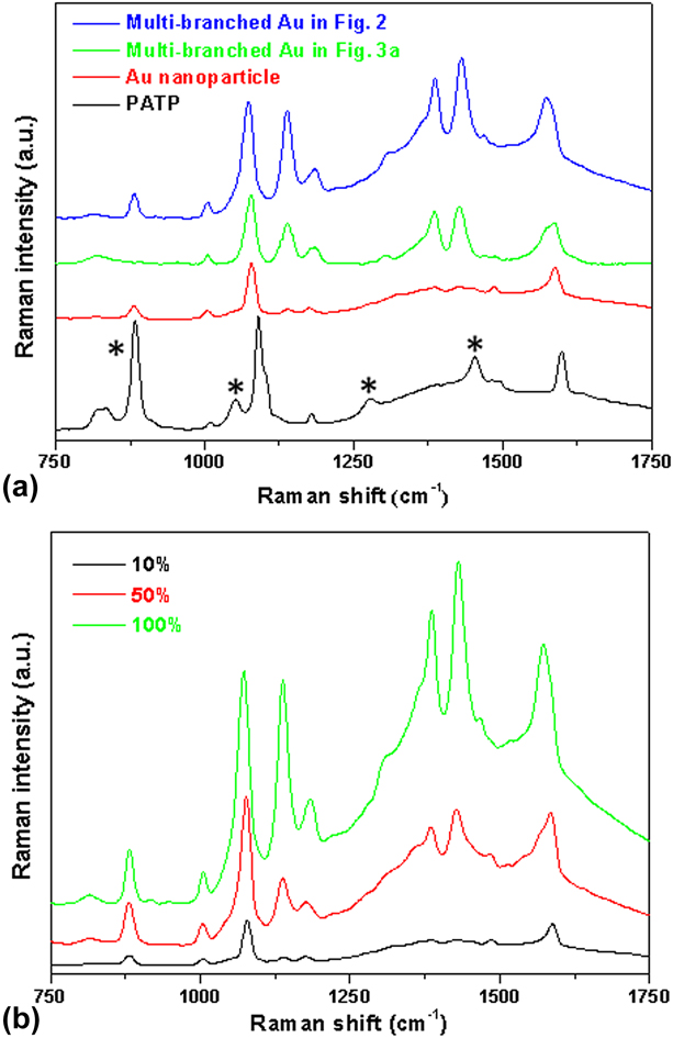
Figure 5(a) shows the normal Raman spectrum of 0.1 M PATP in ethanol and SERS spectra of 5 µM PATP in Au nanoparticle and multibranched Au nanostructure solutions. The peaks marked by asterisks are assigned to the signals of ethanol. The Raman peaks in SERS spectrum of PATP on Au nanoparticles shift to lower wavenumbers compared with that in the normal Raman spectrum of PATP, which maybe due to the strong interaction introduced charge transfer between PATP and Au nanoparticles [Reference Sun, Hu, Zhan, Yan, Liu, Teguh, Yeow, Lee and Shen31, Reference Yan, Qiu, Chen, Liu, Lai, Meng, Song, Liu, Liu and Zhan32]. Multibranched Au nanostructures exhibit the stronger enhancement efficiency than that from spherical Au nanoparticles. Compared with SERS effect of two kinds of multibranched Au nanostructures, the sample of multibranched Au nanostructure in Fig. 2 shows a stronger SERS ability than that of the sample in Fig. 3(a). The stronger enhancement effect is probably because of the SPR of multibranched Au nanostructure (665 nm versus 615 nm) nearer to the excitation wavelength (785 nm) [Reference Li, Kang, Dasari and Barman8]. It is worth noting that the new Raman signals are observed at 1138, 1386, and 1431 cm−1 in the SERS spectrum of multibranched Au nanostructures. There are two explanations about the contribution of these three Raman bonds: (i) ascribe to the ‘‘b2 modes’’ of PATP from the electronic resonance charge transfer between probe molecules and substrates and (ii) originate from the DMAB formed through the catalytic coupling reaction of PATP [Reference Huang, Wu, Zhu, Zhao, Liu, Ren and Tian33]. In order to figure out the reason for the selective Raman enhancement, we performed SERS investigation of PATP absorbed onto multibranched Au nanostructures with different laser power. As shown in Fig. 5(b), the Raman peaks at 1138, 1386, and 1431 cm−1 increased gradually with the increase in the laser power. Our previous study showed that the photoinduced surface catalytic coupling reaction of PATP transformation to cis- and trans-DMAB occurs on the surface of Au nanoparticles at a high power [Reference Cao, Liu and Yang34]. The “hot” electrons generated from surface plasmon have been regarded as the source that induces the surface catalytic coupling reaction. We think it can be rationalized that the conversion of PATP to DMAB was happened under exposure to a higher power of laser during the SERS measurement of PATP in multibranched Au nanostructure solution.
(a) The normal Raman spectrum of 0.1 M PATP in ethanol and SERS spectra of 5 μM PATP in Au nanoparticle and multibranched Au nanostructure solutions. (b) SERS spectra of 5 μM PATP in multibranched Au nanostructure solution acquired at 10%, 50%, and 100% of the laser power.
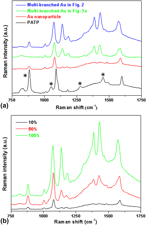
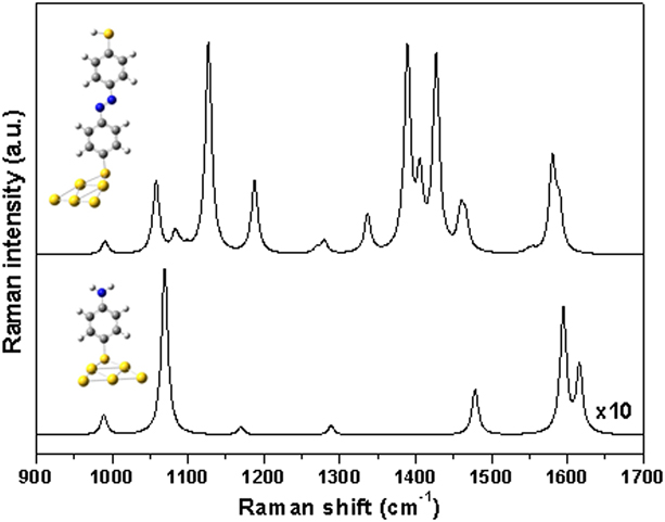
Figure 6 shows the simulated Raman spectra of PATP and DMAB adsorbed on Au5 metal cluster through sulfur atoms. The theoretical Raman spectrum of PATP is almost coincident with the normal Raman and SERS spectra of PATP on Au nanoparticles. The calculated Raman spectrum of DMAB is in good agreement with the SERS spectral feature of PATP in multibranched Au nanostructure solution. From the simulation, it is also clear that the plasmon-driven conversion of PATP to DMAB occurred in multibranched Au nanostructure solution during the SERS measurement.
Calculated spectra and optimized geometries of PATP and DMAB adsorbed on Au5 metal cluster through their S atoms.

Conclusions
In summary, we have presented a facile one-pot and environmentally friendly method for the synthesis of multibranched Au nanostructures. The molar ratio of sodium citrate to HAuCl4 and the concentrations of the reagents play important roles in the formation of multibranched Au nanostructures. Due to the presence of significantly strong electromagnetic field intensity around the sharp tips of the multibranched Au nanostructures, a photoinduced surface catalytic coupling reaction of PATP transformation to DMAB occurs on the surface of multibranched Au nanostructures at a high power during the SERS measurement, which is confirmed by the experimental and simulated results. These multibranched Au nanostructures are expected to find applications in SERS, biosensors, and the photoinduced surface catalytic application fields [Reference Xu, Kang, Mack, Schanze, Han and Wang35, Reference Kim, Kosuda, Van Duyne and Stair36].
Experimental
Materials
Chloroauric acid (HAuCl4·4H2O), sodium citrate (C7H5Na3O7·2H2O), hydrogen peroxide (H2O2, 30%), 4-aminothiophenol (PATP, 97%), and ethanol were commercially purchased from Xi'an Chemical Regent Factory (Xi'an, China). All chemicals were used as received without further purification. Throughout the experimental process, deionized water was used.
Synthesis of multibranched Au nanostructures
All the flasks and stir bars were cleaned with aqua regia, thoroughly rinsed with deionized water, and dried before use. In a typical synthesis, 40 µL of 50 mM chloroauric acid, 0.5 mL of sodium citrate (1 wt%), and 20 mL of DI water were added to a 100 mL flask at room temperature. The solution was stirred at 500 rpm, and 0.1 mL of hydrogen peroxide (30%) was added into the above solution. Within the several minutes, the color of the mixed solution changed from slightly yellow to blue. The reaction was stopped after 30 min. The precipitate was collected by centrifugation for further characterization.
Characterization
TEM, HRTEM, SAED, and EDS were performed on a Tecnai G2 F20 S-TWIN TEM (Thermo Fisher Scientific - FEI, Hillsboro, Oregon) equipped with a field emission gun operated at 200 kV. UV-vis absorption spectra were taken using a UV-1800 UV-Vis spectrometer (Shimadzu Ltd., Shanghai, China). The equal volumes of 10−5 M of PATP solution and Au nanostructure solution were mixed and sealed in a thin glass tube (inner diameter, 0.5 mm) for SERS measurement. The resulting concentration of PATP molecules in SERS examination is 5 μM. In a blank experiment, 5 μM of PATP ethanol solution in a thin glass tube was prepared for the Raman measurement. Raman spectra were collected with a 785 nm excitation line and integration time of 10 s using a Renishaw inVia confocal Raman microscope (Renishaw, Gloucestershire, The United Kingdom).
Model and computational details
The simulated Raman spectra of PATP and DMAB interacting with Au5 clusters through sulfur atoms were performed using a methodology similar to that described in a previous report [Reference Wu, Zhao, Liu, Huang, Huang, Ren and Tian37]. Density functional theory calculations were carried out with the hybrid exchange–correlation functional of B3LYP. The Au atoms were described using the basis set of LANL2DZ. The basis sets of 6-311+G** were used for C, N, S, and H atoms.
Acknowledgments
This work was supported by the National Natural Science Foundation of China (Nos. 21501141 and 21673172) and the Education Commission of Shaanxi Province (Nos. 2015JM2054 and 14JS092). J. Yang thanks Cyrus Tang Foundation for Tang Scholar.