Introduction
Obsessive–compulsive disorder (OCD) has been known by various names since the Medieval period and the suspected cerebral involvement in this disease can be traced back to Magnan's suggestion of obsessions being the psychological stigmata of a degeneration psychosis (Berrios, Reference Berrios1989). Recent meta-analytical studies suggest that OCD is characterized by widespread changes in both the cerebral cortex and subcortical structures (Fontenelle et al. Reference Fontenelle, Harrison, Yücel, Pujol, Fujiwara and Pantelis2009; Radua & Mataix-Cols, Reference Radua and Mataix-Cols2009; Rotge et al. Reference Rotge, Guehl, Dilharreguy, Tignol, Bioulac, Allard, Burbaud and Aouizerate2009, Reference Rotge, Langbour, Guehl, Bioulac, Jaafari, Allard, Aouizerate and Burbaud2010). Volumetric studies that have examined preselected regions of interest (ROIs) support the hypothesis of fronto-striato-thalamo-cortical dysfunction in OCD (Saxena et al. Reference Saxena, Brody, Schwartz and Baxter1998; Huyser et al. Reference Huyser, Veltman, de Haan and Boer2009). The emergence of unbiased whole-brain studies has indicated a prominent role for several cortical regions with widespread reciprocal connectivity and multimodal integrative function, particularly the posterior parietal cortex (Menzies et al. Reference Menzies, Achard, Chamberlain, Fineberg, Chen, del Campo, Sahakian, Robbins and Bullmore2008 a; van den Heuvel et al. Reference van den Heuvel, Remijnse, Mataix-Cols, Vrenken, Groenewegen, Uylings, van Balkom and Veltman2009). Studies of functional connectivity also endorse the importance of distributed cortical networks, involving the anterior insula in particular (Huyser et al. Reference Huyser, Veltman, Wolters, de Haan and Boer2011; Cocchi et al. Reference Cocchi, Harrison, Pujol, Harding, Fornito, Pantelis and Yücel2012; Stern et al. Reference Stern, Fitzgerald, Welsh, Abelson and Taylor2012a, Reference Stern, Welsh, Gonzalez, Fitzgerald, Abelson and Taylorb), in the pathophysiology of OCD.
Numerous studies have investigated the regional grey matter changes across the whole brain using either an ROI or a voxel-based morphometric (VBM) approach. Findings with respect to cortical grey matter have been inconsistent, with both grey matter excesses and reductions reported, sometimes in the same brain regions across different samples (Radua & Mataix-Cols, Reference Radua and Mataix-Cols2009; Rotge et al. Reference Rotge, Guehl, Dilharreguy, Tignol, Bioulac, Allard, Burbaud and Aouizerate2009, Reference Rotge, Langbour, Guehl, Bioulac, Jaafari, Allard, Aouizerate and Burbaud2010). There are several factors that might account for this inconsistency between studies. One of these is that the VBM approach used to characterize structural changes measures grey matter volume (GMV), which is a composite measure of changes in several surface anatomical properties such as thickness, surface area and gyrification. These three properties contribute in variable proportions to the GMV changes in any given brain region (Palaniyappan & Liddle, Reference Palaniyappan and Liddle2011), making it difficult to ascertain whether there is any consistent change in surface anatomy of the cortex in OCD across the different samples. Furthermore, thickness, surface area and gyrification are under diverse genetic influences and have distinct trajectories in the developing brain (Panizzon et al. Reference Panizzon, Fennema-Notestine, Eyler, Jernigan, Prom-Wormley, Neale, Jacobson, Lyons, Grant, Franz, Xian, Tsuang, Fischl, Seidman, Dale and Kremen2009; Winkler et al. Reference Winkler, Kochunov, Blangero, Almasy, Zilles, Fox, Duggirala and Glahn2010). Divergent pathways relating to early cortical neurogenesis and migration determine the thickness and surface area of the human brain (Rakic, Reference Rakic1988). Although surface area is thought to reflect the number and spacing of cortical columns in a region (Casanova & Tillquist, Reference Casanova and Tillquist2008), thickness relates to the neuronal density in at least some brain regions (la Fougère et al. Reference la Fougère, Grant, Kostikov, Schirrmacher, Gravel, Schipper, Reader, Evans and Thiel2011). On the contrary, gyrification is related to the integrity of the cortical and subcortical circuitry of the developing brain (Goldman-Rakic, Reference Goldman-Rakic1980). Between subjects, surface area is found to be independent of thickness (Im et al. Reference Im, Lee, Lyttelton, Kim, Evans and Kim2008), both phenotypically and genetically (Winkler et al. Reference Winkler, Kochunov, Blangero, Almasy, Zilles, Fox, Duggirala and Glahn2010). Total brain surface area is tightly linked to the overall degree of gyrification (Raznahan et al. Reference Raznahan, Shaw, Lalonde, Stockman, Wallace, Greenstein, Clasen, Gogtay and Giedd2011) as a more convoluted cortex could accommodate a larger swath of grey matter. Theoretically, the presence of a highly folded cortex keeps the wiring costs of the brain at a minimum (Im et al. Reference Im, Lee, Lyttelton, Kim, Evans and Kim2008). At a regional level, forces that determine cortical folding can also affect the thickness and laminar morphology (Hilgetag & Barbas, Reference Hilgetag and Barbas2006). Studying these three properties independently will facilitate interpreting the anatomical changes reported in OCD in the context of various proposed mechanisms such as aberrant cortical neurogenesis, abnormal development of cortical circuitry or deviant synaptic pruning. Although cortical thickness has been studied previously in OCD (Shin et al. Reference Shin, Yoo, Lee, Ha, Lee, Lee, Kim, Kim and Kwon2007; Narayan et al. Reference Narayan, Narr, Phillips, Thompson, Toga and Szeszko2008; Nakamae et al. Reference Nakamae, Narumoto, Sakai, Nishida, Yamada, Kubota, Miyata and Fukui2012), the results have been inconsistent, with both cortical thinning (Shin et al. Reference Shin, Yoo, Lee, Ha, Lee, Lee, Kim, Kim and Kwon2007) and increased thickness (Narayan et al. Reference Narayan, Narr, Phillips, Thompson, Toga and Szeszko2008) being observed. A simultaneous characterization of the distinct properties of thickness, cortical folding and surface area will enhance our understanding of the pathophysiology of OCD, as the latter features are likely to reflect early neurodevelopmental influences (Mangin et al. Reference Mangin, Jouvent and Cachia2010). Furthermore, the presence of co-morbid depression has been shown to influence the brain structure in OCD (Radua & Mataix-Cols, Reference Radua and Mataix-Cols2009). Clinical samples studied so far show significant variation in the degree of co-morbidity with depression. In addition, the effect of antidepressant medications on the structure of grey matter in OCD is unclear.
Taking into account the above-mentioned limitations of previous studies exploring the brain structure in OCD, we recruited a sample of 23 unmedicated (including 12 drug-naive) patients with no co-morbidities along with age- and gender-matched healthy controls to investigate the cortical surface anatomy in OCD. We used unbiased whole-brain vertex-wise surface-based morphometric (SBM) methods to evaluate all three surface anatomical properties (cortical thickness, surface area and gyrification) of the grey matter in the two groups. With the emerging importance of the parietal lobe (Menzies et al. Reference Menzies, Williams, Chamberlain, Ooi, Fineberg, Suckling, Sahakian, Robbins and Bullmore2008 b) and the insula in the pathophysiology of OCD, we anticipated that abnormalities in these structures would be revealed using an SBM approach that distinguishes cortical folding, thickness and surface area.
Method
Participants
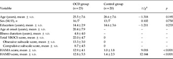
OCD patients were recruited from an out-patient clinic at Shanghai Mental Health Centre, Shanghai, China. We recruited the healthy control subjects through local advertisements. Written informed consent was obtained from all participants involved in the research. The study was approved by the local institutional ethics board. The two groups were all Chinese, right-handed and generally matched for age, gender and educational level (Table 1). Inclusion criteria for patients with OCD were: age between 18 and 54 years; total duration of education ⩾9 years (the minimum duration of compulsory school education in China); a DSM-IV diagnosis of OCD based on detailed clinical interview followed by an assessment using the Mini-International Neuropsychiatric Interview (MINI) for Axis I mental disorders (Sheehan et al. Reference Sheehan, Lecrubier, Sheehan, Amorim, Janavs, Weiller, Hergueta, Baker and Dunbar1998) by a research psychiatrist; and being free of drug treatments for at least 8 weeks before the day of scanning (12 of the 23 OCD patients were drug naive). Exclusion criteria were: co-morbid Axis I psychotic disorders, bipolar disorder, major depression and anxiety disorders other than OCD and a history of neurological disorders. Inclusion criteria for the healthy controls were: age between 18 and 54 years, a total duration of education ⩾9 years and absence of a history of neurological disorders and any DSM-IV Axis I diagnosis.
Demographic and clinical features of the sample

OCD, Obsessive–compulsive disorder; M, male; F, female; Y-BOCS, Yale–Brown Obsessive Compulsive Scale; HAMA, Hamilton Anxiety Rating Scale; HAMD, Hamilton Depression Rating Scale; s.d., standard deviation.
The 24-item Hamilton Depression Rating Scale (HAMD) and the 14-item Hamilton Anxiety Rating Scale (HAMA) were used to assess the depressive and anxiety symptoms of participants respectively. We also used the Yale–Brown Obsessive Compulsive Scale (Y-BOCS) to measure participants' obsessive and compulsive symptoms.
Image acquisition
All imaging studies were performed on a 1.5-T magnetic resonance imaging (MRI) scanner (GE Medical Systems Medical Systems, USA) at Ruijin Hospital, Shanghai, China. Head motion was minimized by inserting inflatable pillows between the patients' head and the head coil. For each participant a set of 232, 1-mm-thick contiguous axial T1-weighted MR images encompassing the whole brain was acquired using a three-dimensional (3D) spoiled gradient-recalled echo (SPGR) sequence. Acquisition parameters were as follows: repetition time=8.1 ms; echo time=1.6 ms; flip angle=20°; field of view=24 cm; matrix size=256×192 pixels; number of excitations=2; acquisition time=7 min 14 s.
Cortical thickness maps
Surface extraction was carried out using FreeSurfer version 5.0 (Fischl et al. Reference Fischl, Sereno and Dale1999; http://surfer.nmr.mgh.harvard.edu/). After skull stripping and intensity correction, the grey matter–white matter boundary for each cortical hemisphere was determined using tissue intensity and neighbourhood constraints. The resulting surface boundary was tessellated to generate multiple vertices (and triangles) across the whole brain before inflating. Using a deformable surface algorithm guided by the grey–cerebrospinal fluid (CSF) intensity gradient, the resulting grey–white interface was expanded to create the pial surface. The inflated surface was then morphed into a sphere followed by registration to an average spherical surface for optimal sulcogyral alignment. All surfaces were inspected visually following an automated topology fixation procedure, and remaining minor defects were corrected manually, as recommended by the software guidelines. Cortical thickness values were computed using the methods developed by Fischl & Dale (Reference Fischl and Dale2000). Smoothing at a full-width at half-maximum (FWHM) Gaussian kernel of 10 mm was applied to enable group comparisons at the level of each vertex.
Area expansion/contraction maps
We used the method described by Joyner et al. (Reference Joyner, Roddey, Bloss, Bakken, Rimol, Melle, Agartz, Djurovic, Topol, Schork, Andreassen and Dale2009) and Palaniyappan et al. (Reference Palaniyappan, Mallikarjun, Joseph, White and Liddle2011b) to obtain the areal maps. The size of the triangles on each subject's grey matter surface obtained during initial tessellation using FreeSurfer is fixed but the number of triangles varies according to the brain size. At this stage, each vertex has the same value for surface area calculated from the triangles that surround a vertex. In the next stage, deformation of individual subject's surfaces using a spherical atlas registration procedure was carried out, followed by registration of individual spheres into the common coordinate system. This resulted in a standard number of triangles across each individual's brain surface. As the dimensions of the tessellations around a given vertex were now redistributed due to deformation, relative contraction/expansion maps of the cortical surface area were obtained by computing the average of the area of the six triangles that define a vertex. These maps were smoothed with a FWHM Gaussian kernel of 10 mm.
Cortical gyrification maps
Local gyrification indices (LGIs) reflecting the amount of cortex folded in the locality of numerous vertices on the cortical surface were obtained from the FreeSurfer-based cortical reconstruction using the method of Schaer et al. (Reference Schaer, Cuadra, Tamarit, Lazeyras, Eliez and Thiran2008). This method is a vertex-wise extension of the GI proposed by Zilles et al. (Reference Zilles, Armstrong, Schleicher and Kretschmann1988), which gives a ratio of the inner folded contour to the outer perimeter of the cortex. The method is described in detail elsewhere (Schaer et al. Reference Schaer, Cuadra, Tamarit, Lazeyras, Eliez and Thiran2008; Palaniyappan et al. Reference Palaniyappan, Mallikarjun, Joseph, White and Liddle2011a). In brief, an outer surface is created from the pial surface obtained from the FreeSurfer-based cortical reconstruction. Corresponding circular ROIs with a radius of 25 mm in the outer and pial surface are identified for each of the vertices (>150 000) on the outer surface using a matching algorithm. A distance-weighted mean of the LGI values of the outer vertices that are contained within the circular ROI are assigned to each of the pial vertices. This process results in gyrification maps in which each vertex is assigned a value: an index of 3 means that there is three times more cortical surface buried within the sulci in the surrounding area than the amount of visible cortical surface; an index of 1 means that the cortex is flat in the surrounding area. Group comparisons at the level of each vertex were carried out using the gyrification maps without further smoothing.
Statistical analysis
All statistical analyses were carried out using the Query, Design, Estimate, Contrast (QDEC) statistical interface of FreeSurfer version 5.0. As we were primarily interested in the group differences in the surface anatomy across the three surface variables, three separate group comparisons using a general linear model were carried out for thickness, area and LGI changes for the right and left hemispheric surfaces. To control for global differences in each of the models, intracranial volume was used as a covariate measure. Age and gender were also used as nuisance covariates. To correct for multiple vertex-wise comparisons, we undertook a Monte Carlo permutation analysis (Hagler et al. Reference Hagler, Saygin and Sereno2006). A total of 10 000 simulations were undertaken under the null hypothesis with a threshold of p=0.05 for each surface anatomical measure and the size of the largest cluster was recorded for each simulation. The p value for each cluster in the actual data is given by the proportion of the 10 000 simulated clusters that are of same size or greater. To relate thickness, surface area and LGI to the clinical symptoms score, we extracted the mean value of the corresponding morphometric measures from each of the significant clusters that emerged from group comparisons in all subjects. A linear regression was carried out with the morphometric measures entered simultaneously as predictors for the YBOCS scores in the patients. Age, gender and mean years of education were also entered as covariates in this regression. A similar model was used to predict HAMD scores in patients, with the morphometric measures as predictors and age, gender and education years as covariates. To study the effect of prior exposure to medication in the patient sample, we also undertook a comparison of the morphometric measures (extracted from the clusters) between the 12 medication-naive and 11 previously medicated patients using an ANOVA. We addressed the question of whether the medication status (naive versus previously medicated) affected the contrast with healthy controls.
Results
In the patients, the mean total symptom score on the YBOCS was 22.0 (range 14–32). The clinical features of the sample are shown in Table 1. Among those who were previously medicated (n=11), nine were exposed only to selective serotonergic reuptake inhibitors (SSRIs: paroxetine, sertraline, fluvoxamine or fluoxetine) whereas two had additional exposure to clomipramine. There were no differences (mean±s.d.) between previously medicated and medication-naive patients in terms of age (24.8±4.8 v. 26.0±9.7 years), education (13.0±2.9 v. 13.7±3.0 years) or YBOCS score (21.7±5.5 v. 22.1±4.0).
Cortical thickness
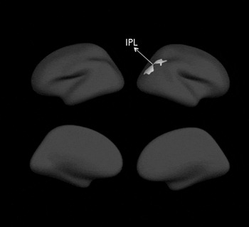
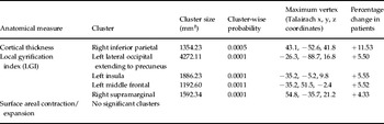
Whole-brain analysis for cortical thickness differences revealed a single cluster of increased thickness (Fig. 1, Table 2) but no regions of relative cortical thinning in patients compared to controls in the left hemisphere. The cluster was located on the left inferior parietal region, predominantly the angular gyrus. On the right hemisphere no significant group differences were found.
Significant increases in cortical thickness in obsessive–compulsive disorder in the right inferior parietal region (IPL). The cluster is displayed on a standard average brain surface (fsaverage).

Clusters showing significant changes in the surface anatomy in obsessive–compulsive disorder (OCD)

Surface area
Areal contraction/expansion maps revealed no significant differences between the patients and the controls in either the right or left hemispheres. No group differences were noted even when the cluster-wise statistical threshold was lowered to p=0.1.
Gyrification
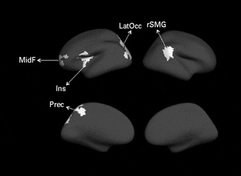
Whole-brain LGI analysis revealed three clusters of increased gyrification (hypergyria) in the left hemisphere and a single cluster of increased gyrification in the right hemisphere in patients compared to controls. The clusters on the left hemisphere included the lateral occipital region, the insula extending to the precentral region and the middle frontal region. The single cluster on the right hemisphere was located on the supramarginal gyrus. The clusters are shown in Fig. 2) and described in Table 2. No regions with reduced gyrification were noted in the patients.
Significant increases in gyrification in obsessive–compulsive disorder in the left lateral occipital region (LatOcc) extending to the precuneus (Prec), left insula extending to precentral region (Ins), left middle frontal region (MidF) and right supramarginal gyrus (rSMG). The clusters are displayed on a standard average brain surface (fsaverage).

Regression analysis
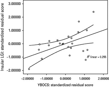
Higher symptom severity was associated with more pronounced gyrification in the insular cluster (β=0.752, t=2.53, p=0.024). None of the other morphometric measures had a significant association with symptom severity. Among the covariates, years of education had a significant association with YBOCS. Patients with less severe OCD symptoms had a longer period of education (β=−0.60, t=−2.68, p=0.018). Insular LGI explained nearly 25.5% of the variation in the YBOCS score (Fig. 3). None of the morphometric measures significantly predicted YBOCS obsessions or compulsions subscale scores.
Significant positive correlation was found between Yale–Brown Obsessive Compulsive Scale (YBOCS) clinical severity scores and local gyrification indices (LGIs) of the insular cluster (r=0.505, p=0.014) in patients with obsessive–compulsive disorder (OCD). Standardized residual scores were generated using age, gender and years of education as covariates.

None of the morphometric measures predicted the HAMD scores although years of education remained as a significant covariate in the regression analysis, with higher HAMD scores being associated with lower levels of education (β=−0.67, t=−3.06, p=0.008).
Effect of medication
There were no significant differences between the previously medicated and medication-naive groups in the thickness of the left inferior parietal cluster or the gyrification of the left insular, lateral occipital or right supramarginal clusters (all p>0.5). All of the clusters showed numerically greater values in the medication-naive group than in the previously medicated group, with the greatest numerical difference noted in the gyrification of left insula (mean ± s.d., controls 4.73±0.30, previously medicated 4.94±0.32, medication-naive 5.03±0.37). None of the observed differences between the two patient groups were statistically significant.
Discussion
Cortical thickness
We observed a significant increase in the thickness of the inferior parietal lobule on the right side, predominantly involving the angular gyrus, extending to the anterior position of the lateral occipital region. We did not find any region with reduced cortical thickness. By contrast, Shin et al. (Reference Shin, Yoo, Lee, Ha, Lee, Lee, Kim, Kim and Kwon2007) observed reduced thickness in several frontal and paralimbic regions and Nakamae et al. (Reference Nakamae, Narumoto, Sakai, Nishida, Yamada, Kubota, Miyata and Fukui2012) noted thinning in a cluster temporo-insular cluster. Narayan et al. (Reference Narayan, Narr, Phillips, Thompson, Toga and Szeszko2008) reported increased rather than decreased cortical thickness in the frontal cortex and posterior middle temporal gyrus in patients. It is possible that these inconsistencies are related to the clinical status of the patients. The patients studied by Shin et al. (Reference Shin, Yoo, Lee, Ha, Lee, Lee, Kim, Kim and Kwon2007) were receiving antidepressant and/or antipsychotic medication. Although the small sample size limited the power to detect differences in our study, the observation that the never-medicated cases exhibited numerically greater abnormalities than the previously medicated cases suggests that the effect of prior medicated should be examined in a larger sample. The patients studied by Narayan et al. (Reference Narayan, Narr, Phillips, Thompson, Toga and Szeszko2008) predominantly included cases with no co-morbid depression (67%), but most were receiving antidepressant medication (81%). Our cases were all unmedicated at the time of scanning and did not have co-morbid depression. Furthermore, our sample had a younger age and a shorter mean illness duration than that of Nakamae et al. (Reference Nakamae, Narumoto, Sakai, Nishida, Yamada, Kubota, Miyata and Fukui2012). Cumulative evidence of VBM studies in major depressive disorder suggests a significant effect of depression on the GMV of multiple brain regions (Bora et al. Reference Bora, Fornito, Pantelis and Yücel2012; Du et al. Reference Du, Wu, Yue, Li, Liao, Kuang, Huang, Chan, Mechelli and Gong2012). It is likely that, in the presence of co-morbid depression, the anatomical changes characteristic of the pathophysiology of OCD become confounded and less apparent.
Our findings are in keeping with the evidence indicating that increased grey matter density in the parietal lobe may be specific to patients without co-morbid depression, as revealed by a meta-regression analysis investigating the anatomical likelihood of VBM changes in OCD (Radua & Mataix-Cols, Reference Radua and Mataix-Cols2009). Studies investigating white matter volume (WMV) and integrity have also reported significant changes in the parietal lobe (Szeszko et al. Reference Szeszko, Ardekani, Ashtari, Malhotra, Robinson, Bilder and Lim2005; Menzies et al. Reference Menzies, Achard, Chamberlain, Fineberg, Chen, del Campo, Sahakian, Robbins and Bullmore2008; Lázaro et al. Reference Lázaro, Castro-Fornieles, Cullell, Andrés, Falcón, Calvo and Bargalló2011). The demonstration of increased cortical thickness in this region adds to the growing body of evidence implicating parietal lobe involvement in the pathophysiology of OCD reviewed elsewhere (Menzies et al. Reference Menzies, Achard, Chamberlain, Fineberg, Chen, del Campo, Sahakian, Robbins and Bullmore2008), and raises the question of whether this increase in thickness is associated with a predisposing developmental abnormality or is a consequence of symptom expression. A focal increase in cortical thickness has been observed following repeated task-related use of specific brain regions as a result of training in healthy participants (Draganski et al. Reference Draganski, Gaser, Busch, Schuierer, Bogdahn and May2004), but such changes are mostly observed following motor learning tasks (Fields Reference Fields2011), are transient (Draganski et al. Reference Draganski, Gaser, Busch, Schuierer, Bogdahn and May2004), and are shown to be related to new learning rather than continued training or use (Driemeyer et al. Reference Driemeyer, Boyke, Gaser, Büchel and May2008). Furthermore, excessive grey matter in the inferior parietal region has been associated with prolonged stop–signal reaction time in both symptomatic patients and asymptomatic relatives with OCD (Menzies et al. Reference Menzies, Achard, Chamberlain, Fineberg, Chen, del Campo, Sahakian, Robbins and Bullmore2007), reducing the likelihood that the increased parietal thickness is secondary to repeated task-related use of parietal cortex consequent to the symptoms of OCD. On the contrary, our observations suggest a disturbance in normal cortical maturation in OCD. Neuroimaging studies investigating normal brain maturation demonstrate a continuous increase in parietal thickness reaching peak levels around age 12, followed by a steady decrease over subsequent decades (Giedd et al. Reference Giedd, Blumenthal, Jeffries, Castellanos, Liu, Zijdenbos, Paus, Evans and Rapoport1999; Gogtay et al. Reference Gogtay, Giedd, Lusk, Hayashi, Greenstein, Vaituzis, Nugent, Herman, Clasen, Toga, Rapoport and Thompson2004). More recently, children with OCD (mean age 13.02 years) have been shown to have a thinner parietal cortex when compared to age-matched healthy controls (Fallucca et al. Reference Fallucca, MacMaster, Haddad, Easter, Dick, May, Stanley, Rix and Rosenberg2011). In this context, our results support the speculation of an abnormal delay in cortical maturation in OCD resulting in a thinner parietal cortex in early adolescence but a thicker cortex in adult life because of the later maturational peak.
Gyrification
We observed hypergyria of the middle frontal region, insula and lateral occipital region extending to the precuneus medially on the left hemisphere. We also observed hypergyric changes in the supramarginal section of the right inferior parietal lobe, anterior to but not overlapping with the region showing increased thickness. Although a direct comparison is not possible because of the methodological differences, the excessive regional cortical folding observed in this sample is in line with volumetric studies that report an increase in GMV in several cortical regions (Valente et al. Reference Valente, Miguel, Castro, Amaro, Duran, Buchpiguel, Chitnis, McGuire and Busatto2005; Song et al. Reference Song, Jung, Jang, Kim, Shim, Park, Choi and Kwon2011; Zarei et al. Reference Zarei, Mataix-Cols, Heyman, Hough, Doherty, Burge, Winmill, Nijhawan, Matthews and James2011). However, such linear relationships between hypergyria and VBM-derived volume seem to be region specific and cannot be generalized to the entire cortex (Palaniyappan & Liddle Reference Palaniyappan and Liddle2011). Indeed, systematic group differences in gyrification can contribute to spurious grey matter changes in VBM (Mechelli et al. Reference Mechelli, Price, Friston and Ashburner2005). To our knowledge there are no previous reports of whole-brain vertex-wise analyses of gyrification in OCD. When sulcal morphology is examined in preselected regions using 2D tracing techniques, focal reduction in cortical folding of the frontal cortex (Wobrock et al. Reference Wobrock, Gruber, McIntosh, Kraft, Klinghardt, Scherk, Reith, Schneider-Axmann, Lawrie, Falkai and Moorhead2010) and the cingulate sulcus is noted in OCD (Shim et al. Reference Shim, Jung, Choi, Jung, Jang, Park, Choi, Kang and Kwon2009). Wobrock et al. (Reference Wobrock, Gruber, McIntosh, Kraft, Klinghardt, Scherk, Reith, Schneider-Axmann, Lawrie, Falkai and Moorhead2010) noted that the effect of prefrontal hypogyria quantified using 2D tracing in OCD became non-significant when a correction for multiple testing was made. Our approach involved using a non-biased whole-brain vertex-wise analysis of gyrification that is relatively unaffected by slice thickness and takes into consideration the significant variation in the LGIs across all of the sulcogyral regions in the cerebral cortex. It is possible that regions with focal hypogyria, which did not survive the rigorous statistical correction involved in whole-brain analysis, are present in the current sample. Nevertheless, our unbiased analysis shows that focal hypergyria is likely to be the predominant gyrification abnormality in OCD.
The significant overlap between OCD and autistic spectrum (Jacob et al. Reference Jacob, Landeros-Weisenberger and Leckman2009), the higher prevalence of neurological soft signs (Karadag et al. Reference Karadag, Tumkaya, Kırtaş, Efe, Alacam and Oguzhanoglu2011) and the predilection towards younger age of onset (Roth et al. Reference Roth, Milovan, Baribeau and O'Connor2005) suggests a neurodevelopmental origin for OCD. Cortical folding is tightly linked to the process of neuronal migration and cortical development during the early years of life. On the basis of tensor-based morphogenetic theory, it is likely that focal regions of disturbed gyrification are related to an underlying disturbance in cortico-cortical or cortico-subcortical connectivity during cortical development. The presence of a widespread aberration in connectivity has been demonstrated in large-scale networks using graph analytical methods in OCD (Zhang et al. Reference Zhang, Wang, Yang, Wu, Li, Chen, Yue, Tang, Yan, Lui, Huang, Chan, Zang, He and Gong2011). Disturbances in cortical gyrification, but not cortical thickness, have been associated with obstetric events that contribute to neurodevelopmental delays (Haukvik et al. Reference Haukvik, Lawyer, Bjerkan, Hartberg, Jönsson, McNeil and Agartz2009, Reference Haukvik, Schaer, Nesvåg, McNeil, Hartberg, Jönsson, Eliez and Agartz2012). Furthermore, poor developmental outcomes in preterm infants are predicted by the presence of gyrification defects (Dubois et al. Reference Dubois, Benders, Borradori-Tolsa, Cachia, Lazeyras, Ha-Vinh Leuchter, Sizonenko, Warfield, Mangin and Huppi2008). This suggests that our observation of focal hypergyria in this sample supports the hypothesis that OCD has neurodevelopmental origins.
In the present sample, the insular LGI significantly predicted the YBOCS symptom scores but not the HAMD scores. Zarei et al. (Reference Zarei, Mataix-Cols, Heyman, Hough, Doherty, Burge, Winmill, Nijhawan, Matthews and James2011) observed a similar relationship with insular volume and YBOCS scores in adolescents with OCD. The fronto-insular region, together with the anterior cingulate cortex, constitutes a salience network (Seeley et al. Reference Seeley, Menon, Schatzberg, Keller, Glover, Kenna, Reiss and Greicius2007) whose function is related to evaluation of stimuli in the context of interoceptive emotional state. This network facilitates switching between self-directed default mode (or resting state) and one of the several task-processing modes such as motor performance, language processing or visual attention (Sridharan et al. Reference Sridharan, Levitin and Menon2008; Menon & Uddin, Reference Menon and Uddin2010). A neuronal state of readiness that leads to updating of expectations and/or generation of actions is associated with the evaluation of internal/external stimulus in the context of the present interoceptive state. This function, termed proximal salience, is ascribed to the salience network, and is hypothesized to be disturbed in psychosis (Palaniyappan & Liddle, Reference Palaniyappan and Liddle2012b). With regard to OCD, aberrant proximal salience may form the basis of (1) a failure to update the expectations associated with external events alongside a recurrent need to generate actions (compulsions) and (2) an inability to switch off from reverberating internal stimuli (ruminations). Huyser et al. (Reference Huyser, Veltman, Wolters, de Haan and Boer2011) reported excessive insular activation during performance of a conflict task in a sample of paediatric patients compared with controls. This effect was most pronounced in the older children. Furthermore, at a follow-up scanning session after 16 sessions of cognitive behavioural therapy, Huyser et al. (Reference Huyser, Veltman, Wolters, de Haan and Boer2011) found that this excessive insular activation was partially ameliorated, with the greatest reduction in the older children. These observations suggest that insular dysfunction is related to expression of symptoms and to treatment response. The functional implication of insular dysfunction needs to be further investigated in OCD.
Although most studies observe an increase in insular volume in OCD (Kim et al. Reference Kim, Youn, Lee, Kim, Kim and Kwon2003; Song et al. Reference Song, Jung, Jang, Kim, Shim, Park, Choi and Kwon2011; Zarei et al. Reference Zarei, Mataix-Cols, Heyman, Hough, Doherty, Burge, Winmill, Nijhawan, Matthews and James2011), there are notable exceptions (Yoo et al. Reference Yoo, Roh, Choi, Kang, Ha, Lee, Kim, Kim and Kwon2008; Rotge et al. Reference Rotge, Langbour, Guehl, Bioulac, Jaafari, Allard, Aouizerate and Burbaud2010). The presence of insular hypergyria could explain the volumetric excess observed in several studies. It is possible that even in samples that show a volumetric reduction, gyrification may be more pronounced in the insula but is accompanied by a reduction in thickness and/or surface area, leading to an overall reduction in grey matter. This discrepancy highlights the importance of studying the surface anatomical properties of gyrification, thickness and surface area separately. We did not observe any significant surface areal contraction and expansion in the present sample of patients with OCD. This morphometric feature is in sharp contrast to the combined areal contraction and reduced gyrification seen in schizophrenia (Palaniyappan et al. Reference Palaniyappan, Mallikarjun, Joseph, White and Liddle2011b; Palaniyappan & Liddle, Reference Palaniyappan and Liddle2012a). The neuronal correlates of the various surface anatomical measures observed using neuroimaging tools are yet to be elucidated. Nevertheless, in terms of neurodevelopmental abnormalities our results may be tentatively interpreted as evidence for a relationship between the expression of OCD and disturbances in factors influencing radial cortical expansion, which influences the thickness rather than being related to the tangential expansion that determines the overall surface area (Rakic, Reference Rakic1988). Speculatively, the brain regions with gyrification defects may represent the earliest neurodevelopmental changes in OCD.
One of the main limitations in the present study is the use of a cross-sectional sample, which precludes a meaningful investigation of the longitudinal course of these alterations. In addition, although the sample size in this study is comparable to many other published investigations in the field, we did not have sufficient power to investigate the surface anatomical correlates of the four factors reported in OCD (Mataix-Cols et al. Reference Mataix-Cols, Wooderson, Lawrence, Brammer, Speckens and Phillips2004). The SBM methods are not suitable for examining deep grey matter nuclei, including the basal ganglia and the amygdala, that are relevant to the symptoms of OCD. Meta-analysis of volumetric studies suggests that increased volume of the putamen is highly specific to OCD when compared to other anxiety disorders (Radua et al. Reference Radua, van den Heuvel, Surguladze and Mataix-Cols2010). As the estimation of cortical thickness and gyrification involves the creation of a surface representation without the need for a template registration, it is unlikely that the observed changes in the insula and other regions are artefacts driven by misalignments arising from volumetric differences in the subcortical structures. The relationship between the subcortical abnormalities and the morphometric changes observed in the insula, frontal and parietal cortices in OCD is currently unknown and warrants further investigation to develop a holistic neuroanatomical model in the pathophysiology of OCD.
Conclusions
In summary, an alteration in the cortical surface anatomy is an important feature of OCD seen in unmedicated samples and relates to the clinical severity of the illness. Our present results can be interpreted as crucial evidence for the involvement of distributed cortical regions with multimodal functions in OCD. The functional relevance of circuits that include these multimodal brain regions and the longitudinal course of the abnormal thickness and gyrification in OCD need further investigation.
Acknowledgements
This study was supported by the Joint Key Project of the National High Technology Research and Development Programme of China (863 Programme; no. 2007AA02Z420), the Key Project from the Science and Technology Commission of Shanghai Municipality (no. 074119520), the Programme for Shanghai Outstanding Academic Leader Plans (no. 08XD14036), the Natural Science Foundation of Shanghai Jiao Tong University School of Medicine (no. 09XJ21023), and the Shanghai Municipal Health Bureau Foundation, China (no. 2010Y028). In part, this work was supported by an Institute of Mental Health (IMH), Nottingham, UK exchange fellowship. No additional external funding was received for this study. The funders had no role in the study design, data collection and analysis, decision to publish, or preparation of the manuscript.
Declaration of Interest
P. F. Liddle has received honoraria for academic presentations from Bristol Myers Squibb. L. Palaniyappan has received a Young Investigator Travel Fellowship from Eli Lilly.