Introduction
Sebaceoma is a rare, benign, adnexal skin neoplasm which is most commonly found in the head and neck region of older individuals.Reference Misago, Mihara, Ansai and Narisawa 1 , Reference Fan, Carr, Sanders, Smith, Lazar and Calonje 2 These neoplasms can occur in any location containing hair and sebaceous glands, and their prevalence on the face and scalp reflects the abundant sebaceous glands and hair in these areas.Reference Eisen and Michael 3 The presence of a sebaceoma has also been linked to Muir–Torre syndrome, an autosomal dominant disorder which predisposes patients to multiple visceral malignancies.Reference Ponti and Ponz de Leon 4
Herein, we report a unique case of sebaceoma of the auricle, and we discuss the relevant differential diagnosis, histopathological characteristics and management of these lesions.
Case report
A 79-year-old man presented with a left auricular lesion which had grown slowly over the past two years. The mass was painless; however, over the past several months increased growth had been noted. The patient had a past medical history of prostate cancer and laryngeal cancer. The family history was negative for malignancy.
Physical examination revealed a firm, 1.2 cm, cauliflower-like mass in the concha, centred at the helical root, without involvement of the external auditory canal. The remainder of the head and neck examination was unremarkable.
The patient underwent wide local excision of the lesion, including a small amount of cartilage removed on the deep aspect, with negative margins. The surgical defect was closed primarily.
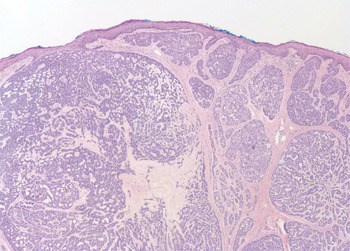
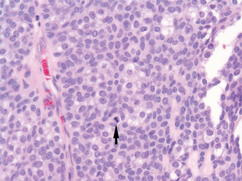
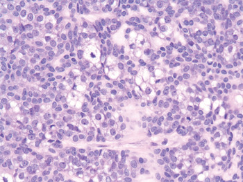
Histopathological evaluation revealed a benign, basaloid, adnexal neoplasm with sebaceous differentiation, consistent with a sebaceoma (Figures 1 to 3).
Photomicrograph showing that the tumour emanates from the undersurface of the epidermis. (H&E; ×100)

Photomicrograph of the tumour showing scattered mitotic foci in the solid areas (arrow). (H&E; ×400)

Photomicrograph showing that the solid area of the tumour is composed of relatively uniform basaloid cells, some with multivacuolated cytoplasm and scalloping of nuclei. (H&E; ×400)

At 36 months' follow up, the patient had no sign of local recurrence or other skin lesions. Due to his newly diagnosed sebaceoma, the patient was considered to be at increased risk of Muir–Torre syndrome and additional malignancy. However, because of the patient's preference and advanced age, genetic testing was not performed. Appropriate surveillance and follow up were instituted; at the time of writing, no further malignancies had been identified.
Discussion
The entire spectrum of cutaneous lesions should be kept in mind when approaching lesions of the auricle. These include benign lesions of epidermal origin, such as seborrhoeic keratoses and verruca vulgaris, as well as common malignancies, such as basal cell carcinoma, squamous cell carcinoma and their variants.Reference Willey, Swanson and Lee 5 Adnexal neoplasms are important in the differential diagnosis, which would include lesions derived from the folliculo-sebaceous-apocrine unit, including sweat gland tumours, tumours derived from hair follicles (e.g. trichoepithelioma) and sebaceous neoplasms (including naevus sebaceous, sebaceous carcinoma and sebaceoma).Reference Alsaad, Obaidat and Ghazarian 6
Sebaceous lesions have been described arising from the nose, face, scalp, neck and external auditory canal.Reference Misago, Mihara, Ansai and Narisawa 1 , Reference Fan, Carr, Sanders, Smith, Lazar and Calonje 2 , Reference Demellawy, Nicholas, Salama and Alowami 7 Sebaceoma typically presents as an asymptomatic, slow-growing, solitary, yellowish papule or nodule with rolled borders.Reference Eisen and Michael 3 Surface ulceration and bleeding has also been noted.Reference Misago, Mihara, Ansai and Narisawa 1 , Reference Eisen and Michael 3 Biopsy and histological assessment is essential for diagnosis, and the treatment is generally local excision to negative margins.
Historically, poor terminology and confusion have surrounded the diagnosis of neoplasms with sebaceous differentiation. In 1984, Troy and Ackerman defined the term and simplified the criteria for sebaceoma.Reference Troy and Ackerman 8 Sebaceoma is defined as a benign tumour involving the basaloid or germinative sebaceous cells, with basaloid, undifferentiated sebocytes mixed with mature sebocytes. It is differentiated from the more aggressive sebaceous carcinoma by its preserved lobulated architecture, lack of nuclear atypia and very infrequent mitotic figures.Reference Fan, Carr, Sanders, Smith, Lazar and Calonje 2 Sebaceoma can be differentiated histologically from other tumours which may display sebaceous differentiation, such as nodular basal cell carcinoma, trichoblastoma and apocrine poroma.Reference Misago, Mihara, Ansai and Narisawa 1 Histological differentiation from basal cell carcinoma can be challenging, and molecular markers such as Ber-EP4 and epithelial membrane antigen (EMA) have been proposed as useful in the histological diagnosis.Reference Fan, Carr, Sanders, Smith, Lazar and Calonje 2
Accurate diagnosis as a sebaceous-derived neoplasm is crucial, given the association of these tumours with Muir–Torre syndrome, which is considered a variant of hereditary nonpolyposis colorectal cancer.Reference Lachiewicz, Wilkinson, Groben, Ollila and Thomas 9 Muir–Torre syndrome is an autosomal dominant disorder with variable penetrance and expression, in which patients exhibit sebaceous neoplasms or keratoacanthomas in addition to visceral malignancy, most often of the gastrointestinal or genitourinary system. Genetic mutations in several DNA mismatch repair proteins and microsatellite instability have been implicated as aetiologies, and immunohistochemical and germline mutation analysis can aid in the diagnosis if Muir–Torre syndrome is suspected.Reference Lachiewicz, Wilkinson, Groben, Ollila and Thomas 9 , Reference Abbas and Mahalingam 10 Interestingly, the sebaceous and visceral tumours found in this syndrome have been noted to be more indolent than their sporadic counterparts, and the visceral tumours may precede the skin lesions or present decades later.Reference Ponti and Ponz de Leon 4
-
• Head and neck skin neoplasms include sebaceous-derived tumours, e.g. sebaceoma
-
• Complete surgical excision of the latter is recommended
-
• Sebaceous neoplasms may arise sporadically or together with Muir–Torre syndrome, with increased risk of visceral malignancy
-
• If this syndrome is suspected, genetic testing and cancer surveillance are needed
While sebaceous neoplasms are most often sporadic, they can be a marker for Muir–Torre syndrome, especially if they present with multiple lesions or in younger patients. The treating physician should maintain a high index of clinical suspicion for the development of visceral malignancy, and the patient should therefore undergo strict surveillance with serial physical examinations, colonoscopies and appropriate imaging.Reference Ponti and Ponz de Leon 4 , Reference Lachiewicz, Wilkinson, Groben, Ollila and Thomas 9 Our patient with an auricular sebaceoma had no further malignancies diagnosed, but did have a history of prostate and laryngeal cancer, which have been described, albeit rarely, in Muir–Torre syndrome.Reference Ponti and Ponz de Leon 4 , Reference Demirci, Nelson, Shields, Eagle and Shields 11 These cases were usually accompanied by colonic involvement. Given the patient's age, sex and previous smoking history, it is reasonable that these tumours had occurred sporadically. Nevertheless, our patient was followed closely for further malignancy, to enable early intervention if needed.
Conclusion
Sebaceoma is a rare, benign, adnexal neoplasm which most commonly presents in the head and neck. It can occur sporadically or as part of Muir–Torre syndrome, an autosomally dominant disorder which is characterised by multiple visceral malignancies and sebaceous neoplasms. Treatment is local excision, and recurrence is uncommon. The management of a patient with sebaceous neoplasms should also include a thorough search for underlying malignancies, and appropriate genetic testing and surveillance should be instituted if the patient is deemed at high risk of Muir–Torre syndrome.