Implications
Sperm glycocalyx plays an important role in reproduction as it is the first interface between the male gamete and the environment. We demonstrated a seasonal variation in the glycosylation pattern of buffalo bull sperm glycocalyx that could explain the lower semen quality during non-mating periods (NMP). Sperm glycocalyx characterization could be exploited in the future for evaluating semen quality. We describe a novel microarray procedure for cell surface glycome profiling. Compared with other methods for in situ cell surface molecular pattern detection, this unbiased methodology can also be used for functional, cell differentiation molecular remodeling, cell identification and clinical studies.
Introduction
Sperm glycocalyx consists of many different glycoconjugates, including glycoproteins, glycolipids and glycophosphatidylinositol-anchored glycoproteins (Tecle and Gagneux, Reference Tecle and Gagneux2015). These glycoconjugates represent the interface between the male gamete and the extracellular environment. Therefore, they play an important role in reproductive biology as it is involved in sperm survival, transport in the oviduct, capacitation, sperm–egg interaction, acrosome reaction, zona binding and fertilization, and immunoprotection in the female tract (Tollner et al., Reference Tollner, Bevins and Cherr2012; Tecle and Gagneux, Reference Tecle and Gagneux2015). Sperm glycocalyx is synthesized during spermatogenesis and is modified as sperm pass through the male urogenital transit (Tecle and Gagneux, Reference Tecle and Gagneux2015).
The composition and structure of glycoconjugates can be decoded using several techniques. Lectins, which are glycan-binding proteins, are particularly well suited for discriminating glycoconjugates because of their specificity and ability to distinguish sugar isomers as well as branching, linkage and terminal modifications of complex glycans (Sharon and Lis, Reference Sharon and Lis2004). Lectins have been used for sperm analysis for many years in several techniques such as MS (Calvete et al., Reference Calvo, Bustos-Obregón and Pastor1994), western blotting (Chandra et al., Reference Chandra, Srinivasan, Jamal, Mehrotra, Singh and Srivastav2008), histochemistry (Bawa et al., Reference Bawa, Govil, Dhingra, Werner and Bains1993; Desantis et al., Reference Desantis, Ventriglia, Zizza, Nicassio, Valentini, Di Summa and Lacalandra2010) and lectin microarray (Xin et al., Reference Xin, Cheng, Diao, Wang, Gu, Wu, Wu, Chen, Zhou, Guo, Shi and Tao2014). Unfortunately, these techniques are very laborious, time-consuming and the possibility to measure large sets of samples is somehow limited. Membrane glycoproteins are known to be difficult to handle, and the immobilization of functional proteins on protein arrays is a real challenge. Therefore, cell microarrays prepared by direct printing of cells on microarray slide seem to be a promising alternative for the characterization of binding molecules that are specific for cell surface antigens (see Accogli et al., Reference Accogli, Desantis, Martino, Dell’Aquila, Gemeiner and Katrlík2016 for reference). More recently, we developed a novel cell microarray method for the in situ profiling of mammalian cell surface glycosylation pattern (Accogli et al., Reference Accogli, Desantis, Martino, Dell’Aquila, Gemeiner and Katrlík2016). This method consists in sperm cell isolation and washing, spotting onto microarray slide, incubation with a panel of biotinylated lectins, reaction with streptavidin fluorescent conjugate and signal intensity detection using a microarray scanner.
The domestic water buffalo (Bubalus bubalis) is an important livestock resource in many Asian, Mediterranean and Latin American countries. This domesticated ruminant species has a seasonal mating cycle. Mating period (MP) takes place from autumn to winter, and there is a long NMP from late spring to the beginning of the autumn (Rossi et al., Reference Roy, Dey and Majumder2014). This seasonal reproduction activity also occurs in Italy as in other countries where the buffalo is bred, and its activity has been found to benefit from a negative photoperiod (Zicarelli, Reference Zicarelli2007; Campanile et al., Reference Calvete, Raida, Sanz, Wempe, Scheit, Romero and Töpfer-Petersen2010).
Seasonal changes have also been found in the reproductive physiology of buffalo bulls. During NMP, a lower testis and epididymis activity can be found (Arrighi et al., Reference Arrighi, Bosi, Groppetti and Cremonesi2010) conversely, during the MP, an enhanced expression of epididymal nitric oxide is present (Scala and Maruccio, Reference Scala and Maruccio2012). The reproductive function is somewhat compromised during the non-mating season, as confirmed by variability in semen quality (Sharma et al., Reference Sharma, Pandey, Nigam, Singh, Saxena and Swain2014). Moreover, it has been recently reported that the buffalo seminal plasma contains seasonal-dependent proteins, which could be used as markers of the semen quality (Sharma et al., Reference Sharma, Pandey, Nigam, Singh, Saxena and Swain2014). To date no reports on seasonal semen glycan pattern profiling are available. Therefore, we aimed to investigate if seasonal glycan profile changes occur in the glycocalyx of buffalo bull spermatozoa during the MP and NMP using our above mentioned innovative cell microarray procedure (Accogli et al., Reference Accogli, Desantis, Martino, Dell’Aquila, Gemeiner and Katrlík2016).
Material and methods
Semen collection, evaluation and preparation
At least two ejaculates were collected from three healthy Italian Mediterranean buffalo bulls (B. bubalis) of proven quality, routinely used for artificial insemination, during the breeding (December) and non-breeding (May) periods. The procedure was performed in the farm ‘Centro Tori Chiacchierini’ (Civitella d’Arna, Perugia, Italy) by means of an artificial vagina. After collection, the ejaculates immediately underwent semen evaluation and cell microarray as well as lectin histochemistry techniques were performed.
Semen evaluation included color of ejaculate, sperm motility and sperm morphology. The normal color of buffalo semen is variable from white, milky to white or creamy, and it was assessed by an experienced operator. The proportion of motile spermatozoa was estimated at 40× magnification with a phase-contrast microscope equipped with an automatic heating stage (37°C). The sperm morphology was evaluated by examining smears (four smears for each ejaculate) of the formalin-fixed spermatozoa under 100× magnification with a phase-contrast microscope on 500 spermatozoa/sample.
Semen preparations were performed as previously described (Desantis et al., Reference Desantis, Ventriglia, Zizza, Nicassio, Valentini, Di Summa and Lacalandra2010). Briefly, the semen was washed twice in 0.01 M phosphate-buffered saline (PBS, pH 7.4) by centrifugation at 800×g for 5 min, fixed in 4% (v/v) buffered paraformaldehyde, pH 7.4, for 45 min at room temperature (RT) (18°C to 22°C) and then pelleted by centrifugation. After supernatant removal, the sperm was washed twice with PBS and stored at 4°C until use.
Cell microarray fabrication
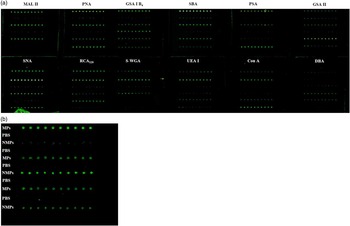
Cell microarray was prepared according to Accogli et al. (Reference Accogli, Desantis, Martino, Dell’Aquila, Gemeiner and Katrlík2016). Briefly, after centrifugation, the pellet was resuspended in ~50 µl of PBS (about 1×10⁶ of spermatozoa) and transferred by spotting (in two/three replicates) onto three-dimensional thin film coated glass slides (Nexterion Slide H; Schott, Jena, Germany). Spotting was performed with a non-contact microarray printing robot sciFLEXARRAYER S1 (Scienion, Berlin, Germany). The spermatozoa were spotted into 12 identical arrays on the slide (Figure 1). Each sample was spotted in 10 replicates/line within each array.
Raw signals of 12 lectin-based cell microarray analysis of mating period spermatozoa (MPS) and non-mating period spermatozoa (NMPS). (a) Each sample was spotted in 10 replicates within each array. (b) Detail of GSA I-B4 signals. PBS=phosphate-buffered saline.

Approximately 500 pl of cell suspension was spotted for each spot. The transfer efficiency and reproducibility of the printing process was assessed with a standard quality control test array performed by the microarrayer and 400 replicates (20×20 spots) were generated in a single run. Printing of the 0.01 M PBS buffer was also performed in order to check the background signal produced by the lectins incubation. The slides were then held in a humidity chamber (50% to 70%) at 37°C for 1 h to ensure sufficient attachment of the spermatozoa to the slide surface. The unoccupied surface of slides was blocked with 1 M ethanolamine dissolved in 0.01 M PBS with 0.05% Tween 20 (PBST) at RT for 1 h. Blocked slides were gently washed with PBST (two rinsing, 2 min each).
Cell microarray: lectin-binding procedure
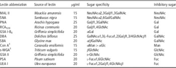
A multi-well incubation chamber was applied onto the surface of the spotted slides, in line with the subarrays that were created during the printing process. The used biotinylated lectins, their concentration and sugar specificity are listed in Table 1. Each lectin (Vector Laboratories, Burlingame, CA, USA) was diluted in PBST at optimized concentrations to have the highest specific signal with the lowest background (Table 1) and loaded directly onto the samples for all 12 subarrays. Samples were allowed to react with 50 µl of lectins solution at RT for 1 h. Then, lectins were gently removed and slides were immediately washed in PBST for 5 min. Subsequently, each subarray was incubated with Cy3-conjugated Streptavidin (Jackson ImmunoResearch Laboratories, Baltimore, PA, USA) at 0.5 μg/ml in PBST for 15 min. Redundant Cy3-conjugated streptavidin was removed and the slides were washed with PBST, and then with distilled water (4 min each). Lastly, residual water was removed using a slides centrifuge at 6000 r.p.m (Arrayit Corporation, Sunnyvale, CA, USA).
Lectin used, their sugar specificities and the inhibitory sugars used in control experiments

NeuNAc=N-acetylneuraminic (sialic) acid; Gal=galactose; GalNAc=N-acetylgalactosamine; Man=mannose; GlcNAc=N-acetylglucosamine; Glc=glucose; Fuc=fucose; s-WGA=succinylated WGA.
1 The used lectins bind terminal sugars except for Con A and s-WGA, which also bind internal residues.
Scanning and data analysis
Images of the stained cell microarray slides were taken using the InnoScan®710 fluorescent scanner (Innopsys, Carbonne, France) set at the appropriate excitation wavelength for Cy3 (570 nm) (Figure 1). The resolution was set at 3 μm for quantification purposes. The slide images obtained in lectin-based cell microarray analysis were evaluated using the Mapix 5.5.0 software (Innopsys, France) and the fluorescence intensity signals of each spot was measured with background subtraction. The raw numeric values, corresponding to the detected intensity of spots from cell microarrays, were normalized and were reported as the value of fluorescence intensity relative to the average number of cells per spot. In addition, the normalized values from each detected sugar residue were grouped to analyze its seasonal expression. Analysis of variance followed by Tukey’s post hoc multiple comparison test was performed on data in order to assess the significant intensity differences between samples using SPSS software (IBM, Italy). The statistical significance was set at P<0.01.
Lectin histochemistry
Microarray results were validated using lectin histochemistry, which is the conventional method used for the in situ detection of carbohydrate residues in glycan chains. The semen was smeared on poly-l-lysine coated glass slides and air-dry fixed. Then, they were incubated for 1 h in the dark with the same panel of lectins used for cell microarray. Lectins (9 fluorescein isothiocyanate and 3 rhodamine conjugated) (Vector Laboratories) were diluted in 0.05 M Tris–HCl-buffered saline (TBS) (pH 7.4) at the same concentration used for cell microarray. Slides were subsequently rinsed in the same buffer and mounted in Vectashield mounting medium (Vector Laboratories). Controls included the followings: (1) incubation of spotted mating period spermatozoa (MPS) and non-mating period spermatozoa (NMPS) with the buffer lacking lectins; (2) incubation of spotted both MPS and NMPS with each lectin in the presence of its hapten sugar (0.05 M in TBS); (3) incubation of spotted buffer (PBS without cells) with each lectin solution (only for cell microarrays). All these control experiments gave negative reactions. Slides were observed and photographed at 40× magnification under a light photomicroscope Eclipse Ni-U (Nikon, Tokyo, Japan) equipped with a digital camera (DS-U3; Nikon). The images were analyzed by the image-analyzing program NIS Elements BR (Version 4.20; Nikon).
Results
Semen evaluation
No significant differences for the assessed parameters of semen quality was observed for each animal between the MP and NMP. The color of ejaculates was milky to white, indicative of normal semen quality. The percentage of motile spermatozoa ranged from 80% to 90%. Less than 1% spermatozoa showed alterations in the head shape or the presence of proximal cytoplasmic droplet and 2% to 3% of spermatozoa exhibited simple distally coiled tail.
Cell microarray method
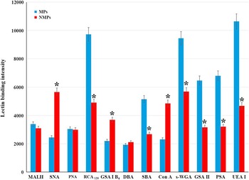
Both MPS and NMPS spotted on the cell microarray slides reacted with all used lectins, although with different signal intensities (Figure 1). The comparison between MPS and NMPS fluorescence intensity signals are displayed in three different patterns (Figure 2). RCA120, SBA, succinylated WGA, GSA II, PSA and UEA I signals were significantly stronger in MPS. On the contrary, SNA, GSA I-B4 and Con A signals were significantly stronger in NMPS. Lastly, MAL II, PNA and DBA did not show significant differences in signal intensities between the two conditions.
Differential fluorescent signals of lectin-based cell microarray of glycocalyx in buffalo bull spermatozoa from mating period (MPS) and non-mating period (NMPS). *Statistical significance (P<0.01) between MPS and NMPS. s-WGA=succinylated WGA.

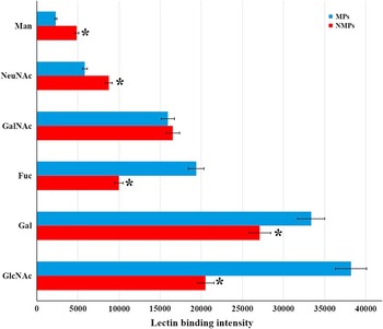
The grouping of signals from the investigated sugar residues demonstrated a progressive increase of mannose (Man), N-acetylneuraminic (sialic) acids (NeuNAc), N-acetylgalactosamine (GalNAc), fucose (Fuc), galactose (Gal) and N-acetylglucosamine (GlcNAc) in MPS (Figure 3). In NMPS it was observed a higher presence of Man and NeuNAc and, on the contrary, a lower expression of Fuc, Gal and GlcNAc when compared with MPS (Figure 3).
Glycome profiling obtained by the summation of fluorescence signals provided by lectins used for the investigated sugar residues in mating period spermatozoa (MPS) and non-mating period spermatozoa (NMPS) buffalo bull spermatozoa. The bar shows the standard error for a given sugar. *Statistical significance (P<0.01) between MPS and NMPS. Man=mannose; NeuNAc=N-acetylneuraminic (sialic) acids; GalNAc=N-acetylgalactosamine; Fuc=fucose, Gal=galactose; GlcNAc=N-acetylglucosamine.

Lectin histochemistry
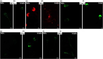
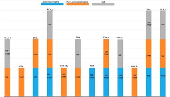
Lectin histochemistry was used to validate cell microarray results, this classical methodology revealed a binding pattern signal on the MPS and NMPS surface comparable with the lectin-based cell microarray. In particular, MPS lectin-binding sites showed a wider distribution of MAL II, succinylated WGA, UEA I and RCA120 (Figure 4a), and a more regional concentration of PNA, GSA I-B4, PSA and SBA (Figure 4c) compared with NMPS (Figure 4b and d). On the contrary, MPS showed a lower distribution GSA II-, Con A- and SNA- (Figure 4e and g) binding sites compared with NMPS (Figure 4f and h). In addition, lectins such as GSA II and DBA (Figure 4i and j) did not show differences in the staining intensity between MPS and NMPS. The regional positivity to lectins is summarized in Figure 5.
Validation of microarray results for mating period spermatozoa (MPS) and non-mating period spermatozoa (NMPS) by means of lectin histochemistry. This figure shows some representative lectins displaying higher (a, c) and lower (e, g) reactivity in MPS when compared with NMPS as well as almost similar lectin staining intensity in both MPS and NMPS (i, j). (a, b, e, f, g, h) fluorescein isothiocyanate-conjugated lectins; (c, d) rhodamine-conjugate lectin.

Scheme representing the regional distribution of lectin-binding sites in mating period (MP) and non-mating period (NMP) sperm surface. s-WGA=succinylated WGA.

Discussion
Mammalian sperm glycocalyx plays a key role in sperm motility, maturation and fertilization (for review see Tecle and Gagneux, Reference Tecle and Gagneux2015). Therefore, the evaluation of the sperm surface glycans could facilitate the general understanding of sperm biology and the changes that occur in different pathological and environmental conditions, including photoperiod and climate (Rossi et al., Reference Roy, Dey and Majumder2014). The buffalo is a domesticated ruminant species that goes through a seasonal mating cycle. In this study, we investigated glycan profile changes that occur in buffalo bull spermatozoa glycocalyx during mating and non-mating seasons.
The evaluated semen quality parameters such as color of ejaculates, sperm motility and sperm morphology did not show differences between the MP and NMP. Microarray technology is a fast, practical and high-throughput way to perform glycome profiling of biological samples (Xin et al., Reference Xin, Cheng, Diao, Wang, Gu, Wu, Wu, Chen, Zhou, Guo, Shi and Tao2014; Accogli et al., Reference Accogli, Desantis, Martino, Dell’Aquila, Gemeiner and Katrlík2016). Among the microarray technologies, cell microarrays prepared by direct printing of cells on microarray slide seem to be a promising alternative in the characterization surface glycoproteins. Here we used, for the first time, cell microarray to perform sperm surface glycan profiling by spotting spermatozoa directly on the microarray slide.
The spermatozoa surface showed lectin-binding signals indicating the presence of both O- and N-linked glycans. Sperm glycocalyx O-glycans contained the core 1 disaccharide (Galβ1,3GalNAc) (named T antigen) (PNA), the Tn antigen (the simplest mucin O-glycan made a single GalNAc linked to serine or threonine) (SBA), the oligosaccharides terminating with GalNAcα1,3(l-Fucα1,2)Galβ1,3/4GlcNAcβ1 (DBA), GlcNAc (succinylated WGA) and the disialyl T sequence NeuNAcα2,3Galβ1,3(±NeuNAcα2,6)GalNAc (MAL II). Sperm glycocalyx N-glycans, on the other hand, were high mannose and biantennary complex types (Con A) as well as asialo complex type terminating with Galβ1,4GlcNAc (RCA120). Buffalo spermatozoa also contained sialo N-glycans terminating with Neu5Acα2,6Gal/GalNAc (SNA). Moreover, signals were observed for core fucose Fucα1,6GlcNAc (PSA), Fucα1,2Galβ1,4GlcNAcβ (UEA I), as well as GlcNAc (GSA II) and galactose-terminating oligosaccharides (GSA I-B4). PNA-, DBA-, WGA-, Con A- and PSA-binding sites have been reported in previous studies on buffalo bull spermatozoa (Bawa et al., Reference Bawa, Govil, Dhingra, Werner and Bains1993; Kaul et al., Reference Kaul, Sharma, Singh and Gandhi2001), whereas the presence of MAL II, SNA, RCA120, GSA I-B4, DBA, SBA, Con A, GSA II and PSA binders has also been reported on the bovine spermatozoa surface (Calvete et al., Reference Calvo, Bustos-Obregón and Pastor1994; Xin et al., Reference Xin, Cheng, Diao, Wang, Gu, Wu, Wu, Chen, Zhou, Guo, Shi and Tao2014), the closest species to buffalo.
The roles of sperm surface glycans are not completely known. O-linked (mucin-type) glycans are important in sperm–zona binding during the process of fertilization. Moreover, the contribution of mucin-type glycans to male fertility has been reported in impaired spermatogenesis correlated to the absence of a functional MUC1 gene (Franke et al., Reference Franke, Kraus, Eiermann, Pauls, Lalani and Bergmann2001). In regard to N-glycans, they greatly contribute to the physicochemical properties of the glycocalyx due to their extraordinary flexibility (high degree of rotation and movement possible about each glycosidic bond) and microheterogeneity (variation in chain length, branching, monosaccharide composition and terminal sialylation) (Treumann et al., Reference Treumann, Lifely, Schneider and Ferguson1995). It has been reported that oligomannosidic chains could be a recognition signal for the elimination of incompetent sperm during their transit through the female reproductive tract (Nardone et al., Reference Nardone, Cerezo and de Cerezo1985), they could also participate in the binding of sperm to zona pellucida in humans (Cheng et al., Reference Cheng, Doncel, Alvarez and Acosta1995). Moreover, sperm with highly fucosylated N-glycans can contribute to immune modulatory activity in the uterus (Pang et al., Reference Pang, Tissot, Drobnis, Sutovsky, Morris, Clark and Dell2007). The importance of N-glycans synthesis in sperm has been demonstrated in both mouse and humans (Huang and Stanley, Reference Huang and Stanley2010). In addition, male basigin knockout mice, characterized by significant reduction of GlcNAc terminated N-glycans, are sterile due to absence of mature sperm in the testis and epididymis (Bi et al., Reference Bi, Li, Sun, Saalbach, Klein, Miller, Hess and Nowak2013). Concerning sialic acids, they contribute to the net negative charge of sperm surface and inhibit the head-to-head autoagglutination of mature spermatozoa in goats (Roy et al., Reference Rossi, Vecchio, Neglia, Di Palo, Gasparrini, D’Occhio and Campanile2014). The presence of sialic acids on the sperm plasma membrane is also indicative of a not yet capacitated sperm in humans and boars (Focarelli et al., Reference Focarelli, Rosati and Terrana1990; Jiménez et al., Reference Jiménez, González-Márquez, Ortiz, Herrera, García, Betancourt and Fierro2003). Moreover, sialic acids protect sperm from immune recognition in the female reproductive tract (Tollner et al., Reference Tollner, Bevins and Cherr2012) and are implicated in the sexual selection by female immunity against paternal antigens (Ghaderi et al., Reference Ghaderi, Springer, Ma, Cohen, Secrest, Taylor, Varki and Gagneux2011).
Differential profiling of MPS and NMPS glycocalyx revealed that the expression of mucin-type glycans terminating with T antigen, disialyl T sequence NeuNAcα2,3Galβ1-3(±NeuNAcα2,6)GalNAc and asialo and fucosylated sequence GalNAcα1,3(l-Fucα1,2)Galβ1,3/4GlcNAcβ1 (PNA, MAL II, DBA, respectively) is not season dependent. However, MPS showed a predominant expression of asialo complex-type N-glycan (RCA120), Tn antigen (SBA), GlcNAc-terminating glycans (succinylated WGA, GSA II), α1,6- and α1,2-linked fucosylated oligosaccharides (PSA, UEA I) compared with NMPS. On the contrary, NMPS expressed a significant higher density of α2,6 sialic acids (SNA) in N-glycan (Con A) as well as Gal-terminating oligosaccharides (GSA I-B4) compared with MPS. These findings were confirmed by lectin histochemistry, which is the conventional method used for the in situ detection of carbohydrate residues in glycan chains. Interestingly, the lectin-based sperm microarray gave more information about the glycocalyx composition compared with lectin histochemistry. In particular, sperm microarray revealed the presence of SNA-binding sites on MPS surface, which were not histochemically detected. Unlike lectin histochemistry, in which the staining intensity is subjectively evaluated by the observers, the lectin-based cell microarray analysis provides an objective evaluation of the sugars expression based on the intensity of the signals detected by the microarray scanner. The quantitative analysis of each sugar residue demonstrated a significant increase of mannose and sialic acids, and a lower expression of fucose, galactose and N-acetylglucosamine residues in NMPS compared with MPS. This study by design does not address the functional role of these variations. However, it has been reported that β-defensin 126, a sialylated glycoprotein (Tecle and Gagneux, Reference Tecle and Gagneux2015), associates with sperm as they transit through the cauda epididymis in cattle and this increases sperm motility (Fernandez-Fuertes et al., Reference Fernandez-Fuertes, Narciandi, O’Farrelly, Kelly, Fair, Meade and Lonergan2016). High sperm motility has been observed during the breeding period in Bhadawari buffalo bulls (Sharma et al., Reference Sharma, Pandey, Nigam, Singh, Saxena and Swain2014). However, the lectin histochemistry revealed a different spatial distribution of the glycocalyx components on MPS and NMPS. The observed glycosylation changes between MPS and NMPS are in line with the idea that the sperm surface glycosylation pattern undergoes seasonal changes related to hormonal state (Desantis et al., Reference Desantis, Ventriglia, Zizza, Nicassio, Valentini, Di Summa and Lacalandra2010). In regard to the buffalo bull, it is well-known that the photoperiod plays an important role in influencing its sexual activity. Specifically, its reproductive behavior is regulated by the retinohypothalamic–pituitary pineal axis that stimulates GnRH secretion by the hypothalamus in short-day breeders (Zicarelli, Reference Zicarelli2007). The comparison with the equine sperm glycan pattern (Desantis et al., Reference Desantis, Ventriglia, Zizza, Nicassio, Valentini, Di Summa and Lacalandra2010) suggests that the seasonal glycosylation changes occur in a species-specific manner. Taking into account the lectin-binding pattern, it can be inferred that the various regions constituting the sperm consist of different glycan domains possibly related to their physiological specialization, which has not been fully illustrated in the several investigated mammals, including the domestic water buffalo (B. bubalis).
The glycocalyx of ejaculated spermatozoa contains glycoproteins secreted by the epithelia of the efferent ducts, epididymis and seminal vesicles and prostate, which binds the spermatozoa surface during transit through the male tract. The extratesticular genital tract exhibits marked functional changes in mammals with seasonal reproduction (Calvo et al., Reference Campanile, Baruselli, Neglia, Vecchio, Gasparrini, Gimenes, Zicarelli and D’Occhio1997; Aguilera-Merlo et al., Reference Aguilera-Merlo, Fogal, Sator, Dominguez, Sosa, Scardapane and Piezzi2009; Chaves et al., Reference Chaves, Aguilera-Merlo, Cruceno, Fogal, Piezzi, Scardapane and Dominquez2012) and this could influence the glycan composition of secreted glycoproteins as observed in non-mammalian species (Labate et al., Reference Labate, Desantis and Corriero1997). Interestingly, bubaline seminal plasma contains season-specific proteins which may be associated with the semen quality (Sharma et al., Reference Sharma, Pandey, Nigam, Singh, Saxena and Swain2014). The seasonal changes of glycocalyx glycopattern could be correlated with poorer semen quality of NMP when compared with MP especially concerning the cryoresistence. Cryopreservation affects the surface glycoconjugates in human spermatozoa (Talaei et al. (Reference Talaei, Esmaeelpour, Aekiyash and Bahmanpour2010)).
In conclusion, this study demonstrated for the first time, the seasonal variation in the glycosylation pattern of buffalo bull sperm glycocalyx. The distinct signals from MPS and NMPS were clearly detected using the cell microarray procedure, which provides a useful, simple and sensitive strategy for cell surface glycome profiling. This methodology is versatile and can be used for different scopes, to quantify differences in the binding pattern of lectin reacting with cell surface glycans, to analyze in contemporary cell surface glycome and the expression of other specific surface molecules of single or different types of spotted cells. In addition, cell printing on microarray slides can also be used for cell surface functional studies, molecular remodeling during cell differentiation, cell identification, as well as in clinical studies.
Acknowledgments
The authors wish to thank Dr Anna Chiacchierini of the Centro Tori Chiacchierini (Perugia, Italy) for the buffalo bull material. This research was supported by grants from the University of Bari Aldo Moro in 2013 (Glycoconjugate pattern in the oviduct, male reproductive system and spermatozoa of the buffalo during breeding and non-breeding seasons), by the project ONEV (Omica e Nanotecnologie applicate agli Esseri Viventi per la diagnosi di malattie) MIUR PONa3 00134–n.254/R&C 18/05/2011 (2013–14), and by the Fondo di Sviluppo e Coesione 2007–13 – APQ Ricerca Regione Puglia ‘Programma regionale a sostegno della specializzazione intelligente e della sostenibilità sociale ed ambientale – FutureInResearch.’