The commonly used term “unroofed coronary sinus” is, in reality, a fenestration between the coronary sinus and left atrium.Reference Raghib, Ruttenberg, Anderson, Amplatz, Adams and Edwards1–Reference Jacobs, Quintessenza, Burke and Mavroudis3 The coronary sinus and left atrium have their own discrete walls, so there are two layers of striated cardiac muscle separating the lumens of the coronary sinus and left atrium.Reference Chauvin, Shah, Haissaguerre, Marcellin and Brechenmacher4, Reference Knauth, McCarthy and Webb5 The coronary sinus, the continuation of the great cardiac vein, receives blood from the middle and smaller cardiac veins, and is located posteriorly and inferiorly in the left atrioventricular groove, with its distal end delineated by the connection to the ligament of Marshall, the embryonic remnant of the left superior caval vein.Reference Nakatani, Katsuki and Miyatake6 The sinus opens into the right atrium.Reference Adatia and Gittenberger-de Groot7 Failure to drain directly into the right atrium can occur either because of either atresia of the orifice, or because one or more fenestrations of previously formed walls provide communication with the left atrium.Reference Knauth, McCarthy and Webb5 Anomalies of the coronary sinus may occur as isolated findings of little functional importance, or they may have greater importance, especially in combination with other congenital defects.Reference Quaegebeur, Kirklin, Pacifico and Bargeron8
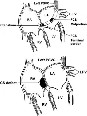
In three-quarters of cases in which the walls of the coronary sinus are fenestated, there is either persistence of the left superior caval vein in the setting of usual atrial arrangement, or persistence of the right superior caval vein when the atrial chambers are mirror-imaged. The usual incidence of persistent superior caval vein is 0.5% in the general population.Reference Adatia and Gittenberger-de Groot7, Reference Becker and Anderson9 Historically, several anatomic variations of fenestrated coronary sinus have been described, ranging from complete absence of the walls between the coronary sinus and left atrium (Fig. 1, lower panel) to one or more partial fenestrations, either in the midportion or terminal portion of the coronary sinus (Fig. 1, upper panel), with or without persistence of the superior caval vein.Reference Quaegebeur, Kirklin, Pacifico and Bargeron8 The so-called “pure form” (Fig. 1, lower panel), with total absence of the walls between the coronary sinus and left atrium, connection of the persistent superior caval vein to the upper ipsilateral corner of the left atrium, and a so-called coronary sinus defect is common in visceral heterotaxy when there is isomerism of the left atrial appendages.Reference Raghib, Ruttenberg, Anderson, Amplatz, Adams and Edwards1, Reference Quaegebeur, Kirklin, Pacifico and Bargeron8 This variant has previously been described in association with isomeric right atrial appendages, but this is a mistake, since the coronary sinus itself is always absent in this setting. Partial fenestrations, as shown in Figure 1, upper panel, are more difficult to diagnose, and cannot be recognized by symptoms and physical examination alone. They may, nonetheless, be of significant clinical importance.
Types of fenestrated coronary sinus. Upper panel, Partial fenestrations of the coronary sinus (FCS). Former “type III” is defined as a defect in the wall in the midportion of the coronary sinus (CS). Former “type IV” is defined as a defect in the terminal portion of the CS. Lower panel, Complete fenestration of the CS resulting in CS defect. These patients were not included in the present series. LA: left atrium; LPV: left pulmonary veins; LV: left ventricle; PSCV: persistent superior caval vein; RA: right atrium; RV: right ventricle. (Modified from Bertram H, Paul T, Kaulitz R, Luhmer I, Kallfelz H-C. Coronary sinus defects: rare form of interatrial communication [German]. Z Kardiol. 1996; 85: 899–905. Used with permission.)

The purpose of our study, therefore, was to analyze the symptoms, diagnostic approach, associated anomalies, and surgical procedures in patients with partially fenestrated coronary sinus, our goal being to enable earlier diagnosis of the condition, and to discuss the surgical options for its repair.
Methods
Patients
We retrospectively searched our institutional database for cardiac surgery to identify patients with anomalies of the coronary sinus who underwent cardiovascular surgery at Mayo Clinic Rochester between January 1958, and December 2003. We excluded patients with complete deficiency of the walls that normally separate the coronary sinus from the left atrium. We also excluded 14 patients lacking an orifice of the coronary sinus at the expected location in the morphologically right atrium, and with blood draining through multiple thebesian veins to the left- or right-sided atrial chambers, 5 patients with an atretic or stenotic orifice of the coronary sinus, and 1 patient with dual orifices for the coronary sinus. Our final cohort included 25 patients. Approval for the study was obtained from the Mayo Clinic Institutional Review Board.
Echocardiography
A complete cross-sectional and Doppler echocardiographic examination was performed on all patients according to the recommendations of the American Society of Echocardiography, including cross-sectionally guided M-mode measurements.Reference Schiller, Shah and Crawford10 All examinations were stored on videotape or acquired in digital format. Preoperative transthoracic echocardiography was performed at our institution in 22 of the 25 patients. The 3 patients who did not have echocardiography were evaluated in 1958, before echocardiography was available, in 1980, when echocardiography was performed elsewhere, and in 1990, when echocardiography was not performed because sedation was not feasible in this infant. Preoperative transoesophageal echocardiography was performed in 2 patients only. Intraoperative transoesophageal echocardiography has been routine since 1992, and was performed in 12 of 13 patients in this group who underwent surgery since then.
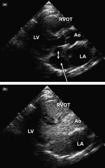
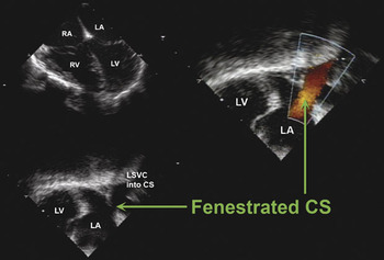
Saline was injected into the left arm whenever there was persistence of the superior caval vein and unroofing of the coronary sinus was suspected. Typical examples of transthoracic echocardiography with and without injections of contrast are shown in Figure 2. After injection into the left arm, the left-sided chambers fill immediately when the coronary sinus is fenestrated. Another typical example of fenestration is shown in Figure 3. Postoperative transthoracic echocardiography was performed in 22 of 24 early survivors before dismissal from the hospital.
Examples of fenestrated coronary sinus (FCS) visualized by transthoracic echocardiography. (a) Parasternal short-axis view of FCS viewed without saline contrast. The clearly dilated coronary sinus (long arrow) and the small communication between the coronary sinus and the left atrium (LA, small double-headed arrow) are shown. (b) Contrast-enhanced image (parasternal long-axis view) of FCS syndrome after saline injection into the patient’s left arm. After injection, contrast is seen immediately in the LA and the left ventricle (LV). Contrast is also seen in the right ventricular outflow tract (RVOT). Ao, aorta.

Transoesophageal echocardiography of a fenestrated coronary sinus (FCS) with a solitary opening in the terminal portion of the coronary sinus (CS) after previous surgery for ventricular septal defect in a 10-year-old boy with a cerebellar abscess due to paradoxical embolism in the presence of FCS. Upper left, The 4-chamber view shows only mildly enlarged right-sided cavities. Lower left, Large CS is shown due to the left superior caval vein (LSVC) draining into the CS. The arrow indicates the FCS. Right, The connection between the large CS and the left atrium (LA) via the FCS is visible with colour Doppler (arrow). LV: left ventricle; RA: right atrium; RV: right ventricle.

Surgical techniques
We used one of 8 methods for repair of the fenestrated coronary sinus.
For patients without persistence of the left superior caval vein, or without persistence of the right vein in cases of mirror-image atrial arrangement:
• Repair by suture or patch to direct the flow through the coronary sinus to the right atrium. This was performed in 2 patients. (Method 1 in Table 1)
• Closure of the orifice of the coronary sinus by suture or patch, leaving the flow through the sinus to drain into the left atrium through the fenestration. This was carried out in 7 patients. (Method 2 in Table 1)
• Placement of an intra-atrial patch or baffle to separate the left atrium from the right atrium and, in most patients, to divert all flow to the left atrium. This was performed in 6 patients, usually as part of the Fontan procedure or repair of atrioventricular septal defect. (Method 3 in Table 1)
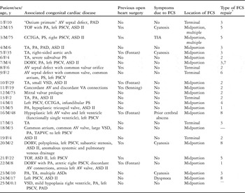
Clinical data.

ASD II: atrial septal defect within the oval fossa; AV: atrioventricular; CCTGA: congenitally corrected transposition; DORV: double outlet right ventricle; FCS: fenestrated coronary sinus; PA: pulmonary atresia; PAD: patent arterial duct; PS: pulmonary stenosis; PSCV: persistent superior caval vein; TA: tricuspid atresia; TAPVC: totally anomalous pulmonary venous connection; TOF: tetralogy of Fallot; VA: ventriculoarterial;VSD: ventricular septal defect.
*Procedures 1-8 in order as described in the Methods.
For patients with bilateral superior caval veins, in some cases we used procedures based on ligation of the persistent superior caval vein if safe, when the increase in pressure was less than 20 millimetres of mercury on temporary occlusion of the vein:
• Ligation of the persistent caval vein and repair of the fenestration by suture or patch in 1 patient. (Method 4 in Table 1)
• Ligation and closure of the orifice of the coronary sinus by suture or patch to leave flow to the left atrium in 3 patients. (Method 5 in Table 1)
• Ligation and placement of an intra-atrial patch or baffle as described above in 1 patient. (Method 6 in Table 1)
Alternatively, a procedure was used based on transfer of the persistent caval vein to the right atrium, the pulmonary artery, or the contralateral superior caval vein. This was achieved by:
• Transfer of the vein to the pulmonary artery on the same side by construction of a bidirectional cavopulmonary shunt in 1 patient, combined with placement of an intra-atrial baffle. (Method 7 in Table 1)
• Anatomic correction of the fenestration by suture or patch directing the flow through the sinus to the right atrium, performed in 3 patients. (Method 8 in Table 1)
In the 2 remaining patients, the fenestration was diagnosed by preoperative catheterization or echocardiography, but was not repaired. In one infant, we placed only a central shunt, whereas another child with additional complex congenital cardiac malformations underwent repair of totally anomalous pulmonary venous connection alone.
Statistical analysis
Descriptive statistics include frequencies and percentages for categorical data and median and range for continuous data.
Results
Clinical characteristics and associated congenital heart disease
Of the 25 patients, 19 (76%) had partial fenestrations in the midportion of the coronary sinus, and 6 (24%) in the terminal portion (Table 1). The latter variant was more common in patients with atrioventricular septal defects. In 2 patients, there were multiple fenestrations (Table 1, patients 2 and 3).
The age at surgery ranged from 0.1 to 75 years, with a median of 8 years. Symptoms thought to be at least partially attributable to the fenestration were present in 7 patients (28%), including cyanosis in 4 patients, and dyspnoea, cerebral abscess, and history of transient ischaemic attack in 1 patient each (Table 1). The other 17 patients had symptoms attributable to the associated severe congenital cardiac malformations.
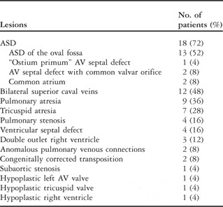
Such associated malformations were present in all 25 patients (Tables 1 and 2). The most common associated lesion was an atrial septal defect, present in 18 patients (72%). Persistence of the right or left superior caval vein was present in 12 patients (48%). In 2 patients, there was only a large atrial septal defect within the oval fossa, with no other associated intracardiac lesion apart from the fenestrated coronary sinus, 1 patient having a persistent left superior caval vein, and a third patient had prolapse of the mitral valve in the absence of a persistent superior caval vein. The other 22 patients had significant associated cardiac malformations. Hypoplasia of the right heart was found in 13 patients, in association with pulmonary atresia in 6, tricuspid atresia in 4, and both tricuspid and pulmonary atresia in 3. One patient had severe hypoplasia of the left atrioventricular valve. Double outlet right ventricle was found in 3 patients. Patient 20 (Table 1) had multiple spleens, but there was neither an echocardiographic or surgical description of the morphology of the atrial appendages in the medical record to exclude or diagnose left isomerism.
Associated cardiac lesions in the 25 patients with fenestrated coronary sinus.

ASD: atrial septal defect; AV: atrioventricular.
Previous cardiovascular surgery
The number of previous cardiovascular operations ranged from 0 to 4, with a median of 1, the procedures including construction of a Blalock-Taussig shunt in 9 patients (36%), creation of the Fontan circulation in 4 patients (16%), and surgery for closure of an atrial septal defect in 4 patients (16%). Other previous surgical procedures included closure of ventricular septal defects, repair of tetralogy of Fallot, pulmonary valvar stenosis, and totally anomalous pulmonary venous connection, construction of an aortopulmonary window, and placement of a conduit from the left ventricle to the pulmonary arteries, with more than 1 of these being performed in some patients. Among these previous cardiovascular interventions, 12 open intracardiac procedures had been performed in 11 patients.
Diagnosis
The diagnosis of fenestration of the coronary sinus was made before surgery in only 15 patients (60%). In 7 of 22 patients, the fenestration was first diagnosed echocardiographically, whereas in 8 of 23 patients who had preoperative catheterization, this technique revealed the fenestration. The diagnosis was first made intraoperatively in 10 patients (40%). Only 3 of the 15 patients (20%) in whom the diagnosis was made preoperatively underwent operation before 1990, and half of the 10 in whom diagnosis was made intraoperatively underwent operation before 1990. In 17 patients (68%), the fenestration had not been noted during previous cardiovascular surgery, with 11 of these patients undergoing open cardiac surgical procedures, 6 at our institution and 5 elsewhere. In 9 patients, the previous procedures had been extracardiac, with 2 shunts created at our institution and 7 elsewhere, some of these patients having more than 1 surgical procedure.
Surgical repair
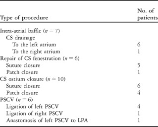
The 8 different methods of surgical repair used in the 23 patients are described in the methods, and are summarized in Tables 1 and 3. Ligation of a persistent left superior caval vein was performed in 4 patients, and of a persistent right superior caval vein in 1 patient with mirror-imaged arrangement of the heart and organs. Death occurred in 1 patient due to cardiac failure on the 18th postoperative day after repair of an atrioventricular septal defect with common valvar orifice, common atrium, and tetralogy of Fallot. No patient had a perioperative complication specifically related to repair of the fenestrated coronary sinus.
Methods of repair for fenestrated coronary sinus and persistent superior caval vein.

CS: coronary sinus; LPA: left pulmonary artery; PSCV: persistent superior caval vein.
Long-term follow-up
The median length of follow-up in the 24 surviving patients was 85 months, with a range from 1 to 238 months. No repeated procedures for repair of the fenestrations were necessary. Death occurred late in 1 patient, but it was unrelated to repair of the fenestration, being due to mediastinitis occurring after a third reoperation for complex congenital cardiac disease. Pulmonary venous obstruction, or stenosis of the coronary sinus, did not occur in any patient after operative repair.
Discussion
Our data show that fenestrations of the coronary sinus are often missed in the preoperative evaluation of congenital cardiac malformations, and also at the time of cardiac surgery. In those with congenital cardiac malformations, especially if these are tricuspid or pulmonary atresia, an atrial septal defect, or persistence of the superior caval vein, fenestration of the coronary sinus should always be considered as a possible additional finding. Such fenestrations can be responsible for clinically significant right-to-left shunting with cyanosis, formation of cerebral abscesses, and rarely dyspnoea. Thus, the diagnosis may be elusive. Once the diagnosis has been made, however, surgical repair is straightforward.
In assembling our series of patients, we excluded those with complete absence of the walls of the coronary sinus, including its orifice in the right atrium, and also those with atresia of the orifice. Fenestrations as defined in our patients were present in 7 of 11 patients in another recently assembled series.Reference Ootaki, Yamaguchi, Yoshimura, Oka, Yoshida and Hasegawa11 The authors of that study also considered the diagnosis of fenestrations in the midportion of the coronary sinus to be difficult, especially in children with complex malformations. They recommended that contrast material routinely be injected into the upper parts of the persistent superior caval vein when this vein was present.Reference Ootaki, Yamaguchi, Yoshimura, Oka, Yoshida and Hasegawa11 We can only further emphasize that, in many types of intracardiac operations, especially those requiring closure of atrial septal defects, the coronary sinus should be probed to exclude fenestrations of its wall. Complete absence of the walls is rarely missed, particularly since this is typically seen in the setting of isomerism of the right atrial appendages.
The coronary sinus can be fenestrated in the setting of tricuspid atresia, where it has been described in up to 3.6% of patients.Reference Matsuwaka, Tomokuni, Ishikawa, Watanabe, Matsushita and Matsuda12 Desaturation occurring after construction of the Fontan circulation by use of intracardiac baffles, or patches to direct systemic venous return to the pulmonary arteries, might be due to fenestrations of the coronary sinus even in the absence of a persistent superior caval vein. Fenestration in association with pulmonary atresia is rare,Reference Freedom, Culham and Rowe2 but a few cases have been reported with pulmonary atresia and an intact ventricular septum.Reference Freedom, Culham and Rowe2, Reference Rose, Beckman and Edwards13 Thus, it is important to exclude abnormalities of systemic venous return in patients being converted to the Fontan circulation.Reference Adatia and Gittenberger-de Groot7 Hypoplasia of the right heart, including tricuspid and pulmonary atresia, was frequent in our patients, suggesting a possible association.
Whenever a persistent superior caval vein is present, it is important to determine the patency of the orifice of the coronary sinus by cross-sectional echocardiographic imaging, and by measuring the direction of the flow in the persistent superior caval vein.Reference Adatia and Gittenberger-de Groot7 If the flow is upward, it indicates that the persistent superior caval vein is the only means of egress of blood from the coronary sinus, prohibiting ligation of this caval vein.Reference Adatia and Gittenberger-de Groot7 It is unwise to rely on dilation of the coronary sinus in the presence of a persistent superior caval vein as an indication of atresia of its orifice.Reference Adatia and Gittenberger-de Groot7
Symptoms at presentation
Haemodynamically, fenestrations of the walls of the coronary sinus may cause a left-to-right shunt and act like an atrial septal defect, but they may also be associated with an atrial septal defect within the oval fossa. It is often unclear, therefore, which symptoms are attributable to the fenestrations. The fenestrations can also cause right-to-left shunting with an increased risk of cyanosis, paradoxical embolus, or cerebral abscess. Failure of the right heart is rare, in contrast to the situation of complete absence of the wall of the coronary sinus. Cyanosis is especially common in patients with high right-sided pressures, especially after creation of the Fontan circulation. None of our patients had right heart failure due to the fenestrations, and haemodynamically relevant right-to-left shunting was rare. In over one-quarter of our patients, nonetheless, symptoms due to the fenestrations included dyspnoea, formation of cerebral abscesses, transient ischaemic attacks, and cyanosis.
Diagnostic techniques
In the past, fenestration of the coronary sinus was often only diagnosed at surgery. In the current era, since the initial description of the typical echocardiographic findings,Reference Chin and Murphy14, Reference Yeager, Chin and Sanders15 echocardiographic techniques have improved, and transoesophageal echocardiography is routinely available preoperatively or perioperatively.Reference Sunaga, Hayashi and Okubo16 As outlined in our results, although the fenestrations are being diagnosed more often preoperatively and intraoperatively than in the past, they may still be missed. The fenestrations are not readily diagnosed in life, despite advances in cardiac imaging, and a high index of suspicion is required.
In transthoracic cross-sectional echocardiography, the coronary sinus can easily be visualized and its size assessed. Transoesophageal echocardiography is even more helpful in diagnosis. If the examiners do not specifically look for the fenestrations, however, they can readily be missed. They are frequently missed in the setting of isomerism of the left atrial appendages due to the frequent concurrence of anomalies of pulmonary and systemic venous return. Of note, the coronary sinus is always absent in the setting of isomerism of the right atrial appendages.Reference Uemura, Ho and Anderson17 We recommend routine assessment of the roof of the coronary sinus by echocardiography or magnetic resonance imaging, therefore, in patients with visceral heterotaxy or persistence of the superior caval vein who are to undergo cardiac surgery, or who have unexplained intracardiac shunting in either direction. Injection of saline contrast into the arm on the same side of patients with persistent superior caval vein is especially helpful, and should be performed in all patients with this diagnosis. In many cases, this appears to be more sensitive in detecting the fenestrations than echocardiography or magnetic resonance imaging.
Intracardiac echocardiography has recently been used in the diagnosis of fenestration, using an 8F 10-MHz transducer to delineate the defect between the left atrium and the coronary sinus.Reference Chen, Hung, Chang, Lo, Chen and Fu18 Intracardiac echocardiography, however, is rarely used in this population of patients.
Rarely, fenestrations can be diagnosed using other techniques. In a patient with cyanosis, dyspnoea, and a systolic murmur, for example, echocardiography revealed complete absence of the walls of the coronary sinus, which was subsequently confirmed by multidetector row computed tomography.Reference Brancaccio, Miraldi, Ventriglia, Michielon, Di Donato and De Santis19 Magnetic resonance imaging is another noninvasive method that can provide excellent images of the fenestrated coronary sinus. During closure of atrial septal defects, surgeons can exclude fenestrations by probing the coronary sinus to exclude any connections with the left atrium.
Surgical repair
The risks associated with repair of the fenestrated walls of the coronary sinus are low, and can be considered comparable to those of repair of atrial septal defect within the oval fossa. In the absence of a persistent superior caval vein, the abnormal haemodynamics produced by the fenestration can be corrected either by closure of the fenestration itself by suture or patching, in which case the flow through the coronary sinus will be to the right atrium, or by closure of the orifice of the sinus itself, again either by suture or patch, in which case the return from the coronary sinus will be directed to the left atrium. In the latter setting, the resulting small right-to-left shunt is haemodynamically insignificant. In both instances, all defects within the atrial septum should be closed by suture or patch. For large interatrial communications via the mouth of the coronary sinus, closure using a patch is preferred in order to avoid damage to the atrioventricular node. Alternatively, for repair of complex anomalies in which baffles are used, as for creation of the Fontan circulation or repair of atrioventricular septal defects, the baffle can be placed in a manner that separates the left atrium from the right atrium and diverts flow through the coronary sinus to the left atrium.
The presence of a persistent superior caval vein adds an additional consideration to repair, and the abnormal haemodynamics can be corrected by several means, based on whether or not it is decided to ligate the persistent superior caval vein. If the option is to ligate, the surgical methods are the same as for the patients without an associated persistent superior caval vein. If, however, it is decided not to ligate the persistent superior caval vein, flow through the vein may be diverted to the right atrium, to the superior caval vein on the opposite side, or to the pulmonary artery on the same side, if technically feasible.Reference de Leval, Ritter, McGoon and Danielson20, Reference van Son, Hambsch and Mohr21 Alternatively, flow through the persistent superior caval vein may be directed to the right atrium by closure of the fenestration, or by diversion using an intra-atrial baffle.
Ideally, choice of a procedure that avoids ligation of a persistent superior caval vein is preferred in order to prevent transient venous hypertension in the head and the arm of that side. If required for technical reasons, or on the basis of safety or expediency, however, our experience shows that it is safe to ligate a persistent superior caval vein in patients undergoing biventricular repairs providing the persistent superior caval vein is the same size or smaller than the superior caval vein on the other side, and if, with temporary occlusion of the persistent superior caval vein, the pressure on the cranial side does not exceed 20 to 25 millimetres of mercury in patients with biventricular circulations.Reference de Leval, Ritter, McGoon and Danielson20
For the 5 patients in our series having a functionally single ventricle, failure to recognize the fenestrations became an issue in 4 late after a Fontan procedure, and in the other intraoperatively after discontinuation of cardiopulmonary bypass. These operations were modifications of the classic Fontan procedure in which intracardiac patches or baffles were used to direct systemic venous return through the right atrium to the pulmonary arteries. Current modifications that use a lateral tunnel or an extracardiac conduit do not require repair of fenestrations of the coronary sinus. If there is an associated persistent superior caval vein, it can be anastomosed to the pulmonary artery on the same side, as was done in 1 patient in our series.
We did not repair the fenestrations in 2 children in our cohort with complex congenital heart disease. In select patients, therefore, it may not be necessary to intervene.
Newer techniques
The Amplatzer device has now been used to occlude fenestrations,Reference Di Bernardo, Fasnacht and Berger22 and may be used in patients with an interatrial communication via the mouth of the coronary sinus, or in rare cases with simple fenestrations. The right atrial orifice of the coronary sinus, of course, functions as an interatrial communication when the fenestrations are significant. The orifice may then be closed with a device, but in most instances the defect is associated with more complex congenital heart disease, making nonsurgical closure less desirable, if not impossible.Reference Ferreira Martins and Anderson23 If closure with a device is an option for those having an atrial septal defect within the oval fossa, prior exclusion of fenestrated coronary sinus by transoesophageal or intracardiac echocardiography is essential.
Limitations
The cohort described in our retrospective study was seen at 1 institution over a period of more than 40 years, during which time there has been a tremendous evolution of echocardiographic and other imaging techniques. Thus, the real value of transthoracic, and especially transoesophageal, echocardiography is likely underestimated. The role of cardiac magnetic resonance imaging and computed tomography are not yet established.
Conclusions
Fenestration of the walls of the coronary sinus is a rare form of congenital heart disease. It occurs most commonly in conjunction with other complex congenital lesions, and is thus often missed during the preoperative evaluation. Prior cardiac surgery does not exclude its presence. It is very rare to find the lesion in isolation, but it may cause symptomatic right-to-left or left-to-right shunting, and should be considered in the differential diagnosis of patients with idiopathic stroke or evidence of volume overload of the right heart.
Comprehensive preoperative imaging, including intraoperative echocardiography, is recommended in all patients with suspected intracardiac shunts that are not readily apparent after initial evaluation. Although the fenestrations can be minor additional findings of no relevance, they can also lead to increased morbidity, especially due to right-to-left shunting. Once the diagnosis is made, repair is straightforward in experienced hands.
Acknowledgment
Editing, proofreading, and verification of the references were provided by the Section of Scientific Publications, Mayo Clinic.