Introduction
The Dyrosauridae represent a monophyletic group of crocodylomorphs that radiated in the marine environment during the Cretaceous. Their earliest record dates from the Late Cretaceous with mentions from the Campanian (Churcher and Russell, Reference Churcher and Russell1992; Lamanna et al., Reference Lamanna, Smith, Attia and Dodson2004; Kear et al., Reference Kear, Rich, Ali, Al-Mufarrih, Matiri, Masary and Attia2008; Salih et al., Reference Salih, Evans, Bussert, Klein, Nafi and Müller2015) or from undetermined Late Cretaceous levels (Sertich et al., Reference Sertich, Manthi, Sampson, Loewen and Getty2006) and perhaps from levels as old as the Cenomanian (Buffetaut and Lauverjat, Reference Buffetaut and Lauverjat1978). The group survived through the end Cretaceous biological crisis and became extinct during the early Eocene, most probably during the Ypresian (Buffetaut, Reference Buffetaut1978a, Reference Buffetaut1982; Hill et al., Reference Hill, Mccartney, Roberts, Bouaré, Sissoko and O'leary2008; Martin et al., Reference Martin, Amiot, Lécuyer and Benton2014). Accounts of dyrosaurid remains in the Lutetian (Pilgrim, Reference Pilgrim1940; Tessier, Reference Tessier1952; Buffetaut, Reference Buffetaut1978a, Reference Buffetautb) should be re-evaluated in view of recent stratigraphic studies (e.g., O'Leary et al., Reference O'Leary, Roberts, Bouare, Sissoko and Tapanila2006) before they can be validated or not. Although the fossil record of dyrosaurids is especially abundant with relatively complete specimens from the late Paleocene (Buffetaut, Reference Buffetaut1980; Jouve, Reference Jouve2007) and early Eocene (Bergounioux, Reference Bergounioux1956; Buffetaut, Reference Buffetaut1978a; Jouve, Reference Jouve2005), fewer representatives from the early Paleocene have been reported so far. They include genera such as Hyposaurus from the Danian of the USA (Troxell, Reference Troxell1925; Denton et al., Reference Denton, Dobie, Parris, Callaway and Nicholls1997), Atlantosuchus from the Danian of Morocco (Buffetaut, Reference Buffetaut1979; Jouve et al., Reference Jouve, Bouya and Amaghzaz2008), and Guarinisuchus from the Danian of Brazil (Barbosa et al., Reference Barbosa, Kellner and Viana2008). The genus Phosphatosaurus has been reported from Mali and Niger and may also be Danian in age according to Buffetaut (Reference Buffetaut1978a). Other fragmentary finds from Bolivia (Buffetaut, Reference Buffetaut1991), Brazil (Cope, Reference Cope1886), Pakistan (Buffetaut, Reference Buffetaut1977), and Senegal (Tessier, Reference Tessier1952) have been attributed a Danian or early Paleocene age. Current data indicate that dyrosaurid diversity was lower during the Late Cretaceous than during the Paleocene (6 genera versus 11, respectively). On the other hand, the diversity of dyrosaurids during the Cretaceous may parallel that of the Paleocene, depending on phylogenetic resolution within Dyrosauridae (Hill et al., Reference Hill, Mccartney, Roberts, Bouaré, Sissoko and O'leary2008). Some authors have proposed that this could be explained by a shift with the extinction of Cretaceous predators such as mosasaurs freeing space for dyrosaurids (Barbosa et al., Reference Barbosa, Kellner and Viana2008; Jouve et al., Reference Jouve, Bouya and Amaghzaz2008). Nevertheless, such diversity counts merge the dyrosaurid fossil record of the entire Paleocene, and dyrosaurid diversity immediately after the K/Pg crisis is poorly known and difficult to evaluate. Records of large vertebrate predators immediately after the K/Pg boundary are equally important as those from the Maastrichtian because they may provide insights into the reorganization of marine food webs following a major biotic disruption.
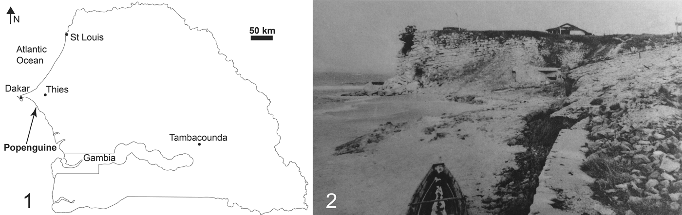
Here, we describe a dyrosaurid specimen collected in the 1930s by two French geologists, Fernand Jacquet and Maurice Nicklès, who both worked for the Service Géologique de l'Afrique Occidentale Française (AOF) in Dakar. Their survey of Paleogene outcrops along the Atlantic coast of Senegal led to the discovery, near the village of Poponguine (Fig. 1), of the presently described specimen, which has been briefly mentioned in several notes (Malavoy, Reference Malavoy1934, p.7, 1935, p. 7; Jacquet, Reference Jacquet1936). The specimen consists of articulated postcranial elements belonging to a single individual, the bones of which were deposited in a low-energy marine environment of Danian age according to associated microfossils.
Location of the dyrosaurid specimen presented in this work. (1) Map of Senegal indicating the locality of Poponguine; (2) the Danian succession at Poponguine where the specimen was discovered as photographed by G. Labitte in the 1920s from Tessier (Reference Tessier1952).

History of the discovery
Before we borrowed the specimen for preparation and study, the following inscription was visible on the surface of the sediment: “Popenguine—Sénégal, Falaise du Bungalow” (Fig. 3). “La Falaise du Bungalow” is a small seaside cliff subjected to wave erosion and made of an alternating sequence of marls and limestones (see geological section). Nowadays, the Presidential residence is sitting on top of it, having replaced the bungalow of the “Gouverneur général de l'Afrique Occidentale Française.”
A note from the head of the Service géologique de l'AOF indicates 1934 as the year of the discovery of the Poponguine specimen, originally identified as a fossil mammal with eight vertebrae, three elongate bones, and six ribs (Malavoy, Reference Malavoy1934). The following year, the same specimen was identified as a crocodile from Poponguine (Malavoy, Reference Malavoy1935). Jacquet (Reference Jacquet1936), reporting on some debris of a “Crocodilien marin”, provided a brief context for the specimen, indicating a provenance from alternating levels of marls and limestones from Poponguine. He attributed the deposits to be early Eocene in age on the basis of their invertebrate content, although more recent studies considered the deposits to be Danian in age (see geological section). Following the tragic loss in 1937 of F. Jacquet during a field survey in Mauritania, M. Nicklès was put in charge of handling the works of his colleague (Legoux, Reference Legoux1937). A few years later, Arambourg and Joleaud (Reference Arambourg and Joleaud1943, p. 63) mentioned that M. Jacquet recovered vertebrae and limb bones belonging to Dyrosaurus in the area of Poponguine. Tessier (Reference Tessier1952) mentioned that this material had been observed by C. Arambourg, citing “1943 Dyrosaurus sp. Arambourg (6), p. 63” and referring to the collection Jacquet of the MNHN (Muséum National d'Histoire Naturelle, Paris). Therefore, the specimen must have been part of the MNHN collections at least until 1952, but it is unclear when the specimen was transferred from Dakar to Paris. From this date, the whereabouts of the specimen became unknown and no mention of it is made in the literature. The specimen re-surfaces much later in Toulouse. The dyrosaurid specimen was stored in the collections of the Institut Catholique de Toulouse from an unknown date until the mid 1990s, when it was donated to the Toulouse Museum (MHNT) on 9 September 1996 (Y. Laurent, personal communication, 2018). Understanding how the specimen transited from MNHN Paris to Toulouse is unclear at this stage, and we can only speculate on potential links between Camille Arambourg (1885–1969), then Professor at MNHN who made the observations on the specimen (see above), and Frédéric-Marie Bergounioux (1900–1983), paleontologist and theologist at the Institut Catholique de Toulouse. Both researchers knew each other, with C. Arambourg having sent to F.-M. Bergounioux several fossil turtle specimens for study (Bergounioux, Reference Bergounioux and Arambourg1952). It is therefore possible that the dyrosaurid specimen arrived at the collections of Toulouse in this way. However, no mention of the Poponguine specimen is made in Bergounioux's monograph, which extensively describes dyrosaurid material from Tunisia (Bergounioux, Reference Bergounioux1956).
Geological setting
Paleocene strata crop out on a narrow littoral stretch between Toubab Dialaw and the southern end of the horst of Diass (Fig. 1). Most of the outcrops are represented by alternating marls and limestones, which comprise the Ndayane Formation. This formation is overlain by discording shelly limestones of the Poponguine Formation, which crop out East of Poponguine. The Ndayane Formation is visible in a cliff, the altitude of which is <10 m above sea level. The Ndayane Formation uncomformably overlies the Maastrichtian gritty limestone containing ammonites with a conglomeratic level (Castelain et al., Reference Castelain, Jardiné and Monciardini1965). A lithological section along the stretch of coastline shows the following units totaling 27 m in thickness.
From the base, gray argillaceous limestone becomes replaced by lenticular limestone (2.5 m thick) with gypsum veins. Most of the cliff (17.5 m thick) is made of limestone beds alternating with yellowish marly limestones with gypsum veins. The limestone beds become thicker in the median section of the cliff and the marly limestone beds become lenticular. Near the summit, limestone beds become thinner whereas marly limestone beds become thicker. Marls containing calcite rosettes show at their base thin lenticular levels of marly limestones (7 m thick).
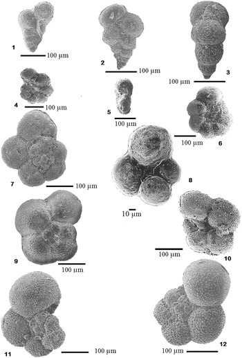
The microfauna of the Ndayane Formation is rich and diverse and includes planktonic and benthic foraminifers as well as ostracodes. These microfaunas have been studied by several authors (Castelain et al., Reference Castelain, Jardiné and Monciardini1965; Faye, Reference Faye1983; Sarr, Reference Sarr1995, Reference Sarr1998; Sarr and Ly, Reference Sarr and Ly1998) who established a middle to upper Danian age for the formation on the basis of the following planktonic foraminifer species, also identified here in sediment associated with MHNT.PAL.2012.0.45: Chiloguembelina morsei (Kline, Reference Kline1943), Morozovella inconstans (Subbotina, Reference Subbotina1953), M. pseudobulloides (Plummer, Reference Plummer1926), M. cf. trinidadensis (Bolli, Reference Bolli1957), and Planorotalites compressa (Plummer, Reference Plummer1926) (Fig. 2). This age is in agreement with that established by Tessier (Reference Tessier1952) on the basis of macrofaunas. The depositional environment corresponds to a middle to external platform with a water depth between 100 m and 150 m, with a muddy bottom covered with algae, receiving episodically coarse material from the infralittoral environment (Sarr, Reference Sarr1998).
Planktonic foraminifers recovered from the sediment of MHNT.PAL.2012.0.45, except in (2), which is from Toubab Dialaw. Chiloguembelina morsei (Kline, Reference Kline1943) in (1) lateral view (PV4); (2) lateral view for comparison (TD90/4); (3) Apertural view (P88/34). Morozovella cf. trinidadensis (Bolli, Reference Bolli1957) in (4) ombilical (P88/23), (5) apertural (P88/23), and (6) spiral (P88/33) views. Planorotalites compressa (Plummer, Reference Plummer1926) in (7) spiral (P88/14) and (8) ombilical (P88/14) views. Morozovella inconstans (Subbotina, Reference Subbotina1953) in (9) spiral (P88/38) and (10) ombilical (P88/38) views. Morozovella pseudobulloides (Plummer, Reference Plummer1926) in (11) spiral (P88/10) and (12) spiral (P88/40) views.

Systematic paleontology
Crocodylomorpha Hay, Reference Hay1930 Dyrosauridae De Stefano, Reference De Stefano1903 Dyrosauridae indet. Figures 3–10
Description
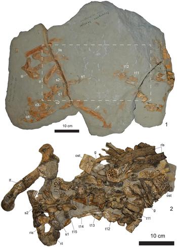
The specimen was preserved as three blocks (Fig. 3.1), which underwent acid preparation (Fig. 3.2). The largest block was prepared so that most elements of the specimen preserve their articulation. Several elements were totally freed from the matrix and could be described individually. The main block preserves the last five thoracal vertebrae (#11 to #15); thoracal vertebra #10 has been freed from the matrix as well as two other thoracal vertebrae (one being heavily weathered and the other only consisting of the centrum), thus totaling eight thoracic vertebrae. The two sacral vertebrae are preserved on the main block. The second sacral vertebra is split in half and is loose from the main block (Fig. 4). Four caudal vertebrae are also available. The right ischium is firmly connected to a fragmentary ilium (Fig. 5). The right femur (Fig. 6) is preserved and is slightly displaced from the pelvic area. The articulated vertebrae are positioned within a mix of ribs, gastralia, and displaced dorsal and ventral osteoderms.
Main block containing the partially articulated dyrosaurid skeleton MHNT.PAL.2012.0.45 from the Danian of Poponguine, Senegal, (1) before as indicated with the stipple area and (2) following preparation. Abbreviations: g = gastrolith; lf = left femur; lil = left ilium; lis = left ischium; ost = osteoderm; rf = right femur; ril = right ilium; ris = right ischium; rls = rib-like structures; c1–c4 = caudal vertebrae; s1–s2 = sacral vertebrae; t11–t15 = thoracic vertebrae.

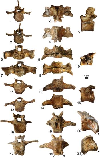
Selected vertebrae of MHNT.PAL.2012.0.45 from the Danian of Poponguine, Senegal. Thoracic vertebra #10 in (1) anterior; (2) posterior; (3) dorsal; (4) ventral and (5) left lateral views; sacral vertebra #2 in (6) anterior; (7) posterior; (8) dorsal; (9) ventral and (10) right lateral views; caudal vertebra #1 in (11) anterior; (12) posterior; (13) dorsal; (14) ventral and (15) left lateral views; caudal vertebra #2 in (16) anterior; (17) posterior; (18) dorsal; (19) ventral and (20) left lateral views; caudal vertebra #3 in (21) right lateral view. The black arrows indicate the position of haemal facets; the white arrow indicates the erosive lesion.

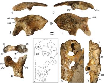
Elements of the sacrum of MHNT.PAL.2012.0.45 from the Danian of Poponguine, Senegal. Left ilium in (1) dorsal; (2) ventral; (3) lateral; (4) medial views. Left ischium in (5) dorsal and (6) lateral views. Right ischium and fragmentary right ilium in lateral view with (7) line drawing and (8) associated photograph; (9) posterior view. Abbreviations: af = acetabular foramen; afi = anterior facet for ilium; afis = anterior facet for ischium; afp = anterior facet for pubis; aip = anterior iliac process; ap = anterior process; fe = femur; ff = facet for femur; ib = ischiac blade; il = ilium; ost = osteoderm; pfi = posterior facet for ilium; pfis = posterior facet for ischium; pip = posterior iliac process; pp = posterior process; ps = posterior scar; sac = supraacetabular crest; s1 = sacral vertebra 1.

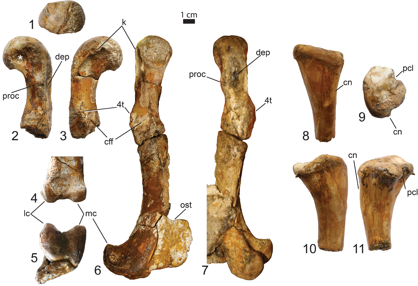
The hind limb of MHNT.PAL.2012.0.45 from the Danian of Poponguine, Senegal. Left femur in (1) proximal view; (2) dorsal view of the proximal third portion; (3) ventral view of the proximal third portion; (4) ventral view of the distal end; (5) distal view; (6) medial and (7) lateral views. Left tibia in (8) lateral; (9) proximal; (10) anterior and (11) posterior views. Abbreviations: cff = caudofemoralis flange; cn = cnemial crest; dep = depressed zone; k = knob; lc = lateral condyle; mc = medial condyle; ost = osteoderm; pcl = posterior cleft; proc = process; 4t = fourth trochanter.

Materials
MHNT.PAL.2012.0.45, postcranial elements of a single specimen from the abdominal and sacral region.
Thoracal vertebrae
The anteriormost thoracal vertebra is complete (Fig. 4.1–4.5) and corresponds to thoracal vertebra #10. In anterior or posterior views, the amphicoelous centrum has a subquadrangular outline, being nearly higher than wide. The posterior margin of the centrum displays a pathological area near its anterolateral corner, which may correspond to an erosive lesion (arrow in Fig. 4.2). The centrum is slightly elongated, being longer than high. In lateral view, the ventral margin of the centrum is faintly concave. The main corpus of the centrum is slightly constricted laterally with thick anterior and posterior margins that define the limits of the anterior and posterior surfaces. The neurocentral suture is visible and runs transversally along the lateral margin of the centrum, in its dorsal half. A distinct and shallow circular pleurocoele spreads over the centrum and neural arch, below the posterior margin of the transverse process. The neural arch is robustly attached to the centrum. The neural spine is relatively short, being nearly as high as the centrum. Its dorsal margin is straight and represents half the length of the centrum. The dorsal surface of the neural spine is vascularized. In anterior and posterior views, the neural arch shows an extensive medial sulcus. The neural canal is heart-shaped in cross-section with its dorsal margin receiving a ventrally projecting lamina at the level of the neural spine. The anterior extent of the prezygapophyses is almost in line with the anterior margin of the centrum, whereas the posterior extent of the postzygapophyses project far beyond the posterior margin of the centrum. The articular faces on the pre- and postzygapophyses are obliquely oriented with respect to the neural spine. The laminar transverse processes are short and comparable in dimensions to the centrum width, but their anteroposterior length is slightly shorter than the length of the centrum. In dorsal view, the distal margin of the transverse processes is unequal with a parapophysis shorter than the diapophysis.
Sacrum
The anterior part of the first sacral vertebra is connected to the rest of the vertebral column and is partly embedded in the sediment. The second sacral vertebra detached during preparation, revealing a strong sutural surface. Much like Hyposaurus (Schwarz et al., Reference Schwarz, Frey and Martin2006), the two sacral vertebrae appear to have not fused together, whereas these bones are fused in Dyrosaurus sp. (Schwarz et al., Reference Schwarz, Frey and Martin2006). The anterior and posterior surfaces of the sacral centra are ovoid in outline, being wider than tall. The sutural surface of the anterior margin of the second sacral vertebra is complex and presents two parallel sulci along its midline (Fig. 4.6). Such similar sulci have previously been reported on the posterior, but not anterior, surface of the second sacral vertebra of D. phosphaticus and of Hyposaurus (YPM 753) by Schwarz et al. (Reference Schwarz, Frey and Martin2006). The second sacral vertebra can be observed in ventral aspect and no obvious shallow sulcus is observed here close to its anterior margin, contrary to the condition in Dyrosaurus sp. and cf. Rhabdognathus (Schwarz et al., Reference Schwarz, Frey and Martin2006). As observed in dorsal view, the first and second sacral ribs are hourglass shaped. The sacral ribs of the first sacral vertebra are about twice as long as the ribs of the second sacral vertebra (Figs. 3, 4.8), as is commonly observed among crocodylians. The first sacral rib occupies the anterior two thirds of the lateral surface of the centrum length, whereas the second sacral rib occupies the entire length of the centrum. On the first sacral vertebra, as observed in posterior view, a distinct circular fossa is visible at the sutural intersection of the neural arch, centrum, and sacral rib. As in Dyrosaurus sp., the proximal insertion of the sacral rib for the second sacral vertebra is restricted to the corpus of the second sacral centrum (Schwarz et al., Reference Schwarz, Frey and Martin2006) and unlike the condition in Rhabdognathus, where the sacral rib overlaps on the centrum of the first caudal vertebra (Langston, Reference Langston, Whitmore and Madden1995). The iliac symphyseal surface of the first rib does not exceed the anterior and posterior margins of the centrum. An anterolateral process is present as in Dyrosaurus sp. and cf. Rhabdognathus (Schwarz et al., Reference Schwarz, Frey and Martin2006). In lateral view, the anterolateral process is thin and shows a concave articular surface; the posterior articular process of the rib is located more medially than the anterolateral process and is nearly as tall as the centrum height, being concave. The iliac symphyseal surface of the second sacral rib is slightly smaller and symmetrical to the first sacral rib, with a tall anterior surface and a thin posterior process. This iliac symphyseal surface exceeds slightly the posterior margin of the second sacral vertebra. The neural spines have weathered out in the described specimen.
Caudal vertebrae
At least three caudal vertebrae were prepared out of the second block. Schwarz et al. (Reference Schwarz, Frey and Martin2006) described shallow bowl-shaped fossae on the anterior and posterior surfaces of the centrum of the caudal vertebrae. Here, such fossae are also observed on both thoracal and caudal vertebrae. The first caudal vertebra possesses no facets for the haemal arches (Fig. 4.14, 4.15). The second caudal vertebra possesses a facet for the haemal arches only on the posteroventral margin of its centrum (Fig. 4.19, 4.20). The third caudal vertebra possesses articular facets for the haemal arches both on the anteroventral and posteroventral margins of its centrum (Fig. 4.21). This pattern of articulation between caudal centra and haemal arches differs from that described in Dyrosaurus sp. and cf. Rhabdognathus, where the facet is present on the anteroventral surface from the second vertebra (Langston, Reference Langston, Whitmore and Madden1995; Schwarz et al., Reference Schwarz, Frey and Martin2006). It is, however, similar to Congosaurus bequaerti Dollo, Reference Dollo1914 where this pattern of articulation starts from the third caudal vertebra (Schwarz et al., Reference Schwarz, Frey and Martin2006). The first caudal vertebra has proportions of the centrum similar to the sacral vertebrae; from the second caudal vertebra, the centrum is longer than high with a quadrangular outline of the anterior and posterior margins; the posteroventral margin is also distinctly lowered than the anteroventral margin (compare Fig. 4.15, 4.20, 4.21).
Ischium
The nearly complete right ischium is preserved in connection with the ilium in the main block (Figs. 3, 5.7–5.9). The left ischium was isolated during preparation; it is eroded on its medial surface and is missing its distal blade (Fig. 5.5, 5.6). The ischium measures ~100 mm in its maximum length, the blade representing ~70% of this length. The anterior and posterior margins of the blade are parallel and do not seem to expand significantly near the distal end of the blade, but this area is damaged. The medial surface of the right element is smooth and gently concave. The broken blade of the left element shows that the anterior margin is thinner than the posterior margin.
The proximal portion of the ischium shows the distinct acetabular foramen, which is bordered anteriorly by the forward-projecting anterior iliac process and posteriorly by the posterior iliac process. The anterior margin of the anterior iliac process is vertical and bears in its lower half an anteriorly oriented pubic process. The pubic process has a short spiny ventral projection, resulting in a markedly concave margin. The facet for accommodating the ilium is flat and obliquely oriented. On the posterior iliac process, the dorsal facet for the ilium is concave, pierced by foramina and bordered by a thin bony rim; its lateral portion is shorter than the medial portion, matching the triangular outline of the connecting ilium. Anterior to this facet, there is a flat surface that descends within the acetabulum and corresponds to the articular area for the femoral head (in lateral view, this gives a truncated outline to the posterior iliac process). The anterior region of the acetabulum, at the level of the anterior iliac process, is anterolaterally depressed. In the same area, but near the medial border, the margin is faintly crested. As seen in posterior view, the posterior iliac process possesses a short but distinct muscle scar near its proximal end (Fig. 5.9)
Ilium
The right ilium measures 67 mm in its maximum height, is connected to the ischium in the main block (Fig. 3), but is heavily damaged. The left ilium is complete, although its internal surface is slightly eroded (Fig. 5.1–5.4). The anterior process of the ilium is massive and projects dorsally whereas it projects anteriorly in Congosaurus. Just below it, the supraacetabular crest is well defined as a thin ridge delimiting the anterodorsal outer margin of the acetabulum. The anterior peduncle of the ilium is mediolaterally thin with the ischial facet, although eroded, facing ventromedially. The posterior peduncle is massive, D-shaped in ventral view, and the ischial facet is here facing fully ventrally. The acetabulum occupies about one third of the lateral surface of the bone and nearly reaches the dorsal surface. The acetabular foramen is large and occupies more space than in Congosaurus. The posterior blade of the ilium is concave on its lateral surface, and in dorsal view, the ilium is sigmoidal. Except near the anterior spine, the dorsal margin of the blade is widely convex (not wasp-waisted), and is indented for muscle attachment. The posteroventral margin of the blade bears strong indentations, especially on the medial surface. The dorsal and ventral margins of the blade merge into a short peduncle projecting posteriorly. The ventral margin between the posterior peduncle and the strong indented area is smooth and concave on the lateral side.
Femur
The right femur preserves much of the femoral head and a proximal portion of the shaft. The left femur is complete and partly connected to the block (Fig. 6). The femur has a general sigmoid curvature with a strongly medially bent femoral head, a thick shaft, is ovoid in section, and terminated distally with a pair of massive and ventrally directed condyles. In proximal view, the femoral head is ovoid being wider than high. On its dorsal surface, an anomalous depression filled with sediment is visible (star in Fig. 6.2). The medial-most outline of the femoral head is distinctly set apart from the main shaft, as originally observed in Hyposaurus (Troxell, Reference Troxell1925). The ventral margin close to the femoral head possesses a small but distinct knob, which sits on the proximal part of an acute ridge that marks the ventrolateral margin of the femoral head. This ridge merges with the lateral margin of the shaft, which is straight and also acute until the level of the fourth trochanter. Dorsal to this ridge, the lateral surface of the femur bears a large depressed zone (Fig. 6.2, 6.7), but is also heavily weathered. At this level, a process is visible on the dorsal surface (Fig. 6.2, 6.7). The fourth trochanter is prominent and is projecting ventrally. Dorsal to it, a shallow ovoid surface corresponds to the caudofemoral flange, which faces fully medially. From this level, and as seen in medial or lateral view, the shaft gently bends ventrally with two massive condyles that project strongly ventrally toward the distal extremity of the shaft. The distal portion of the shaft is largely hidden by an osteoderm. In ventral view, a wide intercondylar fossa is present (Fig. 6.4), which separates a narrow medial condyle from a more massive lateral condyle (Fig. 6.5). The medial surface of the medial condyle is flat and the condyle expands as a narrow outgrowth dorsally, giving to the dorsal surface of the femur an uneven outline. The lateral condyle is sturdy and possesses a slight projection on its convex lateral margin.
Tibia
The proximal epiphysis of a left tibia is preserved and is bent from the shaft in a posteromedial direction (Fig. 6). The proximal end is 45 mm mediolaterally by 41 mm anteroposteriorly, and the oval shaft, as preserved, is much thinner (20 mm mediolaterally by 18 mm anteroposteriorly). It strongly resembles another indeterminate dyrosaurid tibia described from Pakistan (Storrs, Reference Storrs1986, fig. 9). Its proximal articular surface is deeply depressed in the middle as in Congosaurus bequaerti (Schwarz et al., Reference Schwarz, Frey and Martin2006). This surface possesses two articular facets that are continuous on the medial and posterior margins. Both facets are separated by a marked posterior cleft (Fig. 6.9, 6.11). The medial articular facet is about twice as large and markedly concave in comparison to the posterior one. The anterior margin of the articular surface is straight and does not show an articular facet. In anterior view, the medial facet is angled relative to the rest of the proximal margin of the tibia, giving to the shaft a strongly concave medial margin. Conversely, the lateral margin is straight and nearly perpendicular with the articular surface (Fig. 6.10). Here, the cnemial crest is not particularly standing out, being distinct only in its most proximal portion where a step-like process accommodates the proximal margin of the fibula (Fig. 6.9, 6.11). A 9 mm long ovoid depression is observed on the anterolateral corner of the tibia (Fig. 6.8), as previously observed in the tibia of Guarinisuchus munizi Barbosa, Kellner, and Viana, Reference Barbosa, Kellner and Viana2008 (Sena et al., Reference Sena, de Andrade, Bantim, Sayão, Barbosa and de Oliveira2017, fig. 3M).
Calcaneum
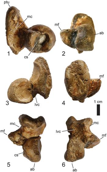
A complete left calcaneum is preserved (Fig. 7). The anterior ball is demarcated from the posterior tuber by an extensive medial channel that runs along most of the medial surface of the posterior tuber, just posterior to the medial flange. As observed in medial view, the calcaneal socket, which accommodates the astragalar peg, is deep. Posterior to it, the anterior face of the medial flange is vast and has a gently concave margin that may have limited rotation of the astragalar peg. The anterior ball is convex in its anterior and dorsal parts, and flat in its plantar margin, as in extant crocodylians. From a lateral view, the anterior ball shows a vertical wall. Ventrally, it is well demarcated from the posterior tuber by a deep lateroventral channel. The posterior tuber shows on its posterior margin a shallow and rugose groove.
The left calcaneum of MHNT.PAL.2012.0.45 from the Danian of Poponguine, Senegal in (1) medial; (2) anterior; (3) lateral; (4) posterior; (5) dorsal; and (6) ventral views. Abbreviations: ab = anterior ball; cs = calcaneal socket; lvc = lateroventral channel; mc = medial channel; mf = medial flange; ptu = posterior tuber.

Radiale
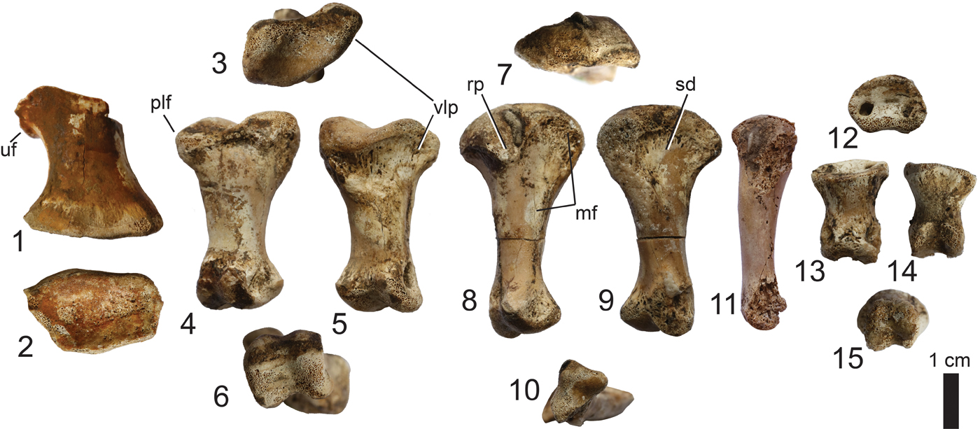
The right radiale is preserved but is missing most of its proximal portion (Fig. 8.1, 8.2) and is heavily eroded on its posterior surface. A portion of the facet for the ulnare is preserved on the lateral margin and distinctly projects laterally from the proximal portion of the bone. The shaft is wasp-waisted. The distal articular surface is concave.
Elements of the manus of MHNT.PAL.2012.0.45 from the Danian of Poponguine, Senegal. Right radiale in (1), anterior and (2), distal views. Right metacarpal I in (3) proximal; (4) dorsal; (5) palmar; and (6) distal views. Right metacarpal II or III in (7) proximal; (8) dorsal; (9) palmar; and (10) distal views. (11) Metacarpal V in lateral or medial view. Phalanx in (12) proximal; (13) dorsal; (14) palmar; and (15) distal views. Abbreviations: mf = medial facet; plf = proximolateral flange; rp = rugose projection; sd = shallow depression; uf = ulnare facet of radiale; vlp = ventrolateral projection. For proximal and distal views, the dorsal aspect is up.

Metacarpals
Three metacarpals are preserved. The right metacarpal I (Fig. 8.3–8.6) is 35 mm long and possesses a straight shaft with proximal and distal portions being nearly as wide and in line with each other. On the proximoventral edge of the bone, a large and wider than long depression is observed on a lateral projection lateral (Fig. 8.3, 8.5) to the main shaft. The proximal articular surface is marked by a distinct dorsoventral sulcus (Fig. 8.3). The distal surface bears two condyles, each possessing deep pits on their medial and lateral surfaces. The right (Fig. 8.7, 8.10) and left metacarpals II or III are preserved. Each bone is 41 mm long with a rod-like shaft and a flared and convex proximal margin. In dorsal view, the proximal epiphysis is rugose and projects briefly along the lateral half of the shaft (rp in Fig. 8.8). Immediately medial to it, an extensive flat facet is inclined toward the medial margin (Fig. 8.8). In palmar view (Fig. 8.9), a wide shallow depression is delimited by a proximomedial bony ridge. The shaft has a rotated axis with the distal condyles ~90° from the proximal portion (Fig. 8.10), as observed in Congosaurus and Hyposaurus (Schwarz et al., Reference Schwarz, Frey and Martin2006). The distal condyles are well individualized and possess marked pits on their medial and lateral edges. The right or left metacarpal V (Fig. 8.11) is partly weathered at both extremities, has a rod-like shaft, and is 38 mm long.
Phalanx
A single carpal phalanx is preserved (Fig. 8.12–8.15). It is 18 mm long, wasp-waisted with a proximal portion nearly as wide as the distal portion. In palmar view, the distal condyles are well demarcated by a marked intercondylar groove.
Osteoderms
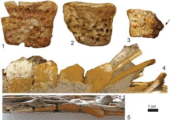
Several osteoderms are associated with the skeleton (Figs. 3, 9). They are all displaced and therefore no clear configuration of the dorsal or ventral shield can be deduced. All dorsal osteoderms lack an external keel (Figs. 3, 9.1, 9.2), a condition identical to that of various hyposaurine dyrosaurids (Schwarz et al., Reference Schwarz, Frey and Martin2006). The dorsal osteoderms attributed to the paravertebral shield are square to trapezoidal in outline being slightly wider than long. Their anterior and posterior margins are slightly vaulted and thin in comparison to the thick median body. As observed in dorsal view, the anterior margin is concave in its median portion and its dorsal surface shows a smooth facet covering the entire width of the osteoderm. A short anterolateral process emerges from this smooth facet. Just posterior to the anterior facet, a row of nearly circular and well-aligned cupules is present. This row is distinct from the rest of the dorsal surface, which is slightly raised and ornamented with slightly larger and randomly distributed cupules. The posterior margin is gently convex. The median and lateral margins are nearly straight. As for the lateral margin, some incipient indentations are sometimes visible and may have contacted a more lateral osteoderm. The ventral surface is perfectly smooth and may exhibit a few foramina.
Isolated ventral osteoderms were also identified and present a wide V-shaped margin and a peg on the opposite margin (Fig. 9.3). Part of the ventral shield is still articulated, but sediment hides much of the organization. A suite of four articulated osteoderms is visible on one side of the specimen and is positioned ventral to the gastralia (Fig. 9.4, 9.5). Ventral osteoderms are smaller than dorsal osteoderms and are distinctly wider than long. Their margins possess interdigitations for suturing to other osteoderms. The medial margin is strongly concave, whereas the lateral margin possesses a pointed process that strongly projects laterally (Fig. 9.3). The external surface is ornamented with a few barely visible shallow circular pits, which do not spread near the margins of the osteoderm. Those ventral osteoderms are gently vaulted, being nearly flat.
Ribs, gastralia, and other structures
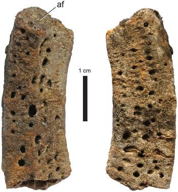
Several elongated elements are preserved in the main block and are intertwined with the locally disarticulated skeletal parts. Their encasing in the sediment and the hiding of articular portions render their identification tentative. Thin, straight rod-like structures with a perfectly circular cross section spread to the left of the thoracic region from the eleventh to thirteenth thoracal vertebrae (Fig. 3). They are <5 mm in diameter. Thicker elements, ~10 mm in diameter, correspond to ribs that are also scattered in this area and offer sigmoid outlines. Another type of morphology is represented by two nearly complete structures observed in this same area (Fig. 3) as well as by some fragments recovered during acid preparation (Fig. 10). These are wide, flat on one side, convex on the other, and gently curved rib-like structures with an articular facet on one end. Remarkably, they display numerous ovoid pits on both sides that are 1–3 mm in diameter. These pits are similar to those observed on the external surface of osteoderms.
The osteoderms of MHNT.PAL.2012.0.45 from the Danian of Poponguine, Senegal. (1, 2) examples of dorsal elements and of a (3) ventral element with an arrow indicating the lateral expansion. Series of four osteoderms from the ventral shield preserving their original organization in (4) ventral and (5) lateral views.

Rib-like element of MHNT.PAL.2012.0.45 from the Danian of Poponguine, Senegal presenting numerous ovoid pits on its surface. Abbreviation: af = articular facet.

Gastroliths
At least five pebbles (1–3 cm size) are scattered adjacent to the thirteenth thoracal vertebra (Fig. 3). Another one is observed near the eleventh vertebra. Another one was freed from the block. The nature of these stones is pure quartz and therefore different than the embedding carbonate matrix. The pebbles are not perfectly rounded and possess smooth angular edges.
Discussion
Taxonomic identification and comparison
The material was initially identified as a suite of vertebrae and limb bones belonging to the genus Dyrosaurus in Arambourg and Joleaud (Reference Arambourg and Joleaud1943). The unprepared block permitted observations of weathered bone surface only (Fig. 3), and although no details are given in Arambourg and Joleaud (Reference Arambourg and Joleaud1943), some osteoderms were obviously identifiable. Ample descriptions of dyrosaurids have been available as early as the first half of the twentieth century, for example, Dyrosaurus (Thévenin (Reference Thévenin1911a, Reference Théveninb), Hyposaurus (Troxell, Reference Troxell1925) or Congosaurus (Swinton, Reference Swinton1950). As noted by Brochu et al. (Reference Brochu, Mamadou, Sissoko, Roberts and O'Leary2002), dyrosaurid taxonomy has been largely established on characters of the rostrum and lower jaw. Most recently erected dyrosaurid species have also been established on the basis of complete skulls (Barbosa et al., Reference Barbosa, Kellner and Viana2008; Hastings et al., Reference Hastings, Bloch, Cadena and Jaramillo2010, Reference Hastings, Bloch and Jaramillo2011, Reference Hastings, Bloch and Jaramillo2015). Isolated skull remains or partially articulated postcranial elements are difficult to assign to a given genus or species, despite extensive description of the postcranial anatomy for Hyposaurinae (e.g., Schwarz et al., Reference Schwarz, Frey and Martin2006). Although the presently described skeleton from Senegal, MHNT.PAL.2012.0.45, lacks those diagnostic characters established on cranial morphology, it is assigned to the Dyrosauridae on the basis of the following postcranial characters in association: (1) amphiplatyan vertebral centra; (2) femoral head being twisted along the shaft by ~30° from the distal condyles; (3) femur with strong beak-like projection of the proximal head (Troxell, Reference Troxell1925); (4) strong posterior cleft on the proximal surface of the tibia; (5) ischium with forward-projecting anterior iliac process; (6) elongated anterior ball and narrow posterior tuber of the calcaneum; (7) absence of external keel on dorsal osteoderms; (8) presence of ventral osteoderms being twice as wide as long. A last character, (9) deeply pitted rib-like structures, may not be used in the diagnosis of Dyrosauridae, but is discussed herein due to a single previous report in a dyrosaurid specimen. These characters are detailed below and anatomical comparisons are made with Alligator mississippiensis (Daudin, Reference Daudin1802), which represents an extant eusuchian.
Amphiplatyan vertebrae widely occur in non-eusuchian crocodylomorphs. In the post-Cretaceous fossil record, only two groups of crocodylomorphs possess such vertebrae: Sebecosuchia, represented by taxa living exclusively in the continental environment; and Dyrosauridae, sharing the marine habitat with longirostrine eusuchians, for which vertebral centra cannot be confounded due to their procoelous intervertebral articulation.
The femur from Poponguine is identical to that of any dyrosaurid in possessing a beak-like projecting head. As such, a right femur with a “strong beak-like curve of the head and the weak fourth trochanter” was described in Hyposaurus (Troxell, Reference Troxell1925, p. 503). The right femur of Congosaurus bequaerti was described by Jouve and Schwarz (Reference Jouve and Schwarz2004) and by Schwarz et al. (Reference Schwarz, Frey and Martin2006). A flattened femur is known for Acherontisuchus guajiraensis Hastings, Bloch, and Jaramillo, Reference Hastings, Bloch and Jaramillo2011, for which “the fourth trochanter is especially large for the family and the paratrochanteric fossa is deeper than the shallow pit seen in Congosaurus bequaerti” (Hastings et al., Reference Hastings, Bloch and Jaramillo2011, p. 1106). Sena et al. (Reference Sena, de Andrade, Bantim, Sayão, Barbosa and de Oliveira2017) described the proximal portion of a right femur for Guarinisuchus munizi. A right dyrosaurid femur was described by Arambourg (Reference Arambourg1952), who noticed that the proximal head is in line with the distal condyles (Arambourg, Reference Arambourg1952, fig. 62), a condition also identified in the specimen from Poponguine (measuring ~30°). On the other hand, the proximal head is arranged perpendicular to the distal condyles in Congosaurus bequaerti and modern crocodylians (Arambourg, Reference Arambourg1952; Schwarz et al., Reference Schwarz, Frey and Martin2006 reported 140° in Congosaurus bequaerti). According to previous works, the morphology of the femur is variable within the family and the specimen from Poponguine most resembles Hyposaurus in sharing with it a weakly developed fourth trochanter and with the dyrosaurid femur described by Arambourg (Reference Arambourg1952) in sharing with it a femoral head in line with the distal condyles.
The tibia of dyrosaurids is distinctive in possessing a marked posterior cleft as well as a strong embayment underlining the cnemial crest, both features being well expressed at the proximal extremity of the bone. Although these features are present in Alligator too, they are not as marked. Overall, the tibia of MHNT.PAL.2012.0.45 is similar to other tibia described for dyrosaurids (Langston, Reference Langston, Whitmore and Madden1995; Schwarz et al., Reference Schwarz, Frey and Martin2006; Sena et al., Reference Sena, de Andrade, Bantim, Sayão, Barbosa and de Oliveira2017).
The acetabular foramen is completely enclosed by both the ischium and the ilium, which contribute to two-thirds and one-third of it, respectively. No clear difference is noticeable between the Senegalese ilium and that of extant eusuchians. The ischium of MHNT.PAL.2012.0.45 is comparable to other dyrosaurid ischia as described for Hyposaurus (Troxell, Reference Troxell1925, fig. 13), for an indeterminate Pakistanese dyrosaurid (Storrs, Reference Storrs1986), for Dyrosaurus sp. (Schwarz et al., Reference Schwarz, Frey and Martin2006), for Dyrosaurus maghribensis Jouve et al., Reference Jouve, Iarochene, Bouya and Amaghzaz2006 (Jouve et al., Reference Jouve, Iarochene, Bouya and Amaghzaz2006), and for Acherontisuchus guajiraensis (Hastings et al., Reference Hastings, Bloch and Jaramillo2011), which all display a strongly projecting anterior ischium process, an overhanging projection of the ischium process that holds the dorsoventrally elongate facet for the pubis, a narrow to blade-like shaft with nearly parallel edges and a distal process that does not particularly widen distally. These characters are also visible in some notosuchians such as Yacarerani boliviensis Leardi et al., Reference Leardi, Pol, Novas and Suárez Riglos2015 (Leardi et al., Reference Leardi, Pol, Novas and Suárez Riglos2015, fig. 13) or Stratiotosuchus maxhechti Campos et al., Reference Campos, Suarez, Riff and Kellner2001 (Riff and Kellner, Reference Riff and Kellner2011, fig. 12). This is contrary to the situation observed in thalattosuchians (e.g., Herrera et al., Reference Herrera, Fernández and Gasparini2013, fig. 5), in extant eusuchians (e.g., Mook, Reference Mook1921, fig. 17) or in coelognathosuchians such as the pholidosaurids Terminonaris robusta Wu, Russell, and Cumbaa, Reference Wu, Russell and Cumbaa2001 (Wu et al., Reference Wu, Russell and Cumbaa2001, fig. 7), Oceanosuchus boecensis Hua et al., Reference Hua, Buffetaut, Legall and Rogron2007 (Hua et al., Reference Hua, Buffetaut, Legall and Rogron2007, fig. 4) or the goniopholidid Anteophthalmosuchus (Martin et al., Reference Martin, Delfino and Smith2016, fig. 15). The shaft in the continuity of the ischiac blade of MHNT.PAL.2012.0.45 is wide but not as wide as that of Dyrosaurus sp. (Schwarz et al., Reference Schwarz, Frey and Martin2006) and the overhanging anterior ischiac process is not as much constricted against the shaft as that seen in Dyrosaurus sp. (compare with Schwarz et al., Reference Schwarz, Frey and Martin2006, fig. 10C).
The only other described calcaneum for a dyrosaurid belongs to Rhabdognathus sp. from the Paleocene of Saudi Arabia (Langston, Reference Langston, Whitmore and Madden1995), which is preserved in connection with an astragalus and is similar in its proportions to MHNT.PAL.2012.0.45. The calcaneum of MHNT.PAL.2012.0.45 differs from that of the extant Alligator in possessing a longer than wide anterior ball and a taller than wide posterior tuber. The calcaneum of Alligator possesses a lateral projection arising from the lateral surface of the posterior tuber; in the dyrosaurid MHNT.PAL.2012.0.45, this process is absent. All other characters described in MHNT.PAL.2012.0.45 are similar to that of Alligator.
The dermal armor of dyrosaurids is composed of both a dorsal and a ventral shield (e.g., Schwarz et al., Reference Schwarz, Frey and Martin2006), and the specimen from Poponguine preserves disarticulated dorsal elements as well as some ventral elements in articulation (Fig. 9). The dorsal elements of MHNT.PAL.2012.0.45 are quadrangular and lack a median keel, a condition seemingly unique to Dyrosauridae (Schwarz et al., Reference Schwarz, Frey and Martin2006; Martin, Reference Martin2015). Among Dyrosauridae, the osteoderms of Anthracosuchus balrogus Hastings, Bloch, and Jaramillo, Reference Hastings, Bloch and Jaramillo2015, are different from those described here or from any other dyrosaurids in being thick, unpitted, and devoid of an imbricating surface (Hastings et al., Reference Hastings, Bloch and Jaramillo2015). Thick osteoderms have also been described in Congosaurus bequaerti (Jouve and Schwarz, Reference Jouve and Schwarz2004; Schwarz et al., Reference Schwarz, Frey and Martin2006), but these are not as thick as in A. balrogus. The dorsal osteoderms of the median series in D. maghribiensis exhibit a laterally projecting peg on their anterolateral margin (Jouve et al., Reference Jouve, Iarochene, Bouya and Amaghzaz2006). None of these characters is seen in the Poponguine specimen, in which osteoderm morphology is typical of hyposaurine taxa (Schwarz et al., Reference Schwarz, Frey and Martin2006). As stated previously, and contrary to Dyrosauridae, the osteoderms of Goniopholididae and Pholidosauridae share a wider than long morphology and the possession of an anterolateral keel (Martin, Reference Martin2015). In MHNT.PAL.2012.0.45, a series of four elements of the ventral shield are preserved in articulation (Fig. 9.4, 9.5). These wider than long elements are not comparable to the hexagonal ventral elements described in goniopholidids (Martin et al., Reference Martin, Delfino and Smith2016) or in the pholidosaurid Terminonaris robusta (Wu et al., Reference Wu, Russell and Cumbaa2001).
Massive, flat, and deeply pitted rib-like structures are present in the Poponguine specimen (Fig. 10). Similar bony structures were briefly described and figured for Rhabdognathus sp. by Langston (Reference Langston, Whitmore and Madden1995) and identified as costal cartilages. At present, these elements cannot be identified with certainty. Their morphology is intriguing and future study on such elements should determine whether they could be pathological or more widespread among dyrosaurids.
The present description of dyrosaurid postcranial elements, although not permitting an identification beyond Dyrosauridae indet., brings some additional knowledge on the morphology of this particular group with details often obscured by compression or articulation when relatively complete specimens are available. A close relationship between dyrosaurids and outgroup taxa such as the Pholidosauridae, Thalattosuchia, or Gavialoidea has often been proposed in phylogenetic works and problems related to branch attraction in phylogenies have been discussed (Pol and Gasparini, Reference Pol and Gasparini2009; Martin et al., Reference Martin, Raslan-Loubatié and Mazin2016). Updating phylogenetic codings with new morphological details, such as those presented here, may help tease apart such problems. For example, the morphology of osteoderms, both dorsal and ventral, differs between these groups of longirostrine forms. Current efforts of describing the postcranial anatomy of various crocodylomorph lineages (e.g., Pol et al., Reference Pol, Leardi, Lecuona and Krause2012; Herrera et al., Reference Herrera, Fernández and Gasparini2013; Leardi et al., Reference Leardi, Pol, Novas and Suárez Riglos2015; Martin et al., Reference Martin, Raslan-Loubatié and Mazin2016) will, among other phylogenetic problems, permit identification of which characters are independent of longirostry.
Presence of gastroliths
The presence and significance of gastroliths or stomach stones in marine reptiles (Taylor, Reference Taylor1993) and more generally among vertebrates (Wings, Reference Wings2007) has been discussed at length. Denton et al. (Reference Denton, Dobie, Parris, Callaway and Nicholls1997) interpreted the presence of gastroliths in the stomachal area of the dyrosaurid Hyposaurus to serve as ballasts, both for diving or to prevent rolling in the waves. Our observations on the Senegalese specimen do not support this view because despite their sizeable dimensions, the total mass of the gastroliths is estimated at ~100 g and is insufficient to account for a role as ballast in comparison to the total weight of the animal. Indeed, the animal total weight cannot be precisely reconstructed here, but may have certainly surpassed 50 kg, which is a highly imprecise estimation. It is nevertheless highly unlikely that 0.2% of this weight would have a ballast effect. Furthermore, previous studies including crocodiles fed experimentally with stones and experimental models revealed that gastrolith masses accounting for less than 2% of body mass do not play a role in buoyancy or stability because the studied crocodiles compensate for added weight by augmenting lung volume (Kirshner, Reference Kirshner1985; Henderson, Reference Henderson2003; Grigg and Kirshner, Reference Grigg and Kirshner2015).
One hypothesis that requires further attention invokes an improvement of dive time thanks to gastrolith weight due to the indirect consequence of increasing lung volume (Seymour, Reference Seymour, Gans and Pough1982 and discussion in Gregg and Kirshner, Reference Grigg and Kirshner2015). Observations on extant crocodiles have shown that pebbles are deliberately swallowed with some animals going into specific areas to collect gastroliths (Cott, Reference Cott1961). The gastroliths preserved in the stomachal cavity of MHNT.PAL.2012.0.45 are clearly allochthonous, the depositional environment where the specimen was recovered consisting exclusively of fine-grained limestone. Dyrosaurids had enhanced capacities for swimming (Schwarz et al., Reference Schwarz, Frey and Martin2006) and may have travelled long oceanic distances. Therefore, the origin of the gastroliths is unknown and their precise function, if any, remain unknown.
Fossil record of Danian dyrosaurids
The Senegalese dyrosaurid described in the present work adds to the limited record of Danian dyrosaurids from Africa. Originally reported as Montian (an equivalent to the Selandian) by Buffetaut (Reference Buffetaut1979), the taxon Atlantosuchus coupatezi Buffetaut, Reference Buffetaut1979 from Sidi Chenane in the Oulad Abdoun Basin of Morocco has been attributed a Danian age based on the selachian assemblage recovered in the sedimentary matrix (Jouve et al., Reference Jouve, Bouya and Amaghzaz2008). Atlantosuchus coupatezi is known from a mandibular symphysis (Buffetaut, Reference Buffetaut1979) and from skull elements including a nearly complete skull as well as some cervical vertebrae (Jouve et al., Reference Jouve, Bouya and Amaghzaz2008). There is no anatomical overlap with the material from Poponguine so no comparison can be made. Another African dyrosaurid of possible Danian age is Phosphatosaurus sp. reported on the basis of a portion of symphysis from Gadamata in the Adar Doutchi of Niger as well as from fragmentary remains collected by René Lavocat in Mali (Buffetaut, Reference Buffetaut1978a). Again, no anatomical overlap exists with the Senegalese specimen.
At about the same latitude as Senegal, dyrosaurids are also known from Danian deposits in Brazil (Cope, Reference Cope1886) and from the early Paleocene of Bolivia (Buffetaut, Reference Buffetaut1991). Given the proximity of coastlines of the recently opened North Atlantic Ocean during the Eocene, faunal exchanges between these two provinces may have been easily covered by marine-dwelling organisms such as dyrosaurids. Reports from the Danian of Brazil including Hyposaurus derbianus (Cope, Reference Cope1886) concerned fragmentary specimens from the Maria Farinha Formation (see review in Hastings et al., Reference Hastings, Bloch, Cadena and Jaramillo2010). Guarinisuchus munizi is known from a complete skull and few postcranial elements, also from the Maria Farinha Formation of Brazil (Barbosa et al., Reference Barbosa, Kellner and Viana2008) and recently, other mandibular and cranial remains have been assigned to this species (Sena et al., Reference Sena, de Andrade, Bantim, Sayão, Barbosa and de Oliveira2017).
Finally, Danian material from Alabama, USA has been revised and assigned to Hyposaurus rogersii Owen, Reference Owen1849 by Denton et al. (Reference Denton, Dobie, Parris, Callaway and Nicholls1997). The material includes various vertebrae, osteoderms, an ischium, part of a femur, and other postcranial fragments (see list in Denton et al., Reference Denton, Dobie, Parris, Callaway and Nicholls1997). Although anatomical overlap with the Poponguine material exists, the Alabama specimens have not been described in detail. Moreover, their attribution to Hyposaurus rogersii needs to be re-evaluated by emending the diagnosis of the species with postcranial material. Presently, the most diagnostic specimen of the species comprises one complete skull from the Maastrichtian of New Jersey (Buffetaut, Reference Buffetaut1976; Denton et al., Reference Denton, Dobie, Parris, Callaway and Nicholls1997).
A revision of H. rogersii is potentially important to better understand dyrosaurid survival across the K/Pg boundary. As presently understood, four species of dyrosaurids (perhaps five if Phosphatosaurus is included) are known from Danian deposits worldwide and include Atlantosuchus coupatezi, Guarinisuchus munizi, Hyposaurus derbianus, and Hyposaurus rogersii. Previous studies have merged post-K/Pg dyrosaurid diversity (i.e., Danian, Selandian, Thanetian) in a whole Paleocene time bin (e.g., Jouve et al., Reference Jouve, Bouya and Amaghzaz2008), which may overestimate diversity counts and not reflect the effects of the K/Pg mass extinction on dyrosaurid survival directly after the event. Therefore, focusing on the Danian diversity through collecting efforts will certainly improve this aspect. Jouve et al. (Reference Jouve, Bouya and Amaghzaz2008) and Barbosa et al. (Reference Barbosa, Kellner and Viana2008) concurred with the idea that dyrosaurids may have been competing with the diverse mosasaurs of the Maastrichtian. Whether the extinction of mosasaurs at the K/Pg boundary freed some ecological space for dyrosaurids during the Paleocene is possible, but still untested (Martin et al., Reference Martin, Vincent, Tacail, Khaldoune, Jourani, Bardet and Balter2017). It is equally possible that dyrosaurid diversity only started to increase later in the Paleocene but admittedly, the dating constraints on the Paleocene dyrosaurid record remains imprecise, despite the availability of some stratigraphic studies, notably from Mali (Hill et al., Reference Hill, Mccartney, Roberts, Bouaré, Sissoko and O'leary2008).
Conclusions
Although the early Paleocene marine deposits of Senegal are less fossiliferous with respect to vertebrate remains than other terrains in northern Africa, such as the phosphate deposits of Morocco, they are potentially important for the fossil record of the Dyrosauridae and other marine reptiles that survived the end-Cretaceous mass extinction events. The present description of a partial dyrosaurid skeleton from Danian deposits adds to the limited record of dyrosaurids of that age worldwide. Thanks to its state of preservation, the specimen also provides an update on the postcranial morphology of the Dyrosauridae and therefore adds new insights into potential characters to consider in future phylogenies.
Acknowledgments
Several people provided insights and information that helped us retrace part of the lost history of the specimen described in this work. We express our sincere thanks to Y. Laurent and F. Duranthon (MHNT, Toulouse), E. Buffetaut (ENS Paris), J. Claude (ISEM, Montpellier), E. Robert and S. Passot (Univ. Lyon), and S. Charbonnier (MNHN, Paris). The specimen was prepared by S. Unal and S. Jiquel (ISEM, Montpellier). We thank S. Jouve, A. Hastings, and E. Frey for their time and effort in reviewing a previous version of this work.