Introduction
Inherent complexity of the sexual reproduction and its implications in evolution
Mammalian reproduction encompasses several highly convoluted phases: gamete production, gamete encounter and fusion, early development, implantation and post-implantation developmental stages. These intertwined phases involve the modification and put on gear thousands of genes. This fact, which is not a posteriori surprising, was nevertheless considerably underestimated before the setting up of pangenomic approaches of gene expression analysis carried out from the early events of sex determination (Nef et al., Reference Nef, Schaad, Stallings, Cederroth, Pitetti, Schaer, Malki, Dubois-Dauphin, Boizet-Bonhoure, Descombes, Parker and Vassalli2005; Beverdam and Koopman, Reference Beverdam and Koopman2006; Cederroth et al., Reference Cederroth, Pitetti, Papaioannou and Nef2007) to implantation (Giudice, Reference Giudice2003; Talbi et al., Reference Talbi, Hamilton, Vo, Tulac, Overgaard, Dosiou, Le Shay, Nezhat, Kempson, Lessey, Nayak and Giudice2006). Any improper regulation in either phase of the highly complex network of genes involved may lead to infertility. The selection pressure applied to reproduction is huge and if fitness is defined as the capability of producing living offspring, then fertility is an obvious target for selection to play with. Such selection pressure is presumably an important reason why, despite the complexity of the reproductive system, things seem to work pretty well.
Cost/benefit of sexual reproduction: the example of high-producer dairy cows
From an evolutionary point of view, two contradictory trends seem to be at work in mammalian reproduction: an irreducible complexity for fabricating male and female gametes in order to allow the fecundation and the vital necessity of the system for the mere survival of the species. In this context, another parameter can be taken into account concerning the instance of energy and resource allotment. Indeed sexual reproduction is a costly method of survival, especially in female mammals, where the normal follow-up of fecundation consists of long-lasting hosting of one or several greedy foetuses. This essential biological and evolutionary condition is made even more severe by the fact that parturition in most mammalian species is followed by the care of the offspring during weeks, months or even years. All these drawbacks are therefore to be counter-balanced by an increased fitness. Recently, the true cost of prolificacy has been illustrated by dairy cows producing ever higher amounts of milk (from 2500 kg in 1945 to almost 9000 kg a year today). This evolution has been particularly rapid in the last 20 years and it is estimated at 3000 kg from 1986 to 2006. In parallel and sometimes controversially, infertility has started to be a significant problem in cattle farming, evocating a potential antagonistic relation opposing fertility and high production, albeit up to now such a negative relationship has not been strictly demonstrated (Lopez-Gatius et al., Reference Lopez-Gatius, Garcia-Ispierto, Santolaria, Yaniz, Nogareda and Lopez-Bejar2006; Garcia-Ispierto et al., Reference Garcia-Ispierto, Lopez-Gatius, Santolaria, Yaniz, Nogareda and Lopez-Bejar2007).
Elucidating the genetic control of reproductive functions is a difficult challenge
Heritability of reproductive parameters
The heritability of reproduction parameters remains relatively elusive but most reports suggest that it is either very low (0.05 or less) or weak (less than 0.20) (Bergmann and Hohenboken, Reference Bergmann and Hohenboken1992; Jamrozik et al., Reference Jamrozik, Fatehi, Kistemaker and Schaeffer2005; Bormann et al., Reference Bormann, Totir, Kachman, Fernando and Wilson2006). Two likely reasons for this may be either a very strong influence of external factors (light, temperature, hormonal and dietary status) on the phenotype, which makes it difficult to isolate the genetic component of the observed variance, or a very high underlying genetic complexity.
General statements on human infertility treatments
In humans, infertility affects up to 10% of the couples in industrialized countries (Feng, Reference Feng2003) and the complexity of the underlying biological disorders remains largely unknown at the molecular level. The responses brought to the patients involve technological approaches based upon two essential techniques: in vitro fecundation (IVF) and intra-cytoplasmic sperm injection (ICSI). These approaches are meant to alleviate or escape the problems encountered during gamete production, gamete encounter and fusion, early development and implantation. Such therapeutics, generally based upon gamete and/or embryo manipulations, bring immediate solution to patient’s infertility but moves the possible genetical basis of the fertility problem to the next generation (Cram et al., Reference Cram, Ma, Bhasin, Arias, Pandjaitan, Chu, Audrins, Saunders, Quinn, deKretser and McLachlan2000; Meschede et al., Reference Meschede, Lemcke, Behre, De Geyter, Nieschlag and Horst2000).
Experimental models for a better understanding of fertility and its control
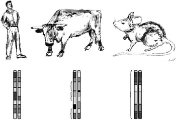
The following two points: (i) poor level of knowledge about the genetic bases of infertility/low fertility in humans and domestic species and (ii) high variation in the measured parameters (litter size, sperm characteristics, etc., inducing a lot of problem while trying to measure heritabilities) lead to the conclusion that a model where genetic variability is limited and where the environment can be tightly controlled has a high potential to make knowledge progress on this difficult subject. Classically, rodent models are consistently exploited to infer mechanisms transposable to other mammals. This is particularly the case for the laboratory mouse, characterized by its short generation time, high prolificacy and small size (Figure 1). The debate of transporting data from mice to humans or other species is controversial and never-ending. However, it should be recognized that many advances on mammalian physiology and genetics originate from mouse studies.
One of the main interests of using mice as animal models is the fact that they are available as strains, meaning that their chromosome content is homozygous at every locus. Intra-strain crosses are largely comparable to the mating of cloned animals, which, genetically speaking, should yield only identical animals, but the rare events of point mutations. By contrast, outbred species such as humans or species, which have been heavily selected for agricultural traits, have a strong level of heterozygosity. In the figure, homozygous regions are shown by brackets. For traits displaying a low heritability, either because they are genetically ill-defined or because environmental factors play a major role, crosses between mice species are very useful, since environmental conditions can be very precisely controlled, and because the genetic diversity is obtained by crossing existing strains, or by analyzing recombinant congenic strains.

Reproduction has not escaped this reality and several research groups around the world use laboratory mice to evaluate fertility parameters. One of the important characteristics of laboratory mice is that they exist as strains, a collection of genetically identical animals. These strains show complete homozygosity at all genomic loci, of course higher than in clones, which can be heterozygous at numerous loci. This situation lessens considerably the potential influences of individual genetic heterogeneity compared with other species.
Different types of mouse models have been constructed corresponding to different strategies for identifying gene(s) governing phenotypic traits: (1) Knocked-out (KO) mice in which a gene of interest has been invalidated. Analyzing the resultant phenotype of these KO mice makes it possible to directly know the function of this gene. (2) Mice in which diverse aleatory mutations have been chemically induced (N-ethyl-N-nitrosourea (ENU) mice). (3) Mice in which a certain degree of polymorphism has been introduced by successive crosses between different mouse strains or species. Indeed, variability is the major tool that geneticist can use; therefore, one of the recurrent problems of lab mice crosses is the low level of genetic diversity of the starting material, generally exclusively congenic homozygous Mus musculus lab mice. This has conducted mice geneticists to perform crosses between different strains in order to restore at least part of the necessary diversity. In a recent study, physiological parameters quantitatively governed by specific genes were studied in a multi-cross generated from eight different lab mouse strains (Valdar et al., Reference Valdar, Solberg, Gauguier, Burnett, Klenerman, Cookson, Taylor, Rawlins, Mott and Flint2006).
Gene invalidation experiments in mice
Most of the information relating to fertility gene effects in mice originate from gene invalidation experiments. A recent review emphasizes the use of gene KO mice for understanding fertility (Roy and Matzuk, Reference Roy and Matzuk2006). KO phenotypes have given categorical data at different levels about numerous pathways involved in fertility. These data may be summarized as follows essentially according to the classification of Roy and Matzuk (Reference Roy and Matzuk2006).
Germ line specifications: Primordial germ cells (PGC) derive from cells of the proximal epiblast after induction by the extra-embryonic ectoderm. More precisely, PGCs originate from the internal wall of the allantois, migrating inside a nascent gonad composed of three independent somatic cell lines (Koopman, Reference Koopman1999). Several molecular actors fixing the fate of these PGCs have been identified by gene invalidation experiments. Among those, transforming growth factor beta (TGFβ) family members appear heavily over-represented. This is the case for ligands such as Bmp4 (Lawson et al., Reference Lawson, Dunn, Roelen, Zeinstra, Davis, Wright, Korving and Hogan1999), Bmp8b (Ying et al., Reference Ying, Liu, Marble, Lawson and Zhao2000) and Bmp2 (Ying and Zhao, Reference Ying and Zhao2001) as well as for their receptors (de Sousa Lopes et al., Reference de Sousa Lopes, Roelen, Monteiro, Emmens, Lin, Li, Lawson and Mummery2004). Defects in TGFβ signalling pathway, by intracellular effectors such as Smad1 and Smad5, also have a reducing effect on the PGC number (Chang and Matzuk, Reference Chang and Matzuk2001; Tremblay et al., Reference Tremblay, Dunn and Robertson2001). The Znf repressor Prdm1 has been shown to have a specifying effect on the PGC (Vincent et al., Reference Vincent, Dunn, Sciammas, Shapiro-Shalef, Davis, Calame, Bikoff and Robertson2005). Systematic analyses of the PGC transcriptome by differential screening revealed genes that contribute to their determination and specificities. To this respect, Fragilis, an interferon-inducible transmembrane protein, seems to play a pivotal function together with the Stella transcription factor (Saitou et al., Reference Saitou, Barton and Surani2002).
PGC proliferation and survival: In the Pin1−/− mice, PGCs were allocated properly at the base of the allantois, but their cell expansion was progressively impaired, resulting in a markedly reduced number of PGCs at 13.5 dpc (Atchison et al., Reference Atchison, Capel and Means2003). Oct4, a major gene of the POU family involved in the inner cell mass/trophectoderm choice, is also involved in PGC proliferation (Niwa et al., Reference Niwa, Miyazaki and Smith2000). Other important genes are Nanos3 (Forbes and Lehmann, Reference Forbes and Lehmann1998), Sdf1 and its receptor Cxcr4 (Molyneaux et al., Reference Molyneaux, Zinszner, Kunwar, Schaible, Stebler, Sunshine, O’Brien, Raz, Littman, Wylie and Lehmann2003; Holt et al., Reference Holt, Jackson, Roman, Aitken, Koopman and McLaughlin2006). PGCs are maintained owing to several growth factors such as LIF and bFGF (Farini et al., Reference Farini, Scaldaferri, Iona, La Sala and De Felici2005). Other less-documented transcription factors may have a very important incidence, such as Zfp148, the loss of which leads to a complete disappearance of PGCs at the heterozygous state (Takeuchi et al., Reference Takeuchi, Mishina, Miyaishi, Kojima, Hasegawa and Isobe2003). It is hypothesized that this gene mediates p53 function by preventing its phosphorylation during germ cell development.
Sex determination: In mice, mutations in many genes such as AMH (Behringer et al., Reference Behringer, Finegold and Cate1994) and its receptor (Jamin et al., Reference Jamin, Arango, Mishina, Hanks and Behringer2002), Wnt4 (Vainio et al., Reference Vainio, Heikkilä, Kispert, Chin and McMahon1999), have been found to lead to at least a partial phenotype of sex reversal, similar either to a persistent Mullerian duct syndrome (PMDS) or to a masculinization with Leydig cells developing in the ovarian context. For a review of these genes, see for instance Vaiman and Pailhoux, (Reference Vaiman and Pailhoux2000) and Vaiman (Reference Vaiman2003).
Preantral folliculogenesis: It may be sometimes difficult to separate genes involved in the early steps of folliculogenesis from genes involved in sex determination. This is illustrated by the recent analysis of the combined double KO of Wnt4 and FoxL2 conducting to XY sex reversal. Interestingly, either gene cannot reproduce such a clear-cut phenotype in mice (Ottolenghi et al., Reference Ottolenghi, Pelosi, Tran, Colombino, Douglass, Nedorezov, Cao, Forabosco and Schlessinger2007). The disruption of Figα (Soyal et al., Reference Soyal, Amleh and Dean2000), Wnt4 (Vainio et al., Reference Vainio, Heikkilä, Kispert, Chin and McMahon1999), Nobox (Rajkovic et al., Reference Rajkovic, Pangas, Ballow, Suzumori and Matzuk2004) and Gdf9 (Dong et al., Reference Dong, Albertini, Nishimori, Kumar, Lu and Matzuk1996) explain to various extents the onset of poor or absent fertility. Other species than mice also helped in the identification of new genes involved in folliculogenesis. This is the case of the sheep model in which different kinds of mutations in FecX (Bmp15) (Galloway et al., Reference Galloway, McNatty, Cambridge, Laitinen, Juengel, Jokiranta, McLaren, Luiro, Dodds, Montgomery, Beattie, Davis and Ritvos2000) and BmpR1B (Mulsant et al., Reference Mulsant, Lecerf, Fabre, Schibler, Monget, Lanneluc, Pisselet, Riquet, Monniaux, Callebaut, Cribiu, Thimonier, Teyssier, Bodin, Cognie, Chitour and Elsen2001) displayed variable phenotypes from total infertility to superfertility in terms of ovulation rate. Furthermore, several important genes for female fertility are located on the X chromosome (for a review, see Vaiman, Reference Vaiman2002).
Spermatogenesis: KO at all steps of spermatogenesis has been shown to disrupt fertility. A relatively exhaustive list was given by Roy and Matzuk (Reference Roy and Matzuk2006). Briefly, for the transition from spermatogonia to spermatids, the role of Kit was early recognized (Yoshinaga et al., Reference Yoshinaga, Nishikawa, Ogawa, Hayashi, Kunisada and Fujimoto1991). More recently, the role of Cks in the differentiation timing between spermatogonia and spermatids was shown (Donovan and Reed, Reference Donovan and Reed2003). Plzf, a zinc finger transcription factor, was shown to be essential for the turnover and renewal of stem cells in the testis (Buaas et al., Reference Buaas, Kirsh, Sharma, McLean, Morris, Griswold, de Rooij and Braun2004). Recently, it was shown that Sohlh1 (testis and ovary expressed basic helix–loop–helix (bHLH) transcription factor) is essential for spermatogonia differentiation (Ballow et al., Reference Ballow, Meistrich, Matzuk and Rajkovic2006). In invalidated testis from Sohlh1 −/− mice, the testis transcription factors Etv5, Taf4b, Zfp148 and Plzf were normally expressed, while the bHLH Tohlh2 transcription factor was increased while Lhx8 and Ngn3 were repressed. These examples illustrate the fact that impaired fertility can result from various breakpoints in the complex cascade of sperm cell differentiation. Defective spermiogenesis is also a potential cause for infertility, relating to alterations of protamine genes (Prm1, Prm2) and transition proteins in the extreme DNA compaction of the sperm nucleus. A recent study made possible the identification by proteomics of not less than 132 proteins important for DNA compaction, chromosome segregation and fertility (Chu et al., Reference Chu, Liu, Nix, Wu, Ralston, Yates and Meyer2006). The final flagellar structure of sperm is the key to normal sperm movement. In a similar manner, many mouse mutants display an asthenozoospermia phenotype. Some genes affecting motility have been described such as Catsper1 (Ren et al., Reference Ren, Navarro, Perez, Jackson, Hsu, Shi, Tilly and Clapham2001), Catsper 2 (Quill et al., Reference Quill, Sugden, Rossi, Doolittle, Hammer and Garbers2003), other members of the same family (Jin et al., Reference Jin, Jin, Zheng, Ro, Tafolla, Sanders and Yan2007), dynein (Neesen et al., Reference Neesen, Kirschner, Ochs, Schmiedl, Habermann, Mueller, Holstein, Nuesslein, Adham and Engel2001) and the SlcA28 anion transporter (Toure et al., Reference Toure, Lhuillier, Gossen, Kuil, Lhote, Jegou, Escalier and Gacon2007), whose mutations induce an abnormal morphology of the flagellum.
Endocrine regulation of gametogenesis: GnrH deletion in mice leads to a condition where neither LH nor FSH are produced (hypogonadism), inducing infertility (Mason et al., Reference Mason, Pitts, Nikolics, Szonyi, Wilcox, Seeburg and Stewart1986). Similarly, KO of Fshb, Fshr, Lhb and Lhr (Kumar et al., Reference Kumar, Wang, Lu and Matzuk1997; Dierich et al., Reference Dierich, Sairam, Monaco, Fimia, Gansmuller, LeMeur and Sassone-Corsi1998) have been produced. Although the disruption of FSH pathways induces the formation of small testes, the males are fertile while the females are sterile. In these KOs, folliculogenesis is blocked before the antrum formation. Contrary to FSH, the LH pathway is essential for both sexes. Other steroid hormones are crucial for reproductive function in mammals since the estrogen beta receptor (Esrb) disruption leads to subfertility in female and males in which serious defects of spermatogenesis are observed (Lubahn et al., Reference Lubahn, Moyer, Golding, Couse, Korach and Smithies1993; Dupont et al., Reference Dupont, Krust, Gansmuller, Dierich, Chambon and Mark2000). The invalidation of the progesterone receptor alpha (Pra) results in uterine defects, preventing normal implantation. Physiologically, once prepared by progesterone, the uterus is able to synthesize catecholestrogens from ovarian estrogens or from the blastocyst, these molecules appearing as essential for a successful implantation (Paria et al., Reference Paria, Lim, Wang, Liehr, Das and Dey1998 and Reference Paria, Song and Dey2001). Numerous studies were performed on the androgen receptor (AR) KO mice (Chang et al., Reference Chang, Chen, Yeh, Xu, Wang, Guillou, Lardy and Yeh2004; De Gendt et al., Reference De Gendt, Atanassova, Tan, de Franca, Parreira, McKinnell, Sharpe, Saunders, Mason, Hartung, Ivell, Denolet and Verhoeven2005), demonstrating its essential role for testosterone metabolism and androgen signalling during spermatogenesis. Consequently, the normal function of this molecule is necessary for male fertility. Additional roles of the AR in female fertility pathways have been shown (Hu et al., Reference Hu, Wang, Yeh, Wang, Xie, Xu, Zhou, Chao, Tsai and Chang2004; Walters et al., Reference Walters, Allan, Jimenez, Lim, Davey, Zajac, Illingworth and Handelsman2007).
This non-exhaustive list emphasizes the importance of gene invalidation in mice for identifying genes involved in controlling fertility. However, many KO mice harbor extreme phenotypes, sometimes missing complete systems such as genitalia, head, spinal cord, etc. In these cases, the KO will act in an on/off manner and induce, of course, a complete sterility when, in fact, many infertility phenotypes appear as quantitative deficiencies. In these cases, KO mice may not be ideal, suggesting that more subtle models could be helpful to deal with the variability observed in humans or livestock.
Genetic mapping techniques for infertility phenotypes in the mouse model
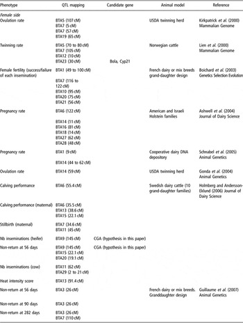
In humans, the identification of genetic loci quantitatively modulating fertility has not been a subject of research. Similarly, in mice, the number of studies aiming at identifying such loci is reduced. Nevertheless, quantitative variations can be important to explain the continuum of inter-individual differences, sometimes leading to infertility. In human male fertility, it is known that the number of spermatozoa is an important parameter, since below a given threshold (around 5 million sperm/ml), human males are almost sterile. The follow-up of quantitative parameters impacting on fertility has been much more consistent in cattle, where the economical relevance of such traits has a great importance. Several recent studies taking into account fertility parameters alongside other production traits illustrate this point (Kirkpatrick et al., Reference Kirkpatrick, Byla and Gregory2000; Lien et al., Reference Lien, Karlsen, Klemetsdal, Vage, Olsaker, Klungland, Aasland, Heringstad, Ruane and Gomez-Raya2000; Boichard et al., Reference Boichard, Grohs, Bourgeois, Cerqueira, Faugeras, Neau, Rupp, Amigues, Boscher and Leveziel2003; Ashwell et al., Reference Ashwell, Heyen, Sonstegard, Van Tassell, Da, VanRaden, Ron, Weller and Lewin2004; Schnabel et al., Reference Schnabel, Sonstegard, Taylor and Ashwell2005). To the best of our knowledge, only four studies in cattle focused exclusively on fertility traits (Holmberg and Andersson-Eklund, Reference Holmberg and Andersson-Eklund2006; Guillaume et al., Reference Guillaume, Gautier, Ben Jemaa, Fritz, Eggen, Boichard and Druet2007; Holmberg et al., Reference Holmberg, Sahana and Andersson-Eklund2007; Schulman et al., Reference Schulman, Sahana, Lund, Viitala and Vilkki2008), while the other studies involve the phenotyping of other production traits. Loci involved mainly in female fertility were identified (see Table 1). Today, no gene could be identified by this preliminary approach of positional cloning. This is explained by the paucity of molecular tools available in ruminants compared with humans and mice.
Quantitative trait loci (QTL) modulating fertility parameters in cattle

Therefore, for the cases where mice quantitative trait loci (QTL) appear at orthologous positions with cattle QTL, it is clear that M. musculus is the experimental species on which to work to identify causal mutation(s). Several different genomic characteristics have been exploited in different mouse models, and always present a combination of advantages and drawbacks. In mice, laboratory strains originate mainly from a small pool of ancestors belonging to the M. musculus species. For this reason, the different strains are genetically highly similar, an issue that sometimes constitutes a serious problem for addressing the genetic bases of complex phenotypes, due to the lack of informative markers. This lack of genetic polymorphisms can be overcome by crossing laboratory mice with wild mice or by creating original mutations with ENU mutagen. The use of such various models has been described for the dissection of several complex traits, such as the genetics of obesity (Brockmann and Bevova, Reference Brockmann and Bevova2002), but most models have not been exploited for the positional cloning of genes involved in fertility.
Since mouse strains are completely homozygous, polymorphism is generally partially restored by crossing two laboratory strains or even two mouse species. The development of inter-specific hybrids strains is made possible by the possibility of getting offspring from mice belonging to two species in the early stages of speciation (Guenet and Bonhomme, Reference Guenet and Bonhomme2003). In this case, generally, the female offspring is fertile whereas the male is sterile, showing a strong disruption of steroidogenic functions (Chubb and Nolan, Reference Chubb and Nolan1987). The origin of this hybrid breakdown has been traced back to meiotic defects (Matsuda and Chapman, Reference Matsuda and Chapman1991; Matsuda et al., Reference Matsuda, Moens and Chapman1992; Hale et al., Reference Hale, Washburn and Eicher1993). To date, nine loci inducing male hybrid sterility have been mapped on chromosomes X (Hstx1, Mhstq2 and Hst3) and 17 (Mhstq1, Hst1, Hst4, Hst5, Hst6 and Hst7). Some of them have been mapped so closely that it is difficult to be certain that they correspond to different genes. In several cases, candidate genes potentially involved in the sterility phenotype have been proposed. For instance, the Hst6 locus encompasses the gene Dnahc8, which seems to be at least partially responsible for a phenotype of sperm tail curvature (Samant et al., Reference Samant, Ogunkua, Hui, Lu, Han, Orth and Pilder2005). This locus is nevertheless complex since in the same region, two candidate genes (Tsga2 and Tctex5) might play synergistic roles in the generation of motility abnormalities and the sperm–egg interaction aberration observed in hybrids. Nevertheless, these features can be regarded as functionally unrelated traits (Pilder et al., Reference Pilder, Lu, Han, Hui, Samant, Olugbemiga, Meyers, Cheng and Vijayaraghavan2007).
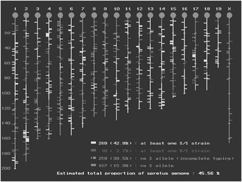
Using more classical QTL mapping approaches, several preliminary localizations of fertility-modulating genes have been achieved in mice, but are still very imprecise in most studies, sometimes an entire chromosome. Such QTL-containing regions have been identified for female (Krewson et al., Reference Krewson, Supelak, Hill, Singer, Lander, Nadeau and Palmert2004; Peripato et al., Reference Peripato, De Brito, Matioli, Pletscher, Vaughn and Cheverud2004; Rocha et al., Reference Rocha, Eisen, Siewerdt, Van Vleck and Pomp2004; Liljander et al., Reference Liljander, Sallstrom, Andersson, Wernhoff, Andersson, Holmdahl and Mattsson2006) and male fertility parameters (Zídek et al., 1998; Le Roy et al., Reference Le Roy, Tordjman, Migliore-Samour, Degrelle and Roubertoux2001; Oka et al., Reference Oka, Mita, Sakurai-Yamatani, Yamamoto, Takagi, Takano-Shimizu, Toshimori, Moriwaki and Shiroishi2004). For instance, two QTLs affecting meiosis on chromosomes 4 and 13, two QTLs for relative testis weight on chromosomes 4 and X and several QTLs for degenerated germ cells and multi-nuclear giant cells (Bolor et al., Reference Bolor, Wakasugi, Zhao and Ishikawa2006). Effects on fertility have been thoroughly analyzed for chromosome X loci in two recent studies based on mouse subspecies (Oka et al., Reference Oka, Aoto, Totsuka, Takahashi, Ueda, Mita, Sakurai-Yamatani, Yamamoto, Kuriki, Takagi, Moriwaki and Shiroishi2004 and Reference Oka, Mita, Sakurai-Yamatani, Yamamoto, Takagi, Takano-Shimizu, Toshimori, Moriwaki and Shiroishi2007) in which parameters of sperm morphology and function were assessed. The results displayed a QTL responsible for testis weight as well as two QTL for sperm head morphology. In the 2007 study, the authors demonstrated the importance of epistatic interactions between various chromosome loci for achieving a normal fertility (Oka et al., Reference Oka, Aoto, Totsuka, Takahashi, Ueda, Mita, Sakurai-Yamatani, Yamamoto, Kuriki, Takagi, Moriwaki and Shiroishi2007). Only one recent study (L’Hote et al., Reference L’Hote, Serres, Laissue, Oulmouden, Rogel-Gaillard, Montagutelli and Vaiman2007), using recombinant congenic strains of mice, made it possible to restrict the gene location to a small interval of 1 to 2 cM. In this study, we proposed using inter-specific recombinant congenic strains (IRCS) in order to rapidly map genes modulating fertility to short chromosome intervals. These mice have been generated over 20 years ago in the Pasteur Institute by an initial cross between two evolutionary distant parental species (M. musculus, C57BL/6 × Mus spretus), followed by two backcrosses on C57BL/6. In total, 118 strains were originally raised by mating mice trios. Then, brother/sister crosses were performed during more than 30 generations. Finally, 53 IRCS strains survived this process. Each strain possesses on average 98% of C57BL/6 genome and 2% of spretus genome fixed at the homozygous state (see Figure 2). The positions of the spretus fragments (1 to 7 per strain) have been precisely determined from the analysis of around 700 single nucleotide polymorphisms (SNPs) and microsatellite markers (Burgio et al., Reference Burgio, Szatanik, Guenet, Arnau, Panthier and Montagutelli2007). Using this model, fertility analysis has been addressed on the male side by measuring several sperm parameters having a known influence on fertility (L’Hote et al., Reference L’Hote, Serres, Laissue, Oulmouden, Rogel-Gaillard, Montagutelli and Vaiman2007). Eight QTL were identified and are currently under scrutiny to identify the underlying genes and potential causal mutations. The female side is addressed on the same model using in vivo high-frequency ultrasonography. Phenotypes of in utero lethality and embryonic resorption have been found and are currently analyzed in more detail searching to clone the relevant endometrial and placental genes (Laissue et al., Reference Laissue, Burgio, L’Hôte, Renault, Marchiol-Fournigault, Fradelizi, Fellous, Serres, Montagutelli, Monget and Vaiman2008). This IRCS model constitutes a unique opportunity to clone new genes involved in fertility in mammals. A summary of fertility-modulating genes is presented in Table 2.
The map status of the 53 interspecific recombinant congenic strains developed at the Pasteur Institute (Paris). Horizontal bars correspond to markers that have been genotyped yellow fragments are present in at least one of the strains in a spretus form. Pink fragments are fragments that can apparently be maintained only at the heterozygous state in at least one strain, despite the number of brother–sister crosses. In average, B6 and spretus genome diverge for one nucleotide every 80 bp, which insures a high level of heterozygosity and the existence of informative markers able to delineate precisely the spretus fragments (http://www.pasteur.fr/recherche/unites/Gfons/ircs/ircshome.htm).

Quantitative trait loci (QTL) modulating fertility parameters in mice

Ethyl-nitrosourea mutagenesis and fertility analysis
Genetic polymorphisms can be induced in laboratory mice by ENU mutational treatment. ENU is an alkylating agent that is a powerful mutagen. In mouse spermatogonial stem cells, it produces single locus mutation at high frequencies (Russell et al., Reference Russell, Kelly, Hunsicker, Bangham, Maddux and Phipps1979; Rinchik et al., Reference Rinchik, Johnson, Margolis, Jackson, Russell and Carpenter1991).
ENU can induce many different types of alleles: Loss of function mutations, viable hypomorphs of lethal complementation groups, antimorphs and gain of function mutations. Mutations induced by ENU are not molecularly tagged, and have to be analyzed by linkage analysis and positional cloning. Mutations can be induced in living males or in embryonic stem cells culture. When the mutation is roughly mapped on a chromosome, different schemes exist to finely map the mutations and establish it at a homozygous state, using a deletion scheme, or balancer chromosomes to isolate mutations.
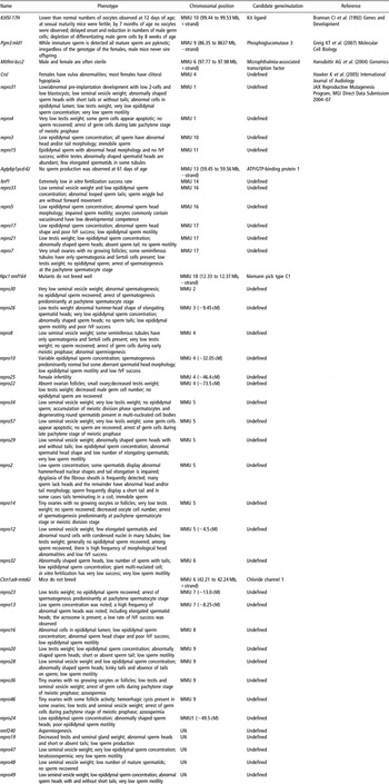
Several consortia have initiated systematic, genome-wide production of mouse mutants. The phenotypic analyses included reproduction traits. For example, the Jackson laboratory has created in 2005 ‘The Reproductive Genomics Program’, and so far has established 40 mutations impairing male or female fertility. Infertility phenotypes include ovaries that develop poorly, ovaries with no oocytes, ovaries whose follicles do not mature properly, oocytes that die inside the follicles, oocytes that cannot complete meiosis and embryos that die before they reach the blastocyst stage. Aberrant male phenotypes include spermatocytes that do not develop defective sperm and sperm with a low fertilization rate. These mice mutants can be obtained from the Jackson laboratory.
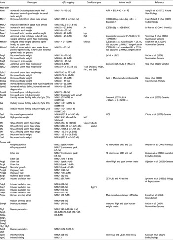
A total of 89 mutations are present in databases, 24 of which have been precisely localized (Table 3) encompassing genes with pleiotropic effects. Consortia tend to submit many mutations with a coarse localization, but with a fine phenotype description. Therefore, while most mutations remain imprecisely mapped, their potential for discovering genes important in mammalian fertility pathways will be very important in the future.
ENU fertility mutant mice

Conclusions
In conclusion, sterility analysis in hybrid animals or hypo-fertility in subspecies crosses revealed some interesting trails for understanding some molecular bases of infertility. One of the major problems of such genetic studies aiming at identifying genes is that QTL locations are generally far from precise, but could be overcome in some cases using IRCS.
In this context and despite the unquestionable progress in the knowledge brought by mice KO analysis, we make the hypothesis that positional cloning approach will be able to give new insights in the complexity underlying mammalian fertility pathways. Several genes identified in mice have seen their application in human cases of infertility (see, for a review, Furnes and Schimenti, Reference Furnes and Schimenti2007). We expect that starting from the phenotype is an efficient way to focus more closely on genes of interest, especially on factors related with important quantitative phenotypic effects, which will be applicable to target species such as humans or domestic ruminants.
Acknowledgments
Paul Laissue is a recipient of a fellowship from the National Research Agency Program ‘Mammifert’. David L’Hôte receives a PhD fellowship by INSERM and University of Limoges. The research program underlying this review is partly financed by the ‘Mammifert’ grant.