Case report
Managing complex congenital cardiac anomalies requires innovative approaches, especially when facing anatomical constraints or limited vascular access. Pulmonary regurgitation, a frequent sequela after right ventricular outflow tract repair, often necessitates reintervention. Percutaneous pulmonary valve implantation has emerged as a preferred alternative to surgery, particularly with the advent of self-expanding valves designed for large or irregular right ventricular outflow tracts. The Venus P-valve system, developed by Venus Medtech (Hangzhou, China), is specifically tailored for these anatomies, offering a flared or straight design to address various morphological challenges. Recent studies have demonstrated its safety, efficacy, and durability in mid- and long-term outcomes. Reference Jin, Long and Zhang1–Reference Morgan, Sivakumar, Promphan, Goreczny, Prachasilchai and Qureshi3 This case highlights a minimally invasive approach that overcomes anatomical and procedural challenges.
The patient was 41-year-old female (140 cm and 42 kg) with left isomerism, interrupted inferior caval vein with azygos continuation to the left superior caval vein, dextrocardia, and repaired tetralogy of Fallot with a transannular patch. She had also undergone transcatheter closure of a patent ductus arteriosus during infancy and ablation for supraventricular tachycardia. No CT-scan was available, and a c-MRI showed severe pulmonary regurgitation (58%), a slightly reduced right ventricular ejection fraction (40%), and preserved left ventricular ejection fraction (60%).
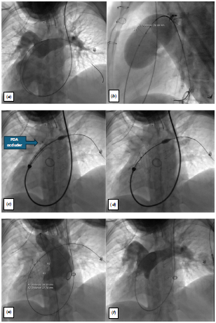
The procedure was performed under general anaesthesia in a biplane lab. Her anatomy precluded femoral venous access, and single venous vascular access was obtained via left jugular vein. Pre-deployment angiography (Panel A, Figure 1) identified a short main pulmonary artery (length 29 mm), and balloon interrogation showed right ventricular outflow waist of 28x30 mm (Panel B, Figure 1) and confirmed that coronaries were not at risk.
Angiograms of the intervention.

A Lunderquist Extra-Stiff Guidewire (Cook Medical, Bloomington, IN, USA) was advanced into the left pulmonary artery to provide support for the delivery system. Due to the anatomy and size of the patient, we were unable to advance a 26 Fr. GORE® DrySeal Flex sheath, and the valve was directly advanced over the guidewire with a smooth positioning of the delivery system though a one curve trajectory. We prioritised minimal length of the valve due to the short pulmonary artery and chose a Venus P-valve measuring 30 by 25 mm in a right ventricular outflow measuring 29 mm of diameter (Panels C and D, Figure 1). Since contrast injections during implantation were not possible, the previously implanted patent ductus arteriosus closure device served as an anatomical landmark for positioning the valve. The valve was implanted as high as possible to stay away from the right ventricle. Post-deployment controls confirmed correct positioning and function of the valve (Panels E and F, Figure 1). The patient experienced transient episodes of supraventricular tachycardia post-procedure, which resolved under betablockade medication. She was discharged on dual antiplatelet therapy after two days. Six months follow-up was uneventful.
Discussion
This case underscores the utility of the Venus P-valve system in addressing complex congenital cardiac anatomies. Reference Góreczny, Szeliga, Lazu, Becht, Mroczek and Qureshi4 Designed to accommodate irregular or dilated right ventricular outflow tracts, the Venus P-valve provides a tailored solution where balloon-expandable systems may be unsuitable. By employing a flared design, it ensures a secure fit and effective sealing even in anatomically challenging scenarios. Reference Jin, Long and Zhang1–Reference Morgan, Sivakumar, Promphan, Goreczny, Prachasilchai and Qureshi3
In this undersized patient with a complex anatomy, the implantation technique was simplified and the valve implanted without a long sheath directly on its delivery system over a one-curve trajectory. The valve was relatively undersized to accommodate a valve as short as possible in a very short pulmonary artery truck. The reliance on anatomical landmarks (such as the previously implanted device) rather than real-time contrast injections further highlights the benefits of individualised planning.
This case highlights the adaptability of the Venus P-valve system and the importance of a patient-specific approach in managing challenging anatomical conditions with successful outcome.
Financial support
All authors have no other potential conflicts of funding or financial relationship to disclose.
Competing interests
Sophie Malekzadeh-Milani is a proctor-consultant for Venus MedTech, for Abbott, and for Edwards.
Ethical standard
The study was conducted in accordance with the Declaration of Helsinki.