Although preventable through vitamin D supplementation, rickets is not eradicated in high-income countries, and several reports suggest that nutritional rickets is a re-emerging disease in the United Kingdom,Reference Allgrove 1 Europe,Reference Beck-Nielsen, Brock-Jacobsen, Gram, Brixen and Jensen 2 and North America, especially in dark-skinned ethnic minorities.
Vitamin D-related hypocalcaemia is a rare cause of dilated cardiomyopathy (DCM), but case reports and case series have been recently published.Reference Maiya, Sullivan and Allgrove 3 – Reference Tomar, Radhakrishnan and Shrivastava 5
Dilated cardiomyopathy associated with Vitamin D-related hypocalcaemia often results in cardiac failure and high mortality, but early diagnosis and proper management can reduce mortality and long-term complications. We report the cases of two children with dilated cardiomyopathy due to vitamin D deficiency-related hypocalcaemia.
Child 1
A 45-day-old Tunisian female child was referred from a peripheral hospital to the Paediatric Cardiology Department because of progressive poor feeding, dyspnoea, and syncope, which had progressively worsened in the last few days and was associated with severe biventricular dysfunction.
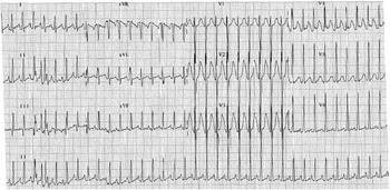
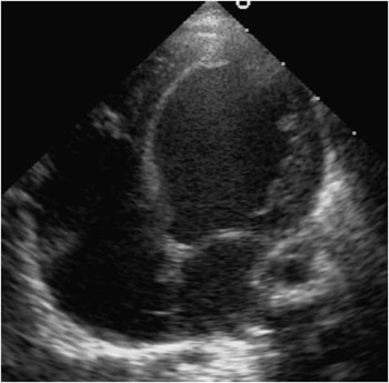
The baby was born by normal delivery after an uneventful gestation and had a normal perinatal history. Family history was unremarkable and parents were not consanguineous. The baby was exclusively breastfed and did not receive vitamin supplements. The mother abstained from dairy products and did not receive vitamin D or calcium supplements during pregnancy or breastfeeding. The baby had no history of recent illnesses or infections. On admission, her temperature was 37.8°C, and she was moderately hypotonic, tachycardic (180 bpm), and tachypnoic (95 breaths/min) with respiratory distress. Oxygen saturation was 98% in room air. Basilar pulmonary crackles were detected bilaterally and the liver edge was palpable 1 cm below the costal margin. Peripheral pulses were palpable, blood pressure was 78/50 mmHg, and she had a good nutritional status (5.150 kg, 75th–90th centile). Electrocardiogram showed sinus tachycardia, ST anomalies, left ventricle hypertrophy, and QTc prolongation (490 ms; Fig 1). Severe cardiomegaly (cardiothoracic ratio 0.75) and pulmonary congestion were observed in the chest X-rays. No anatomic malformation was found on cardiac ultrasonography and the ductus arteriosus was closed; the origin of coronary arteries was normal. Severe biventricular dysfunction (left ventricle ejection fraction 10%) and dilatation of the left ventricle with moderate mitral regurgitation (Fig 2) were present. Markers of inflammation, serological markers of cardiotropic viruses, metabolic investigations, hepato-renal function, and proteins and albumin in blood were normal. Calcium concentration in serum was 6.5 mg/dl (normal range 8.5–10.5 mg/dl). Endomyocardial biopsy was negative for myocarditis. Endocrinological evaluation for hypocalcaemia showed increased levels of parathyroid hormone (494 pg/ml, nr 15–65 pg/ml), osteocalcin (252 ng/ml, nr 7–18 ng/ml), alkaline phosphatase (2010 IU/L, nr 250–915 IU/L), and decreased values of phosphataemia (3 mg/dl, nr 3.8–6.5 mg/dl) and 25-OH vitamin D (4.5 ng/ml, nr 20–55 ng/ml), consistent with vitamin D deficiency rickets causing hypocalcaemic dilated cardiomyopathy.
12-lead electrocardiogram of child 1 at presentation showing sinus tachycardia, left ventricular hypertrophy, and anomalies of ventricular repolarisation; prolongation of QTc to 490 ms.

Transthoracic apical four-chamber view with severe dilatation of the left ventricle.

The baby was placed on inotropic support and furosemide, calcium infusion to correct the hypocalcaemia and enteral vitamin D. Few hours after admission, she experienced two episodes of low cardiac output unresponsive to inotropic support, and was placed in extracorporeal membrane oxygenation. Extracorporeal membrane oxygenation was stopped 10 days later, but evidence of tetraparesis appeared after suspension of analgesic and sedative therapy. Brain ultrasonography and MRI showed severe ventricolomegaly and increase of subarachnoid spaces with mycrocystic lesions of cerebral parenchyma, secondary to severe hypoxic–ischaemic insult.
The child was discharged on treatment with vitamin D, calcium supplements, and oral anti-congestive therapy. After 2 months, cardiac ultrasonography showed a normal left ventricle size with a hypertrophic aspect, normal systolic function, and continence of mitral valve. Chest X-ray reported a significant decrease of the heart size and the electrocardiogram was normal 4 months later. After 6 months, the child had normal cardiac function but was admitted to the neurology department for severe neurological sequelae.
Child 2
A 4-month-old breastfed North African male was referred to the Pediatric Department and then to the Pediatric Cardiology Department with a 1-week history of poor feeding and dyspnoea and clinical signs of cardiac failure.
He was born from consanguineous healthy parents after a normal pregnancy and an uneventful delivery. The family had no history of genetic diseases and the mother could not recall her dietary intake during pregnancy. Medical history was negative for recent illnesses or infections, and his immunisation and developmental progress were satisfactory.
Physical examination revealed an irritable infant with a temperature of 36°C; he was tachycardic (150 bpm) and tachypnoic (52 breaths/min), with a blood pressure of 76/58 mmHg. Oxygen saturation was normal and his lungs were clear on auscultation, but the infant had a gallop rhythm and soft pansystolic murmur. Peripheral pulses were palpable and the liver edge was 2 cm below the right costal margin. Electrocardiogram showed sinus tachycardia, high-amplitude QRS in left leads, and prolonged QTc (QTc 500 ms). Chest X-rays showed a cardiothoracic ratio of 0.58 with clear lung fields. Ultrasonography revealed normal anatomy and origin of coronaries, dilated and hypokinetic left ventricle (EF 23%) with moderate mitral regurgitation, and a closed ductus arteriosus. The child had hypocalcaemia (calcium 6.2 mg/dl), normal phosphorus (5.8 mg/dl), increased alkaline phosphatase (2836 U/L) and parathyroid hormone (845 pg/ml); osteocalcine (71 ng/ml), 25-OH vitamin D (10 ng/ml), and urinary calcium were decreased. The skeletal muscle biopsy showed no evidence of metabolic disease. Cardiotropic virus screening was negative and inflammatory markers were normal.
A diagnosis of dilated cardiomyopathy secondary to vitamin D deficiency rickets was made with medical management of rickets based on calcium and vitamin D, and therapy for congestive cardiac failure (enalapril, furosemide, carvedilol). The patient responded well to the treatment, and 13 days after admission he was discharged with normal chemistry and calcium–phosphorus metabolism. Systolic function improved and he completely recovered after 6 months, when anticongestive medications were discontinued. After 20 months, calcium and vitamin D supplementations were discontinued and dietary intake was fortified with vitamin D. After 6 years, the child had normal growth and psychomotor development.
Discussion
Nutritional rickets can cause hypocalcaemia and represents a re-emerging disease in high-income countries.Reference Allgrove 1 – Reference Beck-Nielsen, Brock-Jacobsen, Gram, Brixen and Jensen 2 Ionised calcium is an inotropic agent with a central role in the excitation–contraction coupling, allowing the actin–myosin interaction via the troponin–tropomyosin complex and playing a role in depolarisation and repolarisation. Hypocalcaemia can cause contractility deficiency and conduction delays of the cardiac impulse. Long-standing hypocalcaemia can cause cardiac dilatation and dysfunction in newborns, especially if the mother has vitamin D deficiency, in infants, and in young children. Parathyroid hormone level is increased in nutritional rickets. In the general population, chronic exposure to high levels of parathyroid hormone has been shown to be related to cardiovascular disease, predisposing to metabolic syndrome, activating the renin–angiotensin system, and stimulating systemic and vascular inflammation. To support the pathogenic role of parathyroid hormone excess, it has been shown that surgical parathyroidectomy for primary hyperparathyroidism and renal transplantation for secondary hyperparathyroidism reduce parathyroid hormone-related cardiovascular risk. The rarity of hypocalcaemic cardiomyopathy can be due to vitamin D supplementation and early diagnosis and treatment of rickets. However, cardiac involvement is not constantly associated with severe hypocalcaemia and some patients are more susceptible to hypocalcaemic myocardic damage than others. Genetic predisposition with relative insensitivity to vitamin D can affect some populations of the Middle EastReference Cox, Sleeper and Lowe 7 and European descendants.Reference Wagner, Taylor, Dawodu, Johnson and Hollis 8
Out of a total of 42 patients diagnosed with dilated cardiomyopathy between 2000 and 2009 in our Paediatric Cardiology Department, two were associated with rickets. As described in the literature, these patients belonged to a high-risk population – infants from dark-skinned mothers with low dairy product intake, exclusively breastfed, not receiving vitamin D supplements, or significant exposure to sunlight.
As previously reported,Reference Maiya, Sullivan and Allgrove 3 – Reference Uysal, Kalayci and Baysal 6 our two cases show that clinical presentation of dilated cardiomyopathy due to nutritional hypocalcaemic rickets can be severe ranging from congestive cardiac failure to cardiogenic shock, especially in younger infants. In both patients, QTc interval was prolonged on admission and normalised after correction of serum calcium levels. QTc prolongation is secondary to hypocalcaemia via prolongation of plateau phase of cardiac action potential, and it is oftenReference Brown, Nunez, Russell and Spurney 4 – Reference Tomar, Radhakrishnan and Shrivastava 5 but not alwaysReference Maiya, Sullivan and Allgrove 3 present in left ventricle dysfunction due to hypocalcaemia.
Once aetiological diagnosis is made and proper treatment is added to anticongestive therapy, clinical status and ventricle function improve, generally needing 1 to 10 months to recover. Nevertheless, irreversible complications can occur: Maiya et al reported neurological damage in one patient and exitus in 3 out of 16 reported cases. Our two cases had completely different outcomes regardless of the improvement of cardiac function: one child had severe brain damage, whereas the other one fully recovered.
Dilated cardiomyopathy in children is known to be mostly idiopathic or genetic, but other aetiologies need to be considered – metabolic diseases, viral infection, muscular disorders, coronary anomalies, cardiotoxic drugs.Reference Cox, Sleeper and Lowe 7 Clinicians need to include nutritional rickets in the differential diagnosis of dilated cardiomyopathy in the presence of hypocalcaemia, especially in high-risk groups, even in apparently healthy children in high-income countries. It has been recently speculated that lack of calcium and vitamin D supplementation during pregnancy and lactation represents a risk factor for rickets in the child and that maternal ultraviolet B exposure during pregnancy influences the skeletal bone development of the child, type 1 diabetes, and asthma, suggesting that in utero exposure to vitamin D can affect different health aspects of the child.Reference Wagner, Taylor, Dawodu, Johnson and Hollis 8 , Reference Baroncelli, Bereket and El Kholy 9 , Reference Wang, Zhang and Richards 10 Prevention is the main pillar for the management of ricketsReference Wagner and Greer 11 and guidelines stress the need for vitamin D supplementation in infants and children and emphasise the importance of weaning children to a diet adequate in calcium and vitamin D supplementation.Reference Holick, Binkley and Bischoff-Ferrari 12
Although often reversible, the prognosis of hypocalcaemic dilated cardiomyopathy depends on the severity at the time of presentation, the promptness of diagnosis, and the availability of intensive medical/surgical support. The two cases presented here had very different onset and outcomes, and the better outcome in the second child could be due to his earlier stage of rickets (hypocalcaemic and normophosphataemic stage) than the first child (hypocalcaemic and hypophosphataemic stage).
Prompt recognition and proper treatment of patients with dilated cardiomyopathy due to hypocalcaemic rickets can lead to a better outcome and reduce long-term complications.
Acknowledgements
This work did not receive grant or financial support.