Introduction
Spodoptera frugiperda, Smith, is a very devastative insect that attacks over 353 crop species worldwide (Montezano et al., Reference Montezano, Sosa-Gómez, Specht, Roque-Specht, Sousa-Silva, Paula-Moraes, Peterson and Hunt2018) and is widely distributed to Africa (Goergen et al., Reference Goergen, Kumar, Sankung, Togola and Tamò2016), America (Nagoshi et al., Reference Nagoshi, Fleischer, Meagher, Hay-Roe, Khan, Murúa, Silvie, Vergara and Westbrook2017), India (Kalleshwaraswamy et al., Reference Kalleshwaraswamy, Asokan, Swamy, Maruthi, Pavithra, Hegbe, Navi, Prabhu and Goergen2018), Philippines (Navasero et al., Reference Navasero, Navasero, Burgonio, Ardez, Ebuenga, Beltran, Bato, Gonzales, Magsino and Caoili2019), and Indonesia (Herlinda et al., Reference Herlinda, Suharjo, Sinaga, Fawwazi and Suwandi2021). Major hosts include cereal crops such as wheat (Triticum aestivum L.), maize (Zea mays L.) (He et al., Reference He, Wang, Chen, Ge, Wyckhuys and Wu2021), soybean (Glycine max L.) (Wang et al., Reference Wang, He, Zhang, Liu, Jing and Zhang2020), sorghum (Sorghum vulgare Pers.) (Goergen et al., Reference Goergen, Kumar, Sankung, Togola and Tamò2016), and coix seeds (Coix lacryma-jobi L.) (Chen et al., Reference Chen, Chen, Yang and Liu2022). Spodoptera frugiperda damaged 30% and 70% of pearl millet and sorghum plants in the field, respectively. Early instars scratch the chlorophyll, which leads to white elongated patches. Older instar larvae cut down the leaves causing foliar damage (Venkateswarlu et al., Reference Venkateswarlu, Johnson, Narasimhulu and Muralikrishna2018). In ginger (Zingiber officinale Roscoe), S. frugiperda larvae damage leaves by making holes and feeding on leaf margins (Shankar and Adachi, Reference Shankar and Adachi2019). Larvae consume maize leaves entirely, making whorls and leaving fresh frass in the whorls (Khan et al., Reference Khan, Ali, Farooq, Asif, Gill and Khalique2020), while larvae feed inside the stem and damage the tissues of potato crops. Due to over-feeding on stems, the potato tillers eventually decline (Mustafa et al., Reference Mustafa, Butt, Shoakat, Nawaz, Yousaf, Zack, Hanif, Rashid, Bilal and Mustafa2021).
Management of S. frugiperda primarily relies upon synthetic insecticides. Various synthetic insecticides such as avermectin, methyl parathion, endosulfan, lindane, emamectin benzoate, spinetoram, chlorantraniliprole, chlorfenapyr, lufenuron, cypermethrin, and spinosad are commonly used for controlling S. frugiperda (Akram et al., Reference Akram, Asi, Mehfooz-ul-haq and Saleem2013; Bruck et al., Reference Bruck, Bolda, Tanigoshi, Klick, Kleiber, Defrancesco, Gerdeman and Spitler2011; El-Sheikh, Reference El-Sheikh2015; Nboyine et al., Reference Nboyine, Kusi, Yahaya, Seidu and Yahaya2021). Improper consecutive insecticide exposures contaminate the environment, a major risk for all forms of life, in addition to causing insecticide resistance in the target pest (Naeem et al., Reference Naeem, Freed, Jin, Akmal and Mehmood2016). In order to mitigate these problems, eco-friendly and safe techniques, including entomopathogenic fungi (EPF), are desired (Ahmed and Freed, Reference Ahmed and Freed2021). Cordyceps fumosorosea is extensively known in tropical and temperate areas owing to its varied host range and has been commercialised for use against various insects (Stauderman et al., Reference Stauderman, Avery, Aristizábal and Arthurs2012). Due to their negligible negative impacts on the environment and considerable efficacy against insect pests (Nussenbaum and Lecuona, Reference Nussenbaum and Lecuona2012; Silva-Santana et al., Reference Silva-Santana, Alves, Ferreira and Bonini2022), it is vital to devise viable alternative management tactics, including EPF (Akutse et al., Reference Akutse, Subramanian, Maniania, Dubois and Ekesi2020).
Potential effects of EPF on S. frugiperda have been known (Akutse et al., Reference Akutse, Kimemia, Ekesi, Khamis, Ombura and Subramanian2019). Conventional methods for determining the lethal concentration of microbial pesticides only focus on assessing mortality over the short term, but data on their transgenerational life table parameters are inadequate (Kaur et al., Reference Kaur, Kaur, Kaur and Kaur2011). Fungal pathogens may degrade to lethal and sub-lethal doses in the field (Darbro et al., Reference Darbro, Johnson, Thomas, Ritchie, Kay and Ryan2012). Sub-lethal doses have both positive and negative effects that may be passed to the next generation (Shikano, Reference Shikano2017). Insects exposed to these doses can trigger many immune responses, which may be due to detoxification enzymes such as acetylcholinesterase (AChE), glutathione S-transferases (GST), and esterases (EST) (Li et al., Reference Li, Schuler and Berenbaum2007). These enzymes have key roles in detoxification of xenobiotics, and their elevated activities are an important factor in the development of resistance against Beauveria bassiana in insects (Zibaee et al., Reference Zibaee, Bandani and Tork2009). The elevated activities of detoxification enzymes have been reported to cause resistance in many insects, such as Rhynchophorus ferrugineus (Olivier) (Ahmed et al., Reference Ahmed, Freed, Naeem and Akmal2022) and Locusta migratoria (Linnaeus) (Jia et al., Reference Jia, Cao, Li, Tu, Wang, Nong, Whitman and Zhang2016) after fungal infection.
Considering the significant damage caused by S. frugiperda and the negative effect of synthetic pesticides, the current research was planned to assess the transgenerational effects of sub-lethal and lethal concentrations of C. fumosorosea on S. frugiperda, and in addition, the impacts on the detoxification enzymes post-infection by fungi were also checked to determine the possible role of these enzymes in resistance development to fungi.
Materials and methods
Spodoptera frugiperda
Larvae of S. frugiperda (n = 250) were collected from a maize crop at Bahauddin Zakariya University (BZU), Multan, Pakistan, and reared on an artificial diet in plastic Petri dishes under controlled laboratory conditions at temperature 26 ± 1°C and humidity 60% (Lekha et al., Reference Lekha, Swami, Vyas and Ahir2020). The pupae were shifted to Petri plates and kept in plexiglass cages (40 × 40 × 45 cm) for adult emergence, using a cloth sleeve in the front and mesh screens on opposite sides of the cages. In these cages, maize leaves and tissue papers were used for oviposition and were changed every 24 h. Cotton soaked with a 10% honey solution was used as a diet for moths, which was changed daily until the death of all individuals. Additionally, the newly laid eggs were collected and placed separately for hatching under the same conditions as stated earlier.
Cordyceps fumosorosea
The native isolate of C. fumosorosea (Cf-02) was taken from the Laboratory of Insect Microbiology and Biotechnology, BZU, Multan, Pakistan, and inoculated on PDA plates kept at temperature 26 ± 1°C and humidity 60%. Spores of C. fumosorosea were collected into Tween-80 (0.05%) solution after 15 days. The conidial concentration of the stock solution was determined by counting spores on a hemocytometer, and a serial dilution method was used to prepare the required concentrations.
Pathogenicity bioassay
Our fungal isolate was evaluated for effectiveness against the 2nd larval instar of S. frugiperda. Larvae were immersed for 10–15 s in one of six concentrations (4 × 10⁸, 2 × 10⁸, 1 × 10⁸, 4 × 10⁷, 2 × 10⁷, and 1 × 10⁷ spores mL−1) of C. fumosorosea suspended in 0.05% Tween-80, whereas Tween-80 without spores was used for the control treatment by immersion. Thereafter, larvae were put on tissue paper to soak up the extra liquid (Ahmed and Freed, Reference Ahmed and Freed2021). For each concentration, four replicates were conducted with 20 larvae per replicate (80 larvae total per treatment). Larvae (n = 560) were treated including the control, which were later kept in Petri dishes with artificial diet. The diet was based on chickpea flour and included agar, yeast, methyl-4-hydroxybenzoate, sorbic acid, ascorbic acid, linseed oil, a vitamin mix, an antibiotic (Amoxil), and distilled water (Yooboon et al., Reference Yooboon, Bullangpoti and Kainoh2023). Mortality was observed on day 7th, and larvae displaying no motion were recorded as dead. Data were used to determine concentrations of C. fumosorosea to use in subsequent experiments.
Effect of C. fumosorosea on parental generation (F0)
Second instar larvae (F0) of S. frugiperda (n = 450) were immersed in the LC15 (2.09× 10⁶) and LC50 (2.17×10⁷) spores mL−1 concentrations determined from the above bioassays. The LC15 was used to obtain a significant proportion of living larvae to permit evaluation of sublethal effects. The surviving larvae from the control and infected groups were permitted to develop into adults. F0 adults (one pair per plastic jar) were paired. The jars were covered with a white muslin cloth. Honey-soaked cotton was kept in the plastic jars as the adult diet. Plastic jars with white tissue papers and fresh maize leaves were placed as an egg-laying substrate. Longevity and fecundity of the parental generation were noted until the end of the experiment. Spodoptera frugiperda (F1) eggs were collected from all treatment groups and kept separately in Petri plates provided with larval diet.
Effect of C. fumosorosea on first filial generation (F1)
To examine the transgenerational impacts of lethal and sub-lethal concentrations of C. fumosorosea on S. frugiperda, eggs were collected as F1 from F0 adults and kept singly in Petri dishes with an artificial diet. This method was replicated 40 times for C. fumosorosea and the control treatment, considering each egg as an individual replication. The changes in development from larvae to adults including larval and pupal duration, male and female longevities, adult pre-oviposition period (APOP), total pre-oviposition period (TPOP), net reproductive rate (R 0), chances of survival of a freshly laid egg (l x), mean fecundity (mx), intrinsic rate of increase (r), finite rate (λ), mean generation time (T), age-stage life expectancy (exj), and reproductive values (vxj) were observed after 24 h until death by age-stage, two-sex life tables. One male and one female were then put together (n = 14, 12, and 16 pairs in the LC50, LC15, and control treatments, respectively) in plastic jars for recording the fecundity and survival data mentioned earlier.
Sample preparation for investigating enzymatic activities
For biochemical analysis, the method of Serebrov et al. (Reference Serebrov, Gerber, Malyarchuk, Martemyanov, Alekseev and Glupov2006) was used to prepare samples treated with C. fumosorosea. Briefly, 2nd instar larvae (n = 4) from each LC50- and LC15-treated and control groups were collected after the 7th day of treatment and crushed with 0.15 M NaCl in an Eppendorf tube. The samples were centrifuged at 10,000 rpm for 10 min at 4°C. The supernatants were shifted into new Eppendorf tubes, kept at −20°C and used for determining enzymatic activities. Protein concentrations were determined by Bradford assay (Bradford, Reference Bradford1976) using bovine serum albumin as the standard, and absorbance was observed using a spectrophotometer (UV3000, O.R.I. Germany).
Enzyme activity assay
GST activity
The activity was computed using substrates of 1 mM CDNB (1-chloro-2,4-dinitrobenzene) and 5 mM GR (glutathione reductase) in 0.1 M Tris-HCl buffer (Kristensen, Reference Kristensen2005). The reaction mixture consisted of ddH2O 954 µL, CDNB 1 µL, GR 5 µL, Tris-HCL 10 µL, and supernatant 30 µL. The absorbance was recorded at λ = 340 nm for 4 min on a spectrophotometer (UV3000, O.R.I. Germany).
AChE activity
The AChE activity was examined by using the Ellman et al. (Reference Ellman, Courtney, Andres and Featherstone1961) method using a mixture of 5-dithiobis-2-nitrobenzoic acid (DTNB), 0.75 mM acetylthiocholine iodide (AChI), and 0.1 M sodium phosphate buffer at pH = 8.0. The working reaction mixture consisted of sodium phosphate buffer 950 µL, DTNB 30 µL, AChI 5 µL, and sample 15 µL. Absorbance was recorded at λ = 412 nm for 4 min on a spectrophotometer (UV3000, O.R.I. Germany).
EST activity
EST was examined using the methodology of Karunaratne et al. (Reference Karunaratne, Jayawardena, Hemingway and Ketterman1993) with 50 mM phosphate buffer and 1 mM p-nitrophenyl acetate (PNPA). The reaction mixture consisted of phosphate buffer 980 µL, PNPA 10 µL, and supernatant 10 µL. Absorbance was recorded at λ = 450 nm for 4 min on a spectrophotometer (UV3000, O.R.I. Germany).
Data analysis
Mortality data of S. frugiperda larvae treated with C. fumosorosea were processed using POLO-PC software (LeOra, Reference LeOra2003) to determine the LC₁₅ and LC₅₀ values, including their 95% fiducial limits, slopes, standard errors (SEs), and chi-squared (χ2) statistics. To assess sublethal effects in the F₀ generation, average adult longevity (for both sexes) and fecundity were evaluated using one-way analysis of variance (ANOVA) in Statistix 8.1, with treatment considered as the independent variable. When significant differences were identified, Tukey’s honestly significant difference (HSD) test was used for pairwise comparisons at a significance level of 0.05. The F₁ generation was analysed using the same method. Adult longevity and fecundity were again compared across treatment groups using one-way ANOVA and Tukey’s HSD test. Other biological traits in the F₁ generation were also subjected to one-way ANOVA followed by post hoc analysis using the Tukey HSD method. The life-history data of S. frugiperda (F1) exposed to sub-lethal and lethal concentrations of C. fumosorosea and the control group were analysed using an age-stage two-sex life table (Chi, Reference Chi1988; Chi and Liu, Reference Chi and Liu1985). Mean ± SE was computed using the bootstrap method (n = 100,000) as described by Meyer et al. (Reference Meyer, Ingersoll, McDonald and Boyce1986), while SigmaPlot (version 14.0) was used to make graphs of the first filial generations.
The following equation was used to calculate R 0 (net reproductive rate) at a given age level, which shows an individual’s total number of eggs throughout its adult stage (Farrokhi et al., Reference Farrokhi, Gharekhani, Iranipour and Hassanpour2017):
 \begin{equation*}{R_0} = \sum \limits_{x = 0}^\infty {l_x}{m_x}\end{equation*}
While lx (survival of freshly laid eggs to x age) was approximated as follows:
 \begin{equation*}{l_x} = \sum \limits_{j = 1}^k {s_{xj}}\end{equation*}
The following equation was used to compute the mx (age-specific fecundity) of female insects at age x.
 \begin{equation*}{m_x} = \frac{{\mathop \sum \nolimits_{j = 1}^k {s_{xj}}{f_{xj}}}}{{\mathop \sum \nolimits_{j = 1}^k {s_{xj}}}}\end{equation*}
Intrinsic rate of increase (r) was calculated and updated using the age-indexed Euler–Lotka condition using the iterative bisection method (Goodman, Reference Goodman1982):
 \begin{align*}\sum\limits_{x = 0}^{\infty} {e^{ - r\left( {x + 1} \right)}}{l_x}{m_x} = 1\end{align*}
The following equation demonstrates a finite rate of increase:
 \begin{equation*}\lambda = {e^r}\end{equation*}
The time needed for a population to develop to R 0-fold extent (average time between the two consecutive generations) is known as the mean generation time (T), and it is measured as follows:
 \begin{equation*}T = ({\text{ln}}\left. {{R_0})} \right|r\end{equation*}
Age-stage life expectancy exj was calculated by using the equation (Chi and Su, Reference Chi and Su2006):
 \begin{equation*}{e_{xj}} = \sum\limits_{i = x}^\infty {\sum\limits_{y = j}^\beta {S{'_{iy}}} } \end{equation*}
Reproductive value of age-stage vxj was determined according to Tuan et al. (Reference Tuan, Lee and Chi2014):
 \begin{equation*}{v_{xj}} = \frac{{{e^{r\left( {x + 1} \right)}}}}{{{s_{xj}}}} \sum\limits^\infty _{i = x} {e^{ - r\left( {i + 1} \right)}}\sum\limits^k_{y = j} S_{iy}^\prime{f_{iy}}\end{equation*}
The specific activities of three detoxification enzymes were recorded on the 7th day post treatment and compared by one-way ANOVA, and means that were significantly different were separated by Tukey’s test. The graphs of the enzymatic studies were prepared with Prism 6.02 software.
Results
Pathogenicity of C. fumosorosea to S. frugiperda
The lethal concentrations of C. fumosorosea against S. frugiperda were determined and are presented in table 1. The LC15 (2.09 × 10⁶) and LC50 (2.17 × 10⁷ spores mL−1) values were used to further assess the sub-lethal, lethal, as well as trans-generational effects of C. fumosorosea on demographic parameters of S. frugiperda.
Concentrations of C. fumosorosea applied against S. frugiperda

FL, fiducial limits; P, probability; SE, standard error; χ 2, chi-squared value; df, degrees of freedom.
Response of C. fumosorosea on longevity and fecundity of S. frugiperda (F0)
The fecundity and adult longevity of F0-tested individuals were reduced when treated with the LC15 or LC50 of C. fumosorosea relative to the control (table 2). The control insects had the greatest male longevity (13.87 days), whereas male adults had shorter life spans (11.03 and 12.07 days) post treatment at the LC50 and LC15, respectively (F = 39.8; df = 2; P < 0.0001). Similar trends were detected in the longevity of females, where the LC50- and LC15-treated females had shorter life spans (10.25 and 11.07 days) than the control group (14.06 days) (F = 37.9; df = 2; P < 0.0001) (table 2). Significant differences were also detected in female fecundity. The average number of eggs laid per female was significantly fewer in the LC15- and LC50-treated females (473.8 and 530.8 eggs per female, respectively) compared to controls (1663.1 eggs per female) (F = 356; df = 2; P < 0.0001) (table 2).
Effectiveness of C. fumosorosea on F0 of S. frugiperda

Male and female longevity = days.
Fecundity = mean eggs per female.
Means in the same rows with the same letter are not statistically different (P > 0.05).
Trans-generational effects of C. fumosorosea on demographic parameters of S. frugiperda (F1)
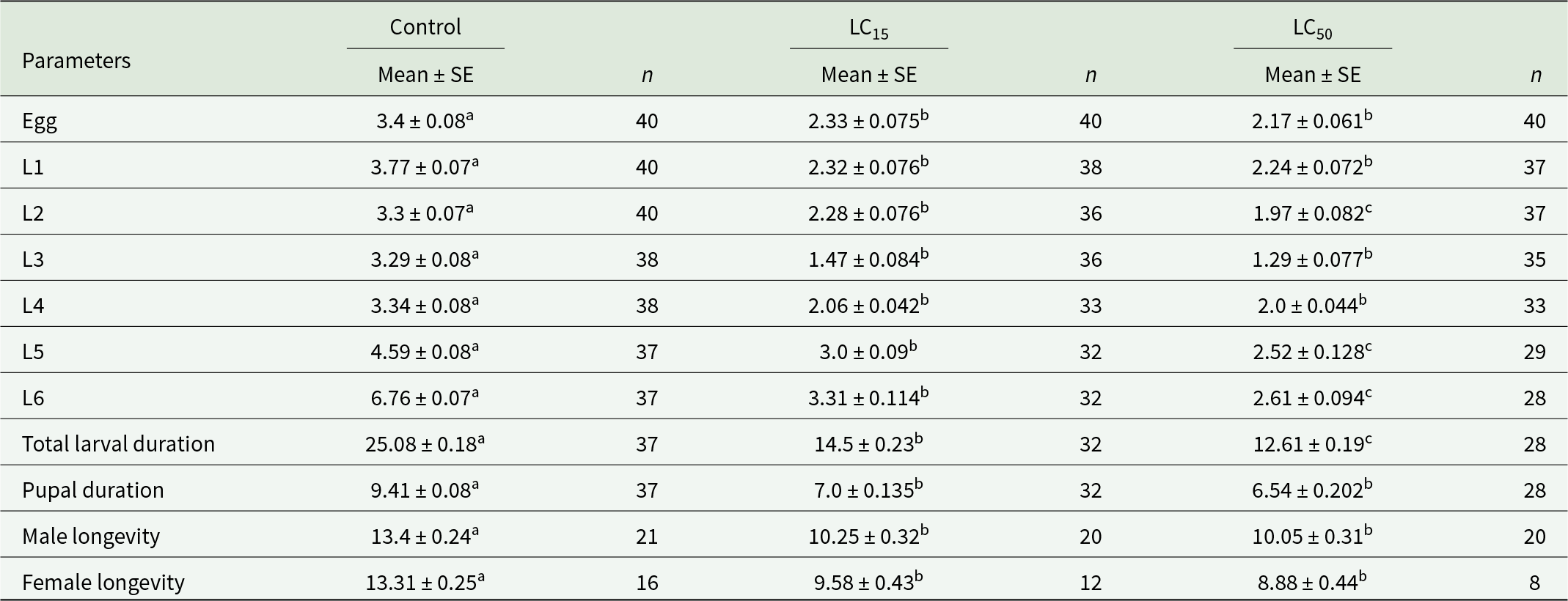
Transgenerational parameters including time required for eggs to hatch, adult longevity, pupal duration, fecundity, pre-oviposition period, and larval duration were evaluated in the F1 of S. frugiperda (table 3). Egg duration in the LC50 group showed early egg hatching (2.17 days), while treatments at the sub-lethal concentration (LC15) (2.33 days) and for the control group (3.4 days) (F = 86.5; df = 2; P < 0.0001) were extended (table 3). Extended larval duration was recorded in both the control and the LC15 treatment. For the control, the maximum larval duration was 25.08 days, while it was 12.61 days for the LC50 group and 14.5 days for the LC15 group (F = 1147; df = 2; P < 0.0001) (table 3). Maximum pupal duration was longer in the control group (9.41 days) than in the LC50- (6.54 days) and LC15-treated insects (7.0 days) (F = 130; df = 2; P < 0.0001) (table 3).
Impacts of C. fumosorosea on demographic parameters of the F1 generation of S. frugiperda

L1–L6 = 1st to 6th instar larvae.
Means marked by the same lowercase letter within the same row are not different (P ˂ 0.05) based on a paired bootstrap test. Standard errors (SEs) are estimated by 100,000 resampling using the bootstrap technique.
All the parameters are shown in the number of days.
The effect of fungal treatment (LC50 and LC15) on fecundity and longevity of S. frugiperda was notable in table 3. F1 male longevity was reduced to 10.05 and 10.25 days post treatment for the LC50 and LC15 treatments, respectively, while maximum male longevity was recorded in the control group (13.4 ± 0.24) (F = 41.9; df = 2; P < 0.0001). A parallel tendency was observed for female longevity, where maximum longevity of the LC50-treated insects was 8.88 ± 0.44 days followed by 9.58 ± 0.43 days for the LC15-treated insects and 13.31 ± 0.25 days for the control group (F = 46.9; df = 2; P < 0.0001) (table 3). Fecundity was also significantly impacted by fungal treatment. The least number of eggs was recorded in the LC50 (379.23 ± 25.018) and LC15 (446.36 ± 24.64) treatments, while the greatest fecundity was recorded in the control group (1452.14 ± 41.91) (F = 299; df = 2; P < 0.0001) (table 3).
Influence of C. fumosorosea on the population parameters of S. frugiperda
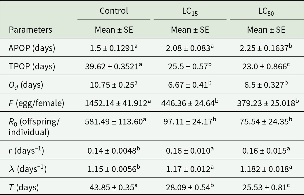
The APOP and the TPOP were significantly different between the treated and control groups (P = 0.0004 and P < 0.0001, respectively) (table 4). A noteworthy difference was noticed in the number of oviposition days (Od) among the LC50- (10.75 days) and LC15 (6.67 days)-infected groups, and the control group (P < 0.0001) (table 4). The r of the LC50 (0.167) and LC15 (0.161) groups differed considerably from the control group (0.144) (P ˂ 0.0001) (table 4). A substantial decrease in R 0 was detected in insects that received the highest concentration of fungus. The greatest net reproductive rate was seen in the control group (581.49 offspring per individual), which was also significantly decreased to 75.54 and 97.11 offspring per adult in the LC50 and LC15 treatments, respectively (P ˂ 0.0001) (table 4). Mean generation time (T) was 43.85 days in the control and significantly declined to 25.53 and 28.09 days in the LC50 and LC15 treatments, respectively (P < 0.0001) (table 4). Mean finite rate of increase (λ) showed non-significant results in the LC50 (1.18 days) and LC15 (1.17 days) treatments, while the control (1.15 days) was significantly different than both treatments (P < 0.0001) (table 4).
Reproduction and life table parameters (mean ± SE) of S. frugiperda

Means marked by the same lowercase letter within the same row are not different (P ˂ 0.05) based on a paired bootstrap test. Standard errors (SEs) are estimated by 100,000 resampling using the bootstrap technique.
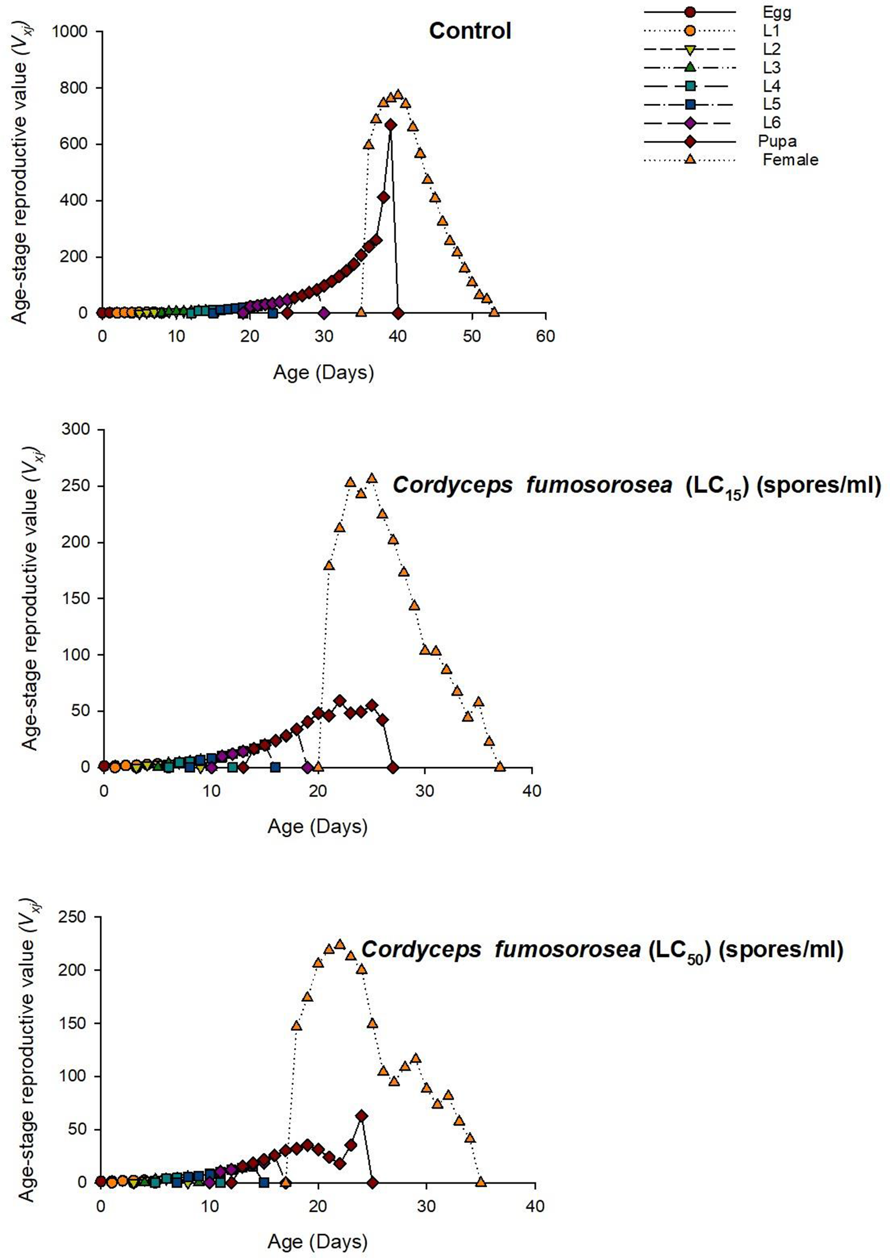
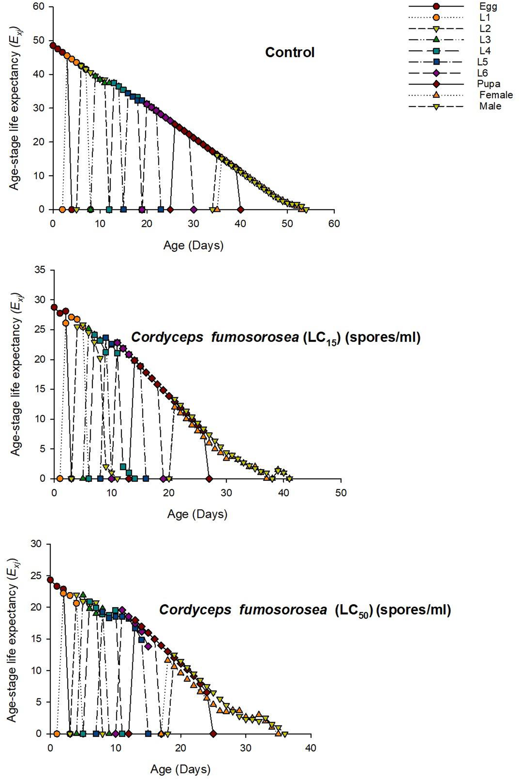
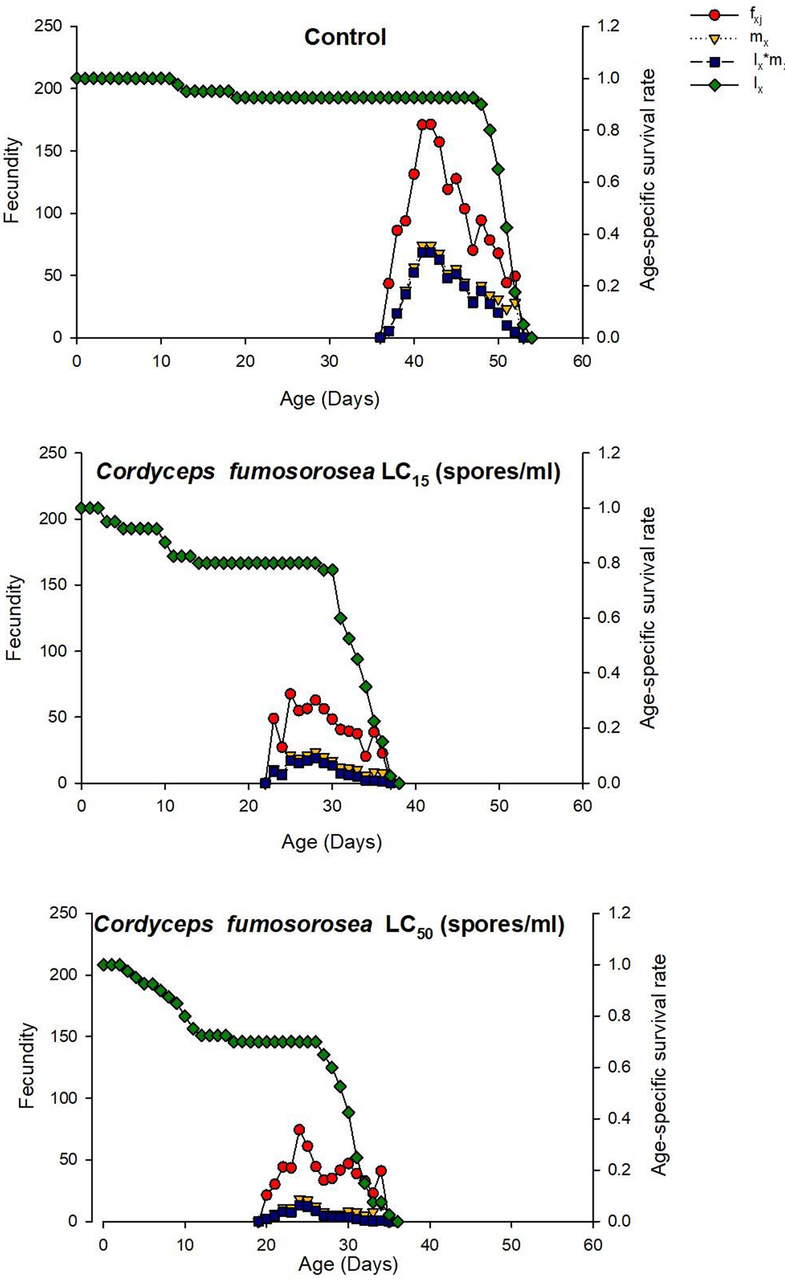
Age-stage survival sxj showed intersecting curves for all stages (fig. 1). The maximum heights of these graphs explained the outlines of differentiation at all stages. After comparison, sxj for females and males extended to the highest values of 0.2 and 0.5, respectively, post treatment with LC50 and 0.3 for female and 0.5 for male post treatment with LC15 compared to the control for female 0.4 and for male 0.52 (fig. 1). The graphs also indicated a delay in the adult’s emergence in the controls. Reproductive curves (vxj) for treated females indicated maximum reproduction at 22 days for the LC50 group and 25 days in the LC15 group compared to 40 days for untreated females (fig. 2). The data also revealed that the treated population had fewer fecundity days than the un-infected population (fig. 2). Age-stage life expectancy exj of S. frugiperda displayed that the L1 controls had longer survival than the treated groups (fig. 3). In general, the overall life period in the control was extended compared to other groups. Adult eclosion occurred later in the untreated groups compared to the infected groups. The peak of mx was higher in the untreated group, while lxmx was higher in the treated groups (fig. 4).
Age-stage survival (sxj) of S. frugiperda infected with C. fumosorosea. Different symbols and colours denote the life stages of S. frugiperda.

Reproductive value of age-stage (vxj) of S. frugiperda infected with C. fumosorosea. Different symbols and colours denote the life stages of S. frugiperda.

Age-stage life expectancy (exj) of S. frugiperda infected with C. fumosorosea. Different symbols and colours denote the life stages of S. frugiperda.

Age-specific life fecundity (mx), age-specific survival rate (lx), and age-specific maternity (lxmx) of C. fumosorosea-treated and -untreated S. frugiperda. Different symbols and colours denote the life stages of S. frugiperda.

Enzymatic activities in C. fumosorosea-treated samples of S. frugiperda
GST activity
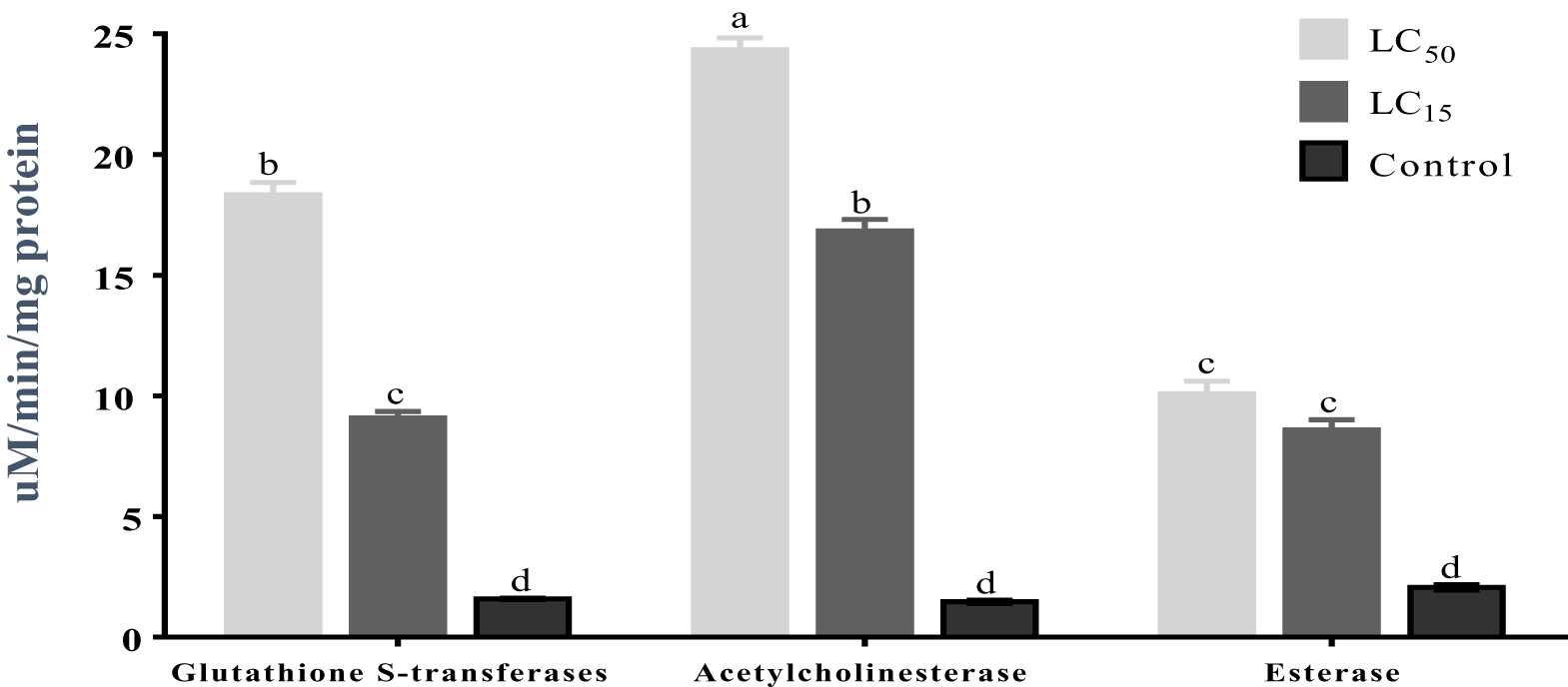
The maximum GST activity (18.34 µmol/min/mg protein) was observed in insects treated with the LC50 (2 × 10⁷ spores mL−1) on the 7th day post treatment (F = 480; df = 8; P < 0.0001), followed by the activity in the larvae treated with the LC15 (fig. 5). Minimum GST activity (1.33 µmol/min/mg protein) was recorded in the control group.
The mean ± SEM activity of GST, EST, and AChE in S. Frugiperda post treatment with C. fumosorosea. Same letters over bars indicate no statistical difference (P ≤ 0.05) in Tukey’s test.

AChE activity
The maximum AChE activity (24.3 µmol/min/mg protein) among treatments was detected in the LC50-treated group on the 7th day post treatment (F = 480; df = 8; P < 0.0001) (fig. 5). The lowest AChE activity (1.46 µmol/min/mg protein) was observed in the controls.
EST activity
The highest maximum EST activity (10.09 µmol/min/mg protein) was noted for the LC50-treated group on the 7th day post treatment (F = 480; df = 8; P < 0.0001) (fig. 5). The minimum EST activity (µmol/min/mg protein) was detected in the controls.
Discussion
The outcomes of this research provide important understandings of the virulence of C. fumosorosea against S. frugiperda and its effect on the demographic parameters of F0 and F1 generations. This information can be useful in evolving Integrated Pest Management plans of S. frugiperda at doses that increase the mortality rate and decrease lethal time. Cordyceps fumosorosea showed maximum larval mortality of 85% against S. frugiperda in the present investigation, which is comparable to the 97.42% mortality of S. frugiperda reported using B. bassiana at a concentration of 9.43 × 10⁸ conidia mL−1 (Montecalvo and Navasero, Reference Montecalvo and Navasero2021). The results of our trial are supported by Ullah et al. (Reference Ullah, Arshad, Abdullah, Khalid, Iftikhar and Zahid2018) who described 73.8% mortality of Diaphorina citri Kuwayama after fungal infection. Mortality rates of 60–90% were reported in other insect pests such as (Dysdercus koenigii Fabricius, Oxycarenus hyalinipennis (Costa), Callosobruchus chinensis (Linnaeus), Culex pipiens (Linnaeus), and Spodoptera litura (Fabricius) owing to infection by C. fumosorosea (Khan et al., Reference Khan, Freed, Zafar and Farooq2014, Reference Khan, Freed, Zafar, Farooq, Shoukat, Ahmad, Li, Zhang, Hua and Shoukat2018; Shoukat et al., Reference Shoukat, Freed and Ahmad2016; Ullah et al., Reference Ullah, Khan, Khan, Jahangir, Habib, Mohammad, Khpalwak, Aziz, Sardar and Qazi2022; Zemek et al., Reference Zemek, Hussein and Prenerová2012), which is also consistent with our results.
Insects have several defences against pathogen ingress. For example, the hydrophobic nature of chitin in an insect’s cuticle serves as a defensive barrier against pathogens. Cordyceps fumosorosea produces hydrophobins that counteract this hydrophobic barrier, which helps fungal spores adhere more effectively to an insect’s cuticle (Dar et al., Reference Dar, Rather and Kandoo2017; Liu et al., Reference Liu, Xie, Xue, Zhang and Zhang2011). EPF affect insects by multiple mechanisms, comprising of mechanical damage by hyphal penetration, nutrient exhaustion, tissue degradation, and the production of secondary metabolites that may contribute to host mortality (Hajek and St, Reference Hajek and St1994). The induced mortality is largely due to the successful penetration of the host cuticle by fungal conidia, followed by vegetative growth within the host haemocoel, leading to systemic damage and death (Mannino et al., Reference Mannino, Huarte-Bonnet, Davyt-Colo and Pedrini2019). Transgenerational impacts of C. fumosorosea were estimated in the F0 and F1 generations of S. frugiperda, which revealed retarded development of different life table parameters such as decreased fecundity in treated insects compared to controls which propose that fungal toxins had affected the developmental parameters of the insect. Previous investigations found similar effects where lower fecundity was recorded for Plutella xylostella (Linnaeus) (Zafar et al., Reference Zafar, Shoukat, Zhang, Freed, Xu and Jin2020), Tetranychus urticae Koch (Seyed-Talebi et al., Reference Seyed-Talebi, Kheradmand, Talaei-Hassanloui and Talebi-Jahromi2012), R. ferrugineus (Gindin et al., Reference Gindin, Levski, Glazer and Soroker2006), Aedes albopictus (Skuse) (Shoukat et al., Reference Shoukat, Zafar, Shakeel, Zhang, Freed, Xu and Jin2020a), S. litura (Kaur et al., Reference Kaur, Kaur, Kaur and Kaur2011), and Musca domestica, Linnaeus (Farooq and Freed, Reference Farooq and Freed2016) after treatment with fungi. Another life table parameter altered by fungal infection in the current experiment was a decline in larval duration of S. frugiperda. Previous studies also showed reduced duration of immature stages (egg to pupae) of Ae. albopictus (Shoukat et al., Reference Shoukat, Hassan, Shakeel, Zafar, Li, Freed, Xu and Jin2020b) and P. xylostella (Zafar et al., Reference Zafar, Shoukat, Zhang, Freed, Xu and Jin2020) after exposure to a different EPF. Our outcomes revealed that infection with sub-lethal and lethal concentrations of C. fumosorosea reduced female and male longevity in the F0 and F1 generations. Plutella xylostella (Zafar et al., Reference Zafar, Shoukat, Zhang, Freed, Xu and Jin2020), Ae. albopictus (Shoukat et al., Reference Shoukat, Zafar, Shakeel, Zhang, Freed, Xu and Jin2020a), T. urticae (Seyed-Talebi et al., Reference Seyed-Talebi, Kheradmand, Talaei-Hassanloui and Talebi-Jahromi2012), and M. domestica (Farooq and Freed, Reference Farooq and Freed2016) showed considerably lower life spans after treatment with M. anisopliae and B. bassiana. Anoplophora glabripennis (Motschulsky) also experienced shorter adult survival post infection of EPF (Dubois et al., Reference Dubois, Hajek, Jiafu and Li2004). A similar trend was recorded for fungus-infected Trogoderma granarium Everts and R. ferrugineus, where adult survival was reduced (Sagheer and Sahi, Reference Sagheer and Sahi2019; Yasin et al., Reference Yasin, Wakil, Ghazanfar, Qayyum, Tahir and Bedford2019).
Fungal infections had a substantial effect on the activity of detoxification enzymes of S. frugiperda. Xenobiotics can be detoxified by enzymes such as GSTs, ESTs, and AChEs, which reduce the toxicity of these compounds and can lead to resistance development in insects, particularly against insecticides (Serebrov et al., Reference Serebrov, Gerber, Malyarchuk, Martemyanov, Alekseev and Glupov2006). These enzymes may also be induced in response to pathogen infection. However, continuous exposure to such compounds can lead to the overactivation of these enzymes, contributing to the development of insecticide resistance in insect populations. In the current research, EST activities were elevated in S. frugiperda larvae after C. fumosorosea infection. Similar results were recorded in D. citri post C. fumosorosea infection (Naeem et al., Reference Naeem, Freed and Akmal2020). Serebrov et al. (Reference Serebrov, Alekseev and Glupov2001) also reported high activity of ESTs in Galleria mellonella (Linnaeus) after M. anisopliae and B. bassiana infection. In our study, AChE activity was also higher post fungal infection on days 7 with the higher concentration of C. fumosorosea. Similar results were recorded in Helicoverpa armigera (Hübner) and L. migratoria post treatment with C. fumosorosea and M. anisopliae (Bilal et al., Reference Bilal, Freed, Ashraf and Muhammad2017; Cao et al., Reference Cao, Jia, Zhao, Wang, Tu, Wang, Nong and Zhang2016). GST activity was also higher in S. frugiperda post C. fumosorosea infection compared to the control. Our findings are comparable with those of Zibaee et al. (Reference Zibaee, Bandani and Tork2009) and Tian et al. (Reference Tian, Diao, Liang, Hao, Arthurs and Ma2015), who reported maximum activity of GST in B. tabaci and Eurygaster integriceps Puton post C. fumosorosea and B. bassiana infection, respectively.
The objective of the current research was to evaluate the impact of both sub-lethal and lethal concentrations of C. fumosorosea on the demographic parameters of S. frugiperda. Exposure to C. fumosorosea influenced S. frugiperda survival, fecundity, and development. Moreover, changes in the activities of enzymes were correlated with fungal infection and may reflect part of the insect’s physiological response to stress, but do not directly demonstrate the mechanisms underlying mortality caused by C. fumosorosea. Further studies involving gene knock-down or enzyme inhibition would be required to establish a causal relationship. These outcomes highlight the potential of C. fumosorosea as a microbial agent for managing S. frugiperda populations. However, further research is needed to assess C. fumosorosea under field conditions and its impact on non-target organisms, including potential natural predators for their incorporation in integrated pest management strategies.
Author contributions
S.R. and S.F. conceived and designed the research, analysed the data, and wrote the manuscript. S.R., H.S., and M.A. conducted experiments. A.N. and R.A. analysed the data. All authors approved the paper for submission.
Competing interests
The authors declare no competing interests.