Hypertrophic cardiomyopathy is an autosomal dominant, heterogeneous disorder characterized by unexplained ventricular hypertrophy, impaired diastolic function, and an increased risk for sudden death, especially in children and young adult.Reference Maron, Gottdiener and Epstein1–Reference Maron3 Over 200 mutations in at least 10 genes encoding sarcomeric proteins have been identified.Reference Chung, Tsoutsman and Semsariuan4–Reference Moolman, Corfield and Posen6 Left ventricular hypertrophy closely mimicking hypertrophic cardiomyopathy may be related to mutations in other genes causing multisystemic disorders, such as Noonan or LEOPARD syndromes, two allelic diseases also frequently due to genetic mutations in the RAS cascade.Reference Tartaglia, Mehler and Goldberg7–Reference Tartaglia, Pennacchio and Zhao10
Several clinical and echocardiographic studies have addressed the morphological and functional findings in patients with hypertrophic cardiomyopathy. Less is known regarding these features of left ventricular hypertrophy when seen in the setting of the Noonan or LEOPARD syndromes. Tissue Doppler imaging has proven to be a useful diagnostic tool for monitoring diastolic function in children with hypertrophic cardiomyopathy.Reference Nagueh, Middleton and Kopelen11–Reference Rajiv, Vinereanu and Fraser14 Particularly, the ratio between the peak velocity of transmitral early filling (E) and the peak velocity (Ea) of early diastolic mitral annular displacement, the E/Ea ratio, has been shown to correlate significantly with left ventricular end-diastolic pressure, and to predict adverse clinical outcomes, such as death, cardiac arrest, ventricular tachycardia and significant cardiac symptoms in young patients with hypertrophic cardiomyopathy.Reference McMahon, Nagueh and Pignatelli15 As far as we know, however, there are no studies comparing the assessment of left ventricular function in children with hypertrophic cardiomyopathy and those with Noonan or LEOPARD syndromes and left ventricular hypertrophy. Our study, therefore, was undertaken non-invasively to evaluate the morphological and functional echocardiographic features potentially affecting symptoms and clinical outcome in suitably chosen groups of these children.
Materials and methods
We enrolled 62 consecutive patients with left ventricular hypertrophy followed up in two different centres, namely the Ospedale Monaldi, Naples, and Ospedale Bambino Gesù, Rome, with the diagnosis of hypertrophic cardiomyopathy. According to clinical features of the Noonan and LEOPARD syndromes, we based the diagnosis of Noonan’s Syndrome on the Noonan Syndrome Scoring System,Reference Van der Burgt, Berends and Lommen16 whereas the diagnosis of LEOPARD Syndrome was based on the criterions established by Voron and Digilio with their respective colleagues.Reference Voron, Hatfield and Kalkhoff17, Reference Digilio, Sarkozy and de Zorzi18 On this basis, we were able to divide the patients into groups with idiopathic hypertrophic cardiomyopathy on the one hand, these being aged from 1 to 16 years, and on the other hand those with Noonan’s or LEOPARD syndromes, also aged from 1 to 16 years. We excluded any patients with systemic hypertension, fixed obstruction of the left or right ventricular outflow tracts, or metabolic disorders. In 7 of the 17 patients with Noonan or LEOPARD syndrome, the clinical diagnosis was confirmed by genetic analysis. Our study was approved by the internal ethical committee of the Monaldi Hospital.
Echocardiographic evaluation
Echocardiographic studies were performed with commercially available instruments (Aplio A; Toshiba-Japan). We characterised the features of hypertrophic cardiomyopathy on the basis of the magnitude and distribution of left ventricular hypertrophy, the degree of left ventricular obstruction, left ventricular diastolic function, right ventricular hypertrophy and obstruction, and the diameters of the coronary arteries. The magnitude and distribution of left ventricular hypertrophy were assessed in parasternal short axis plane by dividing the left ventricle into 4 quadrants, namely the anterior and posterior septal quadrants, and the inferior and antero-lateral mural segments. Left ventricular mural thickness was measured in each of these quadrants in end-diastole in correspondence to the R wave on electrocardiogram at the levels of the mitral valve and the papillary muscles. The mural thickness was also measured at the apex. Maximal thickness was defined as the greatest thickness in any of these segments. In addition, to adjust for age- and growth-related changes in thickness, the maximal thickness was normalised for body surface area and expressed as Z-score relative to its normal distribution. Normal data had already been determined in 101 subjects aged 3 days to 16 years in our echocardiographic laboratory. The Z-score indicates the position of each measurement relative to the normal population expressed as standard deviation from the mean, where both the mean and standard deviations are specific for the age and body surface area. A Z-score of 0 represents the normal mean value, and the normal 95% confidence interval is −2 to +2.
Left ventricular hypertrophy was defined as asymmetric when it mainly involved the ventricular septum, concentric when it involved all the myocardial walls, eccentric when only the antero-lateral wall was involved, and apical, when it was localized at the apex.Reference Shapiro and McKenna19 The maximum gradient across the left ventricular outflow tract was determined in apical long axis view by the continuous wave Doppler and calculated by applying the modified Bernoulli equation. Obstruction in the left ventricular outflow tract was considered significant when the maximum gradient was over 30 mmHg. In addition, pulsed Doppler was used to obtain the pattern of mitral inflow, permitting calculations of peak E, A, and the E/A ratio, at the tip of the mitral valvar leaflets in the apical 4-chamber view. Tissue Doppler imaging was recorded in the apical 4-chamber view to obtain longitudinal annular velocities at the lateral mitral corner, again permitting calculation of Ea, Aa, and the Ea/Aa ratio. All filters and gains were lowered to allow a clear tissue signal, and to minimize background noise. All the measures were averaged over three cardiac cycles. In addition, we calculated the ratio of the velocity of early transmitral left ventricular filling to early diastolic mitral annular velocity, which correlates with left ventricular filling pressure.Reference Nagueh, Lakkis and Middleton12 Right ventricular hypertrophy was evaluated by assessing right ventricular mural thickness in the parasternal long- and short-axis and apical views.Reference McKenna, Kleinebenne and Nihoyannopoulos20 Pulmonary valvar dysplasia was diagnosed in presence of thickening and limited movement of the valvar leaflets.Reference Musewe, Robertson and Benson21 The dynamic right ventricular gradient was determined, in subcostal and parasternal short- axis views, by colour, pulsed, and continuous wave Doppler, again applying the modified Bernoulli equation.Reference Houston, Sheldon and Simpson22, Reference Limongelli, Pacileo and Marino23
The diameters of the coronary arteries, measured in the diastolic frame, were also assessed and deemed to be dilated when their diameters exceeded the 95th centile values for body surface area.Reference De Zorzi, Colan and Gauvreau24 Coronary arterial dilation was considered severe in presence of Z score equal to or greater than +4. Coronary angiography was performed when abnormal dimensions were detected at the echocardiographic evaluation.
Statistical analysis
Statistical analysis was performed with SPSS statistical software (SPSS for Windows 13.1). Data were expressed as mean with standard deviations. Z values were calculated following previous reported data.Reference Henry, Ware and Gardin25 Differences between continuous variables were assessed with the non-parametric Mann-Whitney test. Differences between categorial variables were defined by the chi-squared test. A probability value of p less than 0.05 was considered significant. Inter-observer variability was assessed by analysing 10 digital cross-sectional and Doppler digital frames from different, randomly chosen subjects by 2 independent investigators. For intra-observer variability, 10 cross-sectional and Doppler digital frames were analyzed twice by one investigator within 1 month. The second round of inter-observer measures was blinded to results form initial measures.
Results
Patients
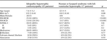
Clinical characteristics of the patients studied are shown in Table 1. A cardioverter defibrillator had been implanted in 2 of the 45 patients with hypertrohpic cardiomyopathy after resuscitation from cardiac arrest. Only one of the patients with Noonan or LEOPARD syndromes had undergone implantation of a cardioverter defibrillator, in this case because of a significantly increased mural thickness, with a Z score of greater than +12, and a hypotensive blood pressure response to exercise test.
Clinical characteristics of the patients studied.

BSA = body surface area; FH = familial history; HCM = hypertrophic cardiomyopathy; SCD = sudden cardiac death; NYHA = New York Heart Association.
Echocardiographic analysis
The distribution of left ventricular hypertrophy was similar in the two groups. Asymmetric hypertrophy was the most common pattern, found in 34 (75.6%) of those with hypertrophic cardiomyopathy and 13 (76.4%) of those with Noonan or LEOPARD syndromes. Hypertrophy was concentric in 9 (20%) of those with hypertrophic cardiomyopathy, and in 3 (17.6%) of the others, and eccentric in 2 (4.4%) of those with hypertrophic cardiomyopathy, and in 1 (6%) of those with Noonan or LEOPARD syndromes. None of the patients had apical left ventricular hypertrophy.
The magnitude of left ventricular hypertrophy, expressed as the Z-score for maximal mural thickness, was significantly higher in those with Noonan or LEOPARD syndromes, at 8.9 + 4.3 as opposed to 6.4 + 3.7 (p = 0.03).
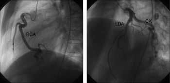
Right ventricular hypertrophy, found in 5 (29.4%) patients with Noonan or LEOPARD syndromes, in absence of fixed obstruction to the right ventricular outflow tract, was not encountered in any of the patients with hypertrophic cardiomyopathy. Dilation of the coronary arteries was also detected in 5 of the 17 patients with Noonan or LEOPARD syndromes, being present in both coronary arteries in 4 patients, and only the right coronary artery in 1 patient (Fig. 1). Coronary angiography, performed in these patients, confirmed the echocardiographic findings, and did not show any stenosis.
Coronary angiography in this 8 year-old boy with Noonan’s syndrome shows significant and diffuse dilation of both coronary arteries, with no evidence of stenosis. Abbreviations: (Cx = circumflex artery; LAD = left anterior descending; RCA = right coronary artery).

Standard Doppler and pulsed tissue Doppler analysis
Significant obstruction of the left ventricular outflow tract was more common in patients with Noonan or LEOPARD syndromes, found in 9 (53%), than in those with idiopathic hypertrophic cardiomyopathy, where it was present in only 7 (15%; p = 0.002). No patient showed a midventricular gradient. Dynamic obstruction of the right ventricular outflow tract was found in 4 of the 5 patients with Noonan or LEOPARD syndromes and right ventricular hypertrophy. There were no significant differences between the groups in terms of transmitral Doppler inflow velocities. Compared to those with idiopathic hypertrophic cardiomyopathy, however, the patients with Noonan or LEOPARD syndromes showed lower early diastolic mitral annular velocities and higher end-diastolic velocities, at Ea 9.1 versus 13.1 centimetres/second (p = 0.02), and Aa 11.6 versus 7.6 centimetres/second (p = 0.02), respectively, a reduced Ea/Aa ratio, at 0.8 versus 1.6; p = 0.05, and a higher E/Ea ratio at 13.7 versus 8.7 (p = 0.01).
Inter- and intra-observer variability
The inter- and intra-observer variability was good both for cross-sectional and Doppler measures, at less than 5%. It is likely that the good agreement is partially due to the high quality ultrasound systems we used, and partially due to the relative young age of our patients.
Discussion
Our data show that right ventricular hypertrophy, dilation of coronary arteries, and more severe left ventricular diastolic dysfunction and hypertrophy are the characteristics of the myocardial changes found in patients with Noonan and LEOPARD syndromes. Previous investigatorsReference Nishikawa, Ishiyama and Shimojo26 did not find any difference in terms of distribution of hypertrophy and incidence of obstructed left ventricular outflow tracts when comparing patients with idiopathic hypertrophic cardiomyopathy and those with Noonan or LEOPARD syndromes. Asymmetric septal hypertrophy was the most common pattern in both our groups, with no cases of apical hypertrophy. In our population, however, gradients across the left ventricular outflow tract were present in more than half of the patients having left ventricular hypertrophy in the setting of Noonan or LEOPARD syndromes, but in only about one-sixth of those with idiopathic hypertrophic cardiomyopathy. The magnitude of left ventricular hypertrophy, expressed as Z-score for maximal mural thickness in order to compare patients with different body surface area, was significantly higher in the patients with Noonan and LEOPARD syndromes. Of interest, a novel mutation in the PTPN11 gene in a patient with LEOPARD syndrome has recently been associated with rapidly progressive left ventricular hypertrophy.Reference Takahashi, Kogaki and Kurotobi27
As documented previously,Reference Pearl28 we found right ventricular hypertrophy in just under one-third of our patients with Noonan or LEOPARD syndromes, even in absence of pulmonary valvar stenosis, but in none of our patients with idiopathic hypertrophic cardiomyopathy. Although a mild increase in the diameter of the coronary arteries is quite common in the latter patients, correlating with the increased left ventricular mass,Reference Krams, Kofflard and Duncker29 we found severe dilation of these arteries in about one-third of our patients with Noonan or LEOPARD syndromes, a finding confirmed by selective angiography.Reference Limongelli, Pacileo and Marino23 Giant coronary aneurysms have recently been described in a patient with Noonan syndrome,Reference Loukas, Dabrowski and Kantoch30 and considered potentially to reflect a vasculitic process superimposed on the connective tissue defect associated with the genetic disorder. Although none of our patients with dilated coronary arteries showed any coronary arterial stenosis, longer follow-up is mandatory better to define their prognostic impact.
In adults with hypertrophic cardiomyopathy, Doppler evaluation of transmitral flow showed lower velocities for the mitral E wave, and higher peak velocities for the A wave, along with a prolonged deceleration time for the E wave and a mitral E/A ratio less than one, suggestive of impaired relaxation. Disorganized aggregation of the myocytes, myocardial fibrosis, and potential ischemia are some of the major determinants of abnormal diastolic function in these patients. As the indexes of mitral inflow are influenced by many factors, such as loading conditions, heart rate and age, the overlap is considerable and many patients have a normal or pseudonormal pattern.Reference Nagueh, Middleton and Kopelen11–Reference McMahon, Nagueh and Pignatelli15 It has recently been shownReference McMahon, Nagueh and Pignatelli15 that, in children with hypertrophic cardiomyopathy, unlike the transmitral E and A wave velocities, the E/Ea ratio, when assessed by Doppler tissue imaging at the mitral annulus, was able to define those patients at major risk of adverse clinical outcome. Data from our patients with Noonan and LEOPARD syndromes showed lower early diastolic, and higher end-diastolic velocities, with a mean Ea/Aa ratio less than one, again suggestive of impaired left ventricular diastolic relaxation. Moreover, our syndromic patients showed higher values of mitral E/Ea ratio, which is suggestive of higher left ventricular filling pressure and decreased compliance.Reference Nagueh, Lakkis and Middleton12 As a consequence, patients with left ventricular hypertrophy in the setting of Noonan and LEOPARD syndromes seem to have more severe diastolic dysfunction, due to either abnormal relaxation and/or reduced compliance. As histopathological studiesReference Burch, Mann and Sharland31 failed to show any difference in terms of myocardial fibrosis and/or disarray between patients with idiopathic hypertrophic cardiomyopathy and Noonan and LEOPARD syndromes, the major degree of left ventricular hypertrophy, expressed as the Z-score for maximal mural thickness, might explain the worse diastolic function found in our patients with Noonan and LEOPARD syndromes. Of note, patients with Noonan syndrome and left ventricular hypertrophy are known to die more often of cardiac failure than sudden death when compared to those with hypertrophic cardiomyopathy.Reference Nishikawa, Ishiyama and Shimojo26 More recently, it has been shown that, in children with hypertrophic cardiomyopathy, the co-existence of Noonan’s syndrome is an independent predictor of death in cardiac failure.Reference Ostman-Smith, Wettrell and Keeton32
In conclusion, compared to children with hypertrophic cardiomyopathy, those with left ventricular hypertrophy in the settings of Noonan and LEOPARD syndromes show greater degrees of left ventricular hypertrophy, expressed as the Z-score for maximal mural thickness, and greater diastolic dysfunction, due to both abnormal relaxation and reduced compliance. They also show an incidence of left ventricular outflow tract obstruction, and dynamic obstruction of the right ventricular outflow tract with right ventricular hypertrophy, as well as an incidence of dilated coronary arteries. These morphological and functional findings should be taken into account when seeking to explain the different symptoms and clinical events, and when defining the most appropriate therapeutic options, in children with left ventricular hypertrophy of different aetiologies.