Introduction
The concept that many adult diseases can have their origins traced back to fetal life has been termed the ‘developmental origins of health and disease’ hypothesis.Reference Barker, Gluckman and Godfrey 1 In addition to altered conditions in fetal life, increasing number of epidemiological and experimental studies indicate that events in early postnatal life can also have life-long consequences for the cardiovascular health of the adult.Reference Nuyt and Alexander 2 – Reference Khorram, Momeni, Desai and Ross 4 Premature birth and low birth weight have been correlated with elevated blood pressure, impaired endothelial function and increased arterial stiffness in children, adolescents and adults.Reference Tauzin, Risso, Buffat, Serra and Simeoni 5 , Reference Cheung, Wong, Lam and Tsoi 6
Experimental data suggest that oxidative stress may be an initiating trigger in programming of adult diseases. We have shown in a previous study that neonatal hyperoxic stress is associated with elevated blood pressure and with vascular dysfunction in conductance arteries of adult rats.Reference Yzydorczyk, Comte and Cambonie 7 However, functional repercussions of the later observations have not been demonstrated. Pulse-wave velocity (PWV) is an indirect measure of the biophysical properties of a vessel (arterial stiffness or distensibility) and a very sensitive marker of cardiovascular health.Reference Safar and Laurent 8 Increased arterial stiffness is now recognized as an independent risk factor for cardiovascular events such as primary coronary events, stroke and mortality.Reference Laurent, Boutouyrie and Asmar 9 Its measurement in rats is mostly based on invasive hemodynamics measurements, prohibiting longitudinal studies particularly relevant in models of developmental programming of cardiovascular dysfunctions. Ultrasound assessment of aortic PWV is a simple and fast alternative. Previous studies have only assessed PWV on a small portion of the thoraco-abdominal length (4–5 cm), and this length was an estimate since the measure was taken on the surface of the animal.Reference Hartley, Taffet, Michael, Pham and Entman 10 , Reference Soucy, Ryoo and Benjo 11 The combination of external measurement and the short distance may be a major source of imprecision. Therefore, we propose to evaluate an adaptation of the technique to measure the aortic PWV on the full length of the aorta. Moreover, to the best of our knowledge, as non-invasive aortic PWV has never been reported in Sprague–Dawley (SD) rats, reference control values are not yet available. The aims of this study were (1) to verify the feasibility and validity of measuring aortic PWV in SD rats by ultrasound; (2) to use the technique to compare aortic PWV in adult rats exposed to oxygen as newborns v. controls; and (3) to identify an anatomic marker that correlates the best with the full length of the aorta, allowing determination of aortic PWV in longitudinal studies.
Materials and methods
Animals
Animals were used according to a protocol of the animal care committee of the Centre Hospitalier Universitaire Sainte-Justine in accordance with the principles of the Canadian Council on Animal Care Guide for the Care and Use of Experimental Animals. SD rat pups (Charles River, St-Constant, Québec, Canada) were maintained in 80% O2 (mixture of medical-grade 100% O2 and room air with an oxycycler A82OCV, Biosherix) from day 3–10 of life (hyperoxia group, n = 3 litters) or in room air (control normoxia group; n = 14 litters). Pups were weaned at 4 weeks of age to regular chow. Survival of the pups, their growth and food intake were similar between groups. Both male and female offsprings (no more than two animals per litter) were studied as adults between 6 and 9 months of age.
Experimental procedures
Measurement of aortic PWV by ultrasound
Animals (n = 6 O2-exposed and n = 6 controls) were lightly anesthetized with isoflurane in order to maintain spontaneous breathing. The animals were placed on a warming pad to maintain normothermia in a prone position. The anterior chest hair was shaved using clippers and three prewired neonatal electrodes (Puppydog, Tyco-Kendall Co., Mansfield, MA, USA) were attached on the right forelimb, left forelimb and left hindlimb. With a Siemens Acuson CV70 ultrasound machine (Siemens Medical Solutions Inc., Mountain View, CA, USA) and a 13–5 MHz linear transducer, Doppler tracings of aortic flow and descending abdominal aorta, immediately above the aortic bifurcation, were recorded at a sweep speed of 200 mm/s simultaneously with the echocardiography (ECG) and stored on a CD-Rom. All Doppler measurements were obtained within 5–10 min after the anesthesia induction; the animals were then allowed to recover from the anesthesia and placed back in their cage.
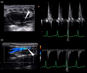
Each tracing was then reviewed with Adobe Acrobat 6.0 Professional software (Adobe Systems Inc., San Jose, CA, USA). After a 300% magnification and off-line calibration, the time elapsed from the peak of the R wave of the QRS to the onset of the flow wave was measured (Fig. 1). For each tracing, at least five measures were performed. The mean value and the coefficient of variation were calculated. For each rat, a time to the onset of the aortic valve (T1) and to the aortic bifurcation (T2) was obtained. The difference (ΔT = T2 − T1) corresponds to the transit time of the pulse wave to travel from the aortic valve to the bifurcation. Five cardiac cycles were used to determine heart rate (HR) during both Doppler recordings. Mean of both values was used for statistical analysis. To assess within-subject inter-sessional variability, measurements were repeated for five cases.
(a) (left) Two-dimensional echocardiography (ECG) of the ascending aorta with the aortic valve clearly identifiable (arrowhead). (right) Doppler and ECG tracings at the level of the aortic valve. Timing lines were drawn at the peak of R wave of ECG and at the onset of the aortic wave (T1). (b) (left) Two-dimensional ECG with color Doppler of the descending aorta with the aortic bifurcation clearly identifiable (arrowhead). (right) Doppler and ECG tracings at the level of the aortic bifurcation. Timing lines were drawn at the peak of R wave of ECG and at the onset of the aortic wave (T2).

Measurement of aortic PWV by intraaortic catheter
Animals (n = 10 controls) were anesthetized with pentobarbital (65 mg/kg i.p.). A polyethylene PE50 catheter was inserted into the aortic arch through the left carotid and another one into the distal abdominal aorta through the left femoral artery. The catheters were connected to a pressure transducer to simultaneously record systolic and diastolic blood pressures at the levels of the aortic arch and abdominal aorta, as well as the HR. Pulse wave was recorded at 500 Hz to define the time difference between the peak pressures at the aortic arch level and the abdominal aorta level. After sacrifice, the distance between the two catheters extremities was measured and divided by the time of propagation wave to calculate PWV (cm/s). For each animal, at least 10 measures obtained at 10 s intervals were recorded and mean was calculated.
Anatomical measures
Rats (n = 6 O2-exposed and n = 18 controls) were sacrificed by aortic exsanguination under general anesthesia (1–4 days after the ultrasound study). Weight, femur length and distance from the manubrium to the anal margin (MA length) were measured. After thoraco- and laparotomy, the aorta was incised immediately (1–2 mm) above the aortic valve and a small wire with a very floppy distal extremity (Arrow Spring-Wire 0.018″ – 45 cm, Arrow Int. Inc., Reading, PA, USA) was gently introduced into the ascending aorta through the aortic arch and pushed until its extremity was visualized by transparency at the aortic bifurcation. A clip was placed on the wire where it entered the aorta. The length of the wire introduced was measured and reported as the length of the full aorta (L): from the aortic valve to the aortic bifurcation. Aortic PWV is calculated as the quotient of the separation distance (L) and the time difference between pulse arrivals, with respect to the R-peak of the ECG (ΔT).
One observer (YM), blinded to the rats group, performed all Doppler and aortic length measurements. For each animal, measures were obtained at least 5 times and showed a high degree of consistency with 5% variability from mean value. On the basis of the anatomic parameter with the greatest correlation with the measured aorta length, an algorithm derived from the regression linear line was established to estimate the aorta length.
Variability measurements
For the non-invasive ultrasound measurement of aortic PWV, to assess intra-observer variability all animals were measured at least five times, and to assess short-term inter-sessional variability five animals had measurements repeated after 30 min. Calculated coefficients of variation were 7.5% for the intra-observer variability and 7% for inter-sessional variability.
Statistical analysis
All values are expressed as means ± s.e. Univariate statistical analysis was performed using the Mann–Whitney test to compare non-parametric features. A linear regression analysis was performed and Spearman's correlation coefficient (R) was calculated to investigate the associations between the throraco-abdominal aortic length (L) and other anatomic parameters measured or calculated. Bland–Altman analysis was used to assess the concordance between the measured and the calculated aorta length. All data were analyzed with commercially available statistical software packages (GraphPad Prism version 5.00 for Windows, GraphPad Software, San Diego, CA, USA). A P-value <0.05 was considered significant.
Results
Control adult rats PWV measured by intraaortic catheters was 413 ± 16 cm/s. When using ultrasound to calculate PWV, the acquisition of the Doppler signal was possible in all rats. The aortic valve was clearly identifiable on a parasternal or suprasternal view of the ascending aorta and Doppler flow waves could be recorded in all cases (Fig. 1). With color Doppler, the abdominal aorta was identified, and either in a transverse or a sagittal view, the aortic bifurcation could be identified in all studies (Fig. 1). The Doppler sample was placed so as to record the flow just above the bifurcation.
Comparison of PWV of O2-exposed v. control adult rats
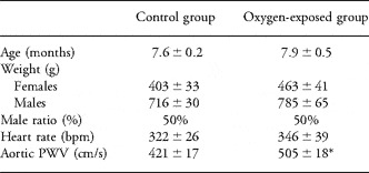
Characteristics of both groups and aortic PWV results are shown in Table 1. There was no significant difference for weight (for each gender), age and HR. Mean aortic PWV was significantly increased in O2-exposed (505 ± 18 cm/s; n = 6) v. control animals (421 ± 17 cm/s; P < 0.01; n = 6). Within each group, there was no difference in age, PWV and HR between genders.
Age, body weight, gender, heart rate and aortic PWV in control and O2-exposed rats.

Aortic PWV, aortic pulse-wave velocity.
All values, except male ratio, are expressed as mean ± s.e.
*P < 0.05.
Correlation between anatomic parameters and aorta length
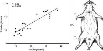
Spearman's correlation coefficient showed the best significant correlation between the aortic and the MA length (R = 0.82, P < 0.0001; n = 24; Fig. 2). Femur length and weight were also significantly correlated but with lower strength (R = 0.77 for both). Therefore, the following algorithm for aortic length estimation on the basis of the MA length was derived from the regression linear line equation:

(right) Full aorta length (L) measured at sacrifice plotted against the length from manubrium to anal margin (MA) measured immediately before euthanasia. (left) Illustration of the measure of the MA length.

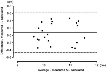
The correlation between the measured and calculated aorta length is shown in Figure 3. The mean difference and the 95% limit of agreement between the two methods were calculated (0.08, −0.47 and 0.63 cm, respectively).
The difference between measured and calculated aorta length (L) plotted against the average of the two corresponding measurements for all studied animals. The mean difference (continuous line) and the limits of agreement (dash line) are shown.

Discussion
In our study, we confirmed that it was feasible to measure the thoraco-abdominal PWV in adult SD rats with a standard ultrasound machine and have validated the method using intraarterial measurements. This ultrasound method allowed us to show a significantly increased aortic PWV in adult rats exposed to high O2 as newborns compared to controls. Finally, we demonstrated that aorta length can be accurately estimated using the manubrium–anal external margin distance measured externally using the equation we derived.
In previous studies measuring PWV by ultrasound, the distance between the two sites of Doppler flow recording was only 4–5 cm and/or was measured externally (i.e. on the surface of the animal) without validation at sacrifice.Reference Hartley, Taffet, Michael, Pham and Entman 10 , Reference Soucy, Ryoo and Benjo 11 The short distance and its external measurement may represent a significant source of error. The aortic valve and the abdominal aortic bifurcation, as selected in this study, are two very easily identifiable anatomic structures localized at both extremities of the aorta allowing aortic PWV assessment on the full length of the aorta.
Comparison with other studies is complex because rat species, age and anesthetic drugs are well recognized to be factors that can influence aortic PWV.Reference Essalihi, Zandvliet and Moreau 12 – Reference Tatchum-Talom, Martel and Marette 14 Indeed, Soucy et al.Reference Soucy, Ryoo and Benjo 11 showed in their study of Wistar rats that PWV increased very significantly with age, from 364 ± 7 cm/s at 3–4 months to 599 ± 19 cm/s at 22–24 months. Only Tatchum-Talom et al.Reference Tatchum-Talom, Martel and Marette 14 performed a study similar to ours with regard to rat species and anesthetic agent. Despite the fact that their rats were probably younger (age not specified, but range weight from 200 to 250 g), they report aortic PWV of 472 ± 42 cm/s in their control group, which is slightly higher than the results in our control group, but nevertheless inferior than what we observed in the O2-exposed group.
The investigation of mechanical and structural properties of arteries is crucial for studies of the mechanisms of developmental origins of cardiovascular disease. Several studies have shown that endothelial-dependent and endothelial-independent vasodilation (a well-recognized precursor of hypertension and atherosclerosis) can be impaired and that flow-mediated dilation is decreased in low-birth weight individuals from childhood to adult life.Reference Franco, Christofalo, Sawaya, Ajzen and Sesso 15 – Reference Martin, Hu, Gennser and Norman 19 PWV has been demonstrated to be an indirect measure of the biophysical properties (arterial stiffness or distensibility) of a vessel, and therefore a promising measure of cardiovascular health. In our study, we noted a significantly greater aortic PWV in adult rats exposed to hyperoxic stress in the neonatal period compared with control rats. This finding is in line with our previous observations that neonatal oxygen exposure is associated with increased blood pressure and impaired endothelium dependent vasodilatation of conductance vessels.Reference Yzydorczyk, Comte and Cambonie 7 Because of technical difficulties, we were not able to record blood pressure simultaneously with aortic PWV, although it is reasonable to assume that the blood pressure was significantly higher in O2-exposed rats v. controls as we have previously reported.Reference Yzydorczyk, Comte and Cambonie 7 As elevated blood pressure can induce increased aortic PWV independent of impaired vascular structure, this raises the following question: what part of the increased aortic PWV is due to increased aortic stiffness and elevated blood pressure?Reference Albaladejo, Laurent and Pannier 20 , Reference Lantelme, Mestre, Lievre, Gressard and Milon 21 However, these two parameters are highly interrelated. Each might be the consequence of the other, but also might result from the same physiopathologic process. Indeed, it has been shown that angiotensin II can (1) elevate blood pressure by enhancing vasoconstriction as well as sensitivityReference Yzydorczyk, Gobeil and Cambonie 22 and (2) increase aortic PWV by promoting structural vascular changes.Reference Simon, Illyes and Csiky 23 Further studies will be needed to clarify this aspect.
The observed increased PWV in adult rats subjected to neonatal hyperoxia are in agreement with studies in humans showing a correlation between altered perinatal conditions (low birth weight, prematurity; both associated with oxidative conditions) and increased indices of arterial rigidity in children as well as adults.Reference Tauzin, Risso, Buffat, Serra and Simeoni 5 , Reference Martin, Hu, Gennser and Norman 19 , Reference Lurbe, Torro, Carvajal, Alvarez and Redon 24 – Reference Oren, Vos and Bos 26 In another animal model, aorta of offsprings of high-fat diet-fed dams have increased arterial stiffness in addition to reduced endothelial cell volume and decreased smooth muscle cell number.Reference Armitage, Lakasing and Taylor 27 Whether conductance vascular structure is altered after neonatal hyperoxic exposure is not known. The proportion of elastin v. rigid collagen is a major determinant of arterial stiffness. Elastin synthesis in the vessels peaks in the prenatal period, mostly approaching term, decreases rapidly after birth and is minimal in the adult aorta.Reference Berry, Looker and Germain 28 In rats, experimentally induced fetal growth inhibition results in lower elastin content of the aorta and persistent changes in aortic wall components.Reference Berry and Looker 29 Composition of the aortic wall and other large conduit arteries is therefore determined relatively early during development, with little possibilities of ‘catch-up’ or compensation later on.
In the perspective to study the evolution of aortic PWV longitudinally, we looked for anatomic measurements that correlate the best with the total aorta length. The best correlation was found with the MA length. The proposed algorithm for aortic length calculation showed a good agreement with the measured aorta length in the 24 rats studied. Indeed, the limits of agreement between the measured and calculated method is about ±0.5 cm (5%). A 0.5 cm variation in aorta length will result in only a 5% variation of the PWV (a 25 cm/s variation for a PWV of 500 cm/s), which could be considered clinically of low relevance. We therefore propose that this method of assessment of total aortic length could be used to assess non-invasively aortic PWV and to monitor the progression of the clinical manifestation of vascular structural changes in different models studying vascular (patho)physiology over time (such as developmental programming of elevated blood pressure as in our studies) in SD rats. To the best of our knowledge, this is the first report of an anatomical marker that could be used as a surrogate of the rat aorta length for the non-invasive measurement of the PWV.
Study limitations
Technical considerations that may be important are the following. First, the ascending aorta was cut as close as possible to the aortic valve; but due to its elasticity, probably the real length of the aorta should be 1 or 2 mm longer. This imprecision represents between 1% and 2% of the mean length of the aorta. Second, because of the rapid velocities, the measured time differences between the onset of the Doppler flow in the ascending and abdominal aorta were relatively short. To minimize this issue, at least five measurements were performed on each Doppler tracings recorded with the highest sweep (200 mm/s) and off-line measurement performed after a 300-times magnification. Despite these technical considerations, the variability of the measures between animals was relatively small considering that statistically significant differences were detected between the groups, despite a relatively small (about 20%) increase in PWV observed in the O2-exposed animals.
Conclusions
Assessment of the aortic stiffness is crucial to monitor cardiovascular health and the effects of preventive strategies and new therapies. In this animal study, PWV, a very sensitive marker of cardiovascular health, could be measured by standard ultrasound. Using this method, we found an increased aortic PWV in adult animals subjected to a brief period of hyperoxic stress as newborns. Whether this observation is associated with structural vascular change remains to be determined. The MA length could be used as a surrogate of the rat aorta length for the non-invasive follow-up of longitudinal studies, providing insight into arterial disease processes, and for monitoring the success of future medical therapies.
Acknowledgments
This work was supported by grants from the Canadian Institutes of Health Research, the Heart and Stroke Foundation of Canada and Pfizer Carviovascular Research Foundation of Canada. Yvan Mivelaz was supported by a research grant from the Swiss National Science Foundation, SICPA Foundation and the Société Académique Vaudoise. Anne Monique Nuyt was supported by a fellowship from the Fonds de la Recherche en Santé du Québec.
Statement of Interest
We have no potential competing interests.