Introduction
Disturbances in redox balance in cells cause oxidative stress, leading to toxic effects on cells through oxidation of plasma membrane lipids, modification of nucleotide bases, breakdown of DNA, and damage to proteins, including enzymes. Oxidative stress is related to the occurrence of various diseases such as Alzheimer’s disease, Parkinson’s disease, cancers, inflammatory diseases, and ageing (Kwiecien et al., Reference Kwiecien, Jasnos, Magierowski, Sliwowski, Pajdo, Brzozowski, Mach, Wojcik and Brzozowski2014; Uttara et al., Reference Uttara, Singh, Zamboni and Mahajan2009; Reuter et al., Reference Reuter, Gupta, Chaturvedi and Aggarwal2010). Reactive oxygen species (ROS) molecules, such as superoxide (O2 −), hydroxyl radical (•OH), and hydrogen peroxide (H2O2), produced by incomplete reduction of oxygen molecules are the main players in cellular oxidative stress. ROS levels increase rapidly in response to extrinsic stress such as ultraviolet rays, heat, and ionizing radiation (Devasagayam et al., Reference Devasagayam, Tilak, Boloor, Sane, Ghaskadbi and Lele2004). The most representative source of ROS under physiological conditions is mitochondrial oxidative phosphorylation. Mitochondria produce ATP through a variety of electron transfer complexes, whereas ROS are produced as a by-product. ROS are removed in the cell by antioxidant enzymes, such as superoxide dismutase, or by antioxidant compounds, such as glutathione (Birben et al., Reference Birben, Sahiner, Sackesen, Erzurum and Kalayci2012). However, the culture of oocytes outside the body for in vitro fertilization raises ROS levels by exposing oocytes to a high oxygen concentration. Severe oxidative stress results in oocyte death, and even moderate oxidation triggers apoptosis or oocyte ageing (Sasaki et al., Reference Sasaki, Hamatani, Kamijo, Iwai, Kobanawa, Ogawa, Miyado and Tanaka2019). The ageing oocyte undergoes time-dependent deterioration of its quality, and the ability to be fertilized is dramatically diminished.
Most mammalian oocytes, including those in mice, arrest at the prophase of the first meiotic cell cycle and feature a large germinal vesicle (GV) before ovulation. When the oocyte resumes the maturation process for ovulation, GV breakdown (GVBD) occurs, during which the nuclear envelope disappears. The cell cycle is arrested again in metaphase II (MII) after the completion of the first polar body extrusion through metaphase I (MI). The oocyte, now called an egg, must reach this stage for fertilization in many organisms including humans and mice (Küpker et al., Reference Küpker, Diedrich and Edwards1998). At the time of fertilization, a cortical reaction occurs, during which the cortical granule (CG) is released to the outside of the egg because of an increase in calcium ions in the egg. This leads to a hardening of the zona pellucida (ZP) to prevent fertilization with multiple sperm. Abnormal hardening of the ZP results in decreased fertilization or causes infertility (Dietzel et al., Reference Dietzel, Floehr, Van de Leur, Weiskirchen and Jahnen-Dechent2017).
Cumulus cells surrounding oocytes transfer various mRNAs and metabolites into oocyte cells through gap junctions to support the maintenance of the oocytes in a healthy state. Additionally, cGMP is generated in cumulus cells and transferred to oocytes to keep the concentration of cAMP high in GV oocytes through the inhibition of phosphodiesterase 3A (Vaccari et al., Reference Vaccari, Weeks, Hsieh, Menniti and Conti2009). When luteinizing hormone (LH) is released from the pituitary gland for ovulation, LH stimulates betacellulin and amphiregulin (Areg) secretion from granulosa and cumulus cells. These signal molecules act on cumulus cells to close gap junctions. Consequently, the cAMP is decreased in the oocyte, and the oocyte maturation process is promoted (Richani et al., Reference Richani, Ritter, Thompson and Gilchrist2013). Therefore, the status of cumulus cells is very important for the maintenance of healthy eggs. Bcl2 inhibits the action of pro-apoptotic protein and promotes cellular survival (Morita et al., Reference Morita, Perez, Maravei, Tilly and Tilly1999). Sirt1 is a deacetylase enzyme, which is known to delay ageing of cells through the deacetylation of ageing-promoting genes (Kwon and Ott, Reference Kwon and Ott2008). Therefore, the expression of both genes encoding these proteins can be used for evaluating the state of the cumulus–oocyte complex (COC).
Coenzyme Q10 (CoQ10) transfers electrons to complex III, known as the cytochrome c reductase, during the oxidative phosphorylation process in mitochondria. Cytochrome c, which is reduced by complex III, transfers electrons to complex IV (cytochrome c oxidase) to reduce oxygen to water (Gendelman and Roth, Reference Gendelman and Roth2012). Therefore, CoQ10 plays a role as a scavenger of free radicals (Prakash et al., Reference Prakash, Sunitha and Hans2010; Linnane et al., Reference Linnane, Kios and Vitetta2007; Navas et al., Reference Navas, Villalba and de Cabo2007). Treatment with CoQ10 restores fertility in Caenorhabditis elegans that had oxidative DNA damage induced by bisphenol A (Hornos Carneiro et al., Reference Hornos Carneiro, Shin, Karthikraj, Barbosa, Kannan and Colaiácovo2020), and CoQ10 enhances blastocyst formation and hatching rates by decreasing ROS and glutathione in ovine oocytes (Heydarnejad et al., Reference Heydarnejad, Ostadhosseini, Varnosfaderani, Jafarpour, Moghimi and Nasr-Esfahani2019b). Additionally, high follicular fluid CoQ10 levels are related to higher pregnancy rates (Akarsu et al., Reference Akarsu, Gode, Isik, Dikmen and Tekindal2017).
In this study, we investigated whether CoQ10 could enhance mouse oocyte quality through the relief of oxidative stress during in vitro maturation and parthenogenesis.
Material and Methods
Animals and oocyte collection
Female C57BL/6 mice aged 3 weeks were purchased from Korea BioLink (Chungbuk, Korea). To induce superovulation in mice, they were treated with 5 IU of pregnant mare serum gonadotropin. After 44–48 h, the ovaries were collected in HEPES minimum essential medium containing 1% penicillin/streptomycin, 0.23 mM sodium pyruvate, 1 mM milrinone, and 3% bovine serum albumin. After the ovaries were torn with a 26G needle, COCs were isolated. COCs in which oocytes had a complete and compact cumulus mass were selected.
Measurement of ROS level
To induce ageing, isolated COCs were cultured in 5% CO2 at 37°C for the indicated durations. The culture medium contained milrinone to block spontaneous maturation. CoQ10 (Sigma-Aldrich, C9538) was dissolved in chloroform, and the indicated concentration of CoQ10 or an identical volume of chloroform was added to the culture medium. After 24 h, the COCs were collected and cumulus cells were removed using a glass pipette. To measure ROS levels, oocytes were incubated with 10 μM dichlorofluorescin-diacetate (DCFH-DA) for 30 min at 37°C. Fluorescence intensity was monitored using a fluorescence microscope (Nikon inverted microscope, TE2000-U) and quantified using ImageJ software (NIH).
In vitro maturation
The effect of CoQ10 on the maturation time and rate was monitored by counting the number of GVBD and MII oocytes after release from cell cycle arrest. Briefly, the COCs were incubated with or without 50 μM CoQ10 for 24 h in Areg-free, milrinone-containing medium, then they were isolated from the COC. The denuded oocytes underwent spontaneous maturation by milrinone removal. Using the disappearance of the germinal vesicle membrane as a readout, the GVBD oocytes were counted every 30 min, and the timing of polar body extrusion was checked every hour. Alternatively, to investigate the effect of CoQ10 on the rate of in vitro maturation of cumulus-surrounded oocytes, COCs were supplemented with 50 ng/ml Areg and incubated for 16 h with or without 50 μM CoQ10. The surrounding cumulus cells were removed using a glass pipette and the number of oocytes that reached the MII stage was counted.
ZP hardening assay
MII stage oocytes were transferred to α-MEM containing 1 mg/ml α-chymotrypsin (Sigma-Aldrich) and 3% BSA and incubated in 5% CO2 at 37°C. Using a real-time cell observation system (JuLI Stage, NanoEntek, Korea), photographs were taken every 5 min, and the time at which 50% of the oocytes had completely lost their ZP was measured.
Distribution of cortical granules
The distribution of CGs in oocytes was measured after fixation in 4% formaldehyde for 30 min followed by incubation in 0.3% BSA and 100 mM glycine/PBS for 1 h at room temperature. After incubation in 0.1% Triton X-100/PBS for 15 min, the CGs in the oocytes were stained for 30 min at 37°C with 50 ng/ml lectin (Sigma-Aldrich) derived from Arachis hypogaea. Hoechst 33258 solution was used for nucleus staining.
Parthenogenesis
COCs were maturated in vitro for 16 h at 37°C in a culture medium supplemented with 50 ng/ml Areg with or without 50 μM CoQ10. MII oocytes were selected and transferred to 10 mM SrCl2 in Ca2+-free CZB culture medium for parthenogenesis. Subsequently, the cells were washed three times with Ca2+-containing CZB medium, and the presence of pronuclei and two-cell embryos was observed every 15 min using the JuLI Stage system (NanoEnTek).
Sirt1 and Bcl2 gene expression
To check the expression of Sirt1, Bcl2, and β-actin genes, total RNA was extracted from cumulus cells using Easy-blue reagent (iNtRON Biotechnology, Korea) and reverse transcribed using Superscript III enzyme (Invitrogen, CA). Real-time polymerase chain reaction (RT-PCR) was performed using a TaqMan gene expression assay kit (Thermo Fisher Scientific, Mm01168521_m1 (Sirt1), Mm00477631_m1 (Bcl2) and Mm02619580_g1 (β-actin)), and a StepOne thermal cycler (Applied Biosystems, MA).
Statistical analysis
Statistical analysis is described within the figure legends. Statistical significance was analyzed using the GraphPad Prism v.5.01 package.
Results
CoQ10 reduces ROS levels in aged oocytes
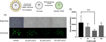
When oocytes are cultured in vitro, they undergo ageing, which results in the accumulation of ROS (Khazaei and Aghaz, Reference Khazaei and Aghaz2017). To determine the activity and potency of CoQ10 in the reduction of ROS accumulation during in vitro maturation of mouse oocytes, COCs were incubated for 24 h in medium containing 30, 50 or 100 μM CoQ10 or chloroform solvent (Fig. 1) (Gendelman and Roth, Reference Gendelman and Roth2012). After the removal of cumulus cells, the level of ROS in oocytes was determined by measuring fluorescence intensity after treatment with DCFH-DA, which is converted into green fluorescent DCF by oxidation. Treatment with up to 50 μM CoQ10 resulted in a gradual decrease in ROS (Fig. 1A). ROS accumulation decreased by 75% upon treatment with 50 μM CoQ10 compared with that in the vehicle-treated oocytes (Fig. 1B). When 100 μM CoQ10 was added, the amount of ROS increased over that in 50 μM CoQ10-treated oocytes (Fig. 1). These results indicated that an adequate concentration of CoQ10 could efficiently reduce the ROS generated during in vitro oocyte culture.
The effect of CoQ10 on the ROS levels in oocytes. (A) ROS levels in mouse oocytes after in vitro culture for 24 h with or without CoQ10 were determined by treatment with 10 μM DCFH-DA. (B) Intensity of ROS fluorescence in oocytes treated with 50 μM CoQ10 or vehicle control was measured using ImageJ software and plotted. Dots indicate the intensity of individual oocytes. Lines represent the average value of all measurements ± standard error of the mean (SEM) from three independent experiments. Statistical significance was determined by conducting an unpaired t-test. ***P < 0.001.

CoQ10 affects on the in vitro spontaneous maturation time of mouse oocytes
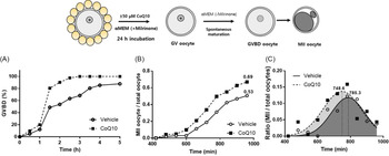
We tested whether the CoQ10 treatment-mediated ROS reduction affects the in vitro oocyte maturation time and rate. Generally, the oocyte maturation timing is checked by counting the number of GVBD oocytes that have undergone the resumption of meiosis. As the GVBD oocytes could not be detected clearly in the COC, the oocyte maturation process of treating Areg to the COC could not applied. Instead, COCs were incubated with or without 50 μM CoQ10 for 24 h in Areg-free, milrinone-containing medium. The oocytes were recovered from the COC and spontaneous maturation was induced by milrinone removal. Every 30 min, the GVBD oocytes were counted and the polar body extrusion rate and time was checked. The CoQ10-treated oocytes underwent GVBD faster than the vehicle-treated oocytes (Fig 2A). Approximately 70% of the oocytes reached the MII stage of the cell cycle in the CoQ10-treated group, while only 53.6% of the oocytes reached the MII stage in the vehicle-treated group (Fig. 2B). The majority of the CoQ10-treated oocytes reached the MII stage at 748.6 min, which was faster than the time observed in the vehicle-treated oocytes (785.3 min; Fig. 2C). Therefore, CoQ10 supplementation enhanced the in vitro maturation rate and timing.
In vitro spontaneous maturation rate and timing of mouse oocytes. The COCs were aged for 24 h with or without 50 μM CoQ10 in Areg-free, milrinone-containing medium for 24 h. The oocytes were denuded and underwent spontaneous maturation by milrinone removal. (A) The number of oocytes with disappearing germinal vesicle was counted every 30 min and the GVBD oocyte numbers were plotted. (B) The total number of oocytes reaching the MII stage was counted every hour and the ratio of MII oocyte to total oocyte were represented on a graph. (C) The number of oocytes newly reaching the MII stage in each hour of observation was analyzed by the Gaussian method using Prism software. Empty circles and black squares show the ratio of newly reaching MII oocytes at the timepoint. The time at which the largest number of oocytes become MII was indicated on the graph.

CoQ10 enhances the in vitro maturation rate of mouse oocytes
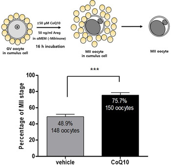
To determine the effect of CoQ10 on in vitro maturation of oocytes surrounded by cumulus cells, COCs were cultured in a medium containing 50 ng/ml Areg with or without 50 μM CoQ10. Oocytes were incubated for 16 h, which is sufficient for oocytes to resume the cell cycle and reach the MII stage. After the isolation of oocytes from COCs, the number of oocytes reaching the MII stage and extruding the polar body was measured. The fraction of CoQ10-treated eggs reaching the MII stage was 75.7% (n = 150), which was higher than that of the vehicle-treated group (48.9%, n = 148) (Fig. 3). Therefore, the addition of CoQ10 was effective in improving the in vitro maturation rate.
In vitro maturation rate of mouse oocytes. Maturation rate was monitored by measuring the number of oocytes in the MII stage after the incubation of COCs with or without 50 μM CoQ10 in Areg-containing medium for 24 h. The denuded oocytes were collected by the removal of cumulus cells. The number of oocytes reaching the MII stage was counted. Data are reported as mean ± SEM using at least five independent experiments. (***P < 0.001, unpaired t-test). The numbers in the bars indicate the numbers of oocytes observed.

CoQ10 treatment modulates the distribution of cortical granules in aged oocytes
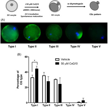
As the distribution of CGs in oocytes influences fertilization efficiency (Liu et al., Reference Liu, Mal, Miao, Liu, Bao and Tan2005), determining the distribution pattern of CGs serves as a measure of the fertilization ability of an egg. We observed MII oocytes and classified the oocytes into five types depending on their CG distribution (Fig. 4). The first type was defined by CGs evenly distributed in the ooplasm and a cortical granule-free domain (CGFD) present on the chromosomal side (Type I in Fig. 4A). This CGFD form of CG distribution is known to be the most ideal for fertilization (Sun et al., Reference Sun, Wang, Xu, Lee, Cui and Kim2011). The ratio of oocytes in type I status increased from approximately 35% to 45% upon 50 μM CoQ10 treatment compared with the vehicle-treated oocytes (Fig. 4B). The second type was defined by CGs that were mainly distributed around the oolemma, the plasma membrane of the oocyte. The third type was defined by CGs that were spread throughout the ooplasm, including the chromosomal region. These types are known to have less fertilization potency than type I. The fractions of oocytes of these two types were decreased by treatment with CoQ10. In the fourth and fifth types, CGs were mainly aggregated around the chromosomes or were absent in oocytes. These forms, which are believed to have very little fertilization potential (Liu et al., Reference Liu, Mal, Miao, Liu, Bao and Tan2005), were detected at very low frequency regardless of CoQ10 treatment. These results indicated that CoQ10 treatment improved fertilization ability by the modulation of GC in in vitro-cultured oocytes.
Distribution of cortical granules in CoQ10-treated mouse oocytes. (A) Representative cortical granule distribution in the five types of mouse MII stage oocytes. (B) Categorization of MII oocytes into five types as shown in (A) after treatment with 50 μM CoQ10 or vehicle for 24 h. Mean ± SEM of at least eight independent experiments is shown. Statistical significance was calculated using an unpaired t-test. *P < 0.05.

CoQ10 has little effect on the hardening of the zona pellucida
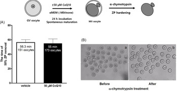
The ZP of aged oocytes is known to undergo hardening, leading to a lower rate of fertilization (Katska et al., Reference Katska, Kauffold, Smorag, Duschinski, Torner and Kanitz1989). To explore whether CoQ10 affects oocyte ZP hardening, the ZP dissolving time was measured using α-chymotrypsin. A harder ZP takes a longer time to be dissolved by α-chymotrypsin (Zhang et al., Reference Zhang, Liu, Chen, Wu, Nie, Gao and Miao2017). After mouse oocytes were maturated in 50 μM CoQ10 medium for 24 h, MII stage oocytes were selected and treated with α-chymotrypsin. The oocytes were observed every 2 min, and the time at which 50% of the total oocytes lost their ZP was recorded. The time required for 50% ZP removal in vehicle-treated oocytes was 56.3 min, and that of CoQ10-treated oocytes was 55 min (Fig. 5). Therefore, CoQ10 did not affect ZP hardening during the maturation of oocytes.
Effect of CoQ10 on ZP hardening. (A) ZP hardening of MII oocytes was monitored after maturation with 50 ng/ml Areg with or without 50 μM CoQ10 and incubation with 1 mg/ml α-chymotrypsin in α-MEM. Incubation time at which half of the oocytes lost the ZP was measured. Oocytes were checked every 2 min for 1 h. Data are reported as mean ± SEM using three independent experiments. (B) The presence of oocyte ZP before (a) and after (b) α-chymotrypsin treatment.

CoQ10 promotes early embryonic development
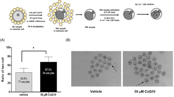
The induction of parthenogenesis is a simple way to indirectly determine the fertility of an egg. After the activation of MII stage oocytes using 10 mM SrCl2, the degree of parthenogenesis was evaluated by measuring the number of two-cell embryos that formed. When CoQ10 was added, 67.3% of the eggs developed to the two-cell stage, whereas only 43.5% of the control oocytes developed to the two-cell stage (Fig. 6). This result indicated that the addition of CoQ10 during in vitro egg maturation enhanced the efficiency of early embryonic development.
In vitro embryonic development of mouse oocytes. (A) MII oocytes selected after COCs were maturated in vitro using 50 ng/ml Areg with or without 50 μM CoQ10 and the activation of parthenogenesis using 10 mM SrCl2. MII oocytes in which a pronucleus appeared and two-cell embryos were generated were counted. Graphs summarize three independent experiments. Data are reported as mean ± SEM. (*P < 0.05, unpaired t-test). The numbers of oocytes observed are shown in the bars. (B) Representative images with arrows indicating two-cell embryos.

CoQ10 enhances anti-ageing and anti-apoptotic gene expression in cumulus cell
Cumulus cells transport mRNA and various metabolites to the oocyte to keep the oocyte in a healthy state. Therefore, the quality of oocytes is related to the state of cumulus cells. The level of specific mRNAs in oocytes is associated with that in cumulus cells as transcription is blocked in GV oocytes (De La Fuente and Eppig, Reference De La Fuente and Eppig2001). Sirt1 is known to be associated with anti-ageing (Kwon and Ott, Reference Kwon and Ott2008), and Bcl2 plays a role in preventing cell death as an anti-apoptotic gene (Morita et al., Reference Morita, Perez, Maravei, Tilly and Tilly1999). Therefore, the levels of Sirt1 and Bcl2 gene expression in cumulus cells were quantified using RT-PCR to check the quality of in vitro-cultured COCs. COCs were incubated in a culture medium containing CoQ10 for 24 h, and cumulus cells surrounding the oocytes were separated. In CoQ10-treated cumulus cells, the expression of Bcl2 and Sirt1 increased 6.7-fold and 1.75-fold over those in solvent-treated COCs, respectively. As a result, CoQ10 prevented both ageing and apoptosis in cumulus cells and may help to maintain the oocyte in a healthy state (Fig. 7).
Bcl2 and Sirt1 gene expression in cumulus cells. Levels of Bcl2 and Sirt1 mRNAs in cumulus cells were measured using the real-time polymerase chain reaction. After COCs were cultured with or without 50 μM CoQ10, the cumulus cells were harvested. All values are presented as relative values compared with the expression level of the gene in COCs cultured without 50 μM CoQ10. Statistical significance was determined by conducting an unpaired t-test. *P < 0.05.

Discussion
ROS are known to have various effects on cells. ROS oxidize many macromolecules, such as plasma membrane lipids and proteins, and cause chromosome changes that can lead to cell cycle arrest, apoptosis, and necrosis (Liou and Storz, Reference Liou and Storz2010; Schieber and Chandel, Reference Schieber and Chandel2014; Panieri and Santoro, Reference Panieri and Santoro2016). This phenomenon is observed in both somatic cells and germ cells. In spermatozoa, oxidative stress is associated with male infertility because of the fragmentation and oxidation of DNA to form 8-hydroxy-2′-deoxyguanosine (8-OHdG) (Wright et al., Reference Wright, Milne and Leeson2014). Additionally, because the ovary is metabolically active, many ROS are produced within granulosa cells, oocytes in growing and Graafian follicles, and the corpus luteum after ovulation (Agarwal et al., Reference Agarwal, Gupta and Sharma2005). Increases in ROS have also been reported in the follicular fluid of preovulatory follicles in humans, swine, and cows (Rizzo et al., Reference Rizzo, Minoia, Trisolini, Mutinati, Spedicato, Jirillo and Sciorsci2009; Freitas et al., Reference Freitas, Neto, Matos, Silva, Ribeiro, Silva-Carvalho and Almeida2017). ROS are generated as a by-product during mitochondrial oxidative phosphorylation. Moreover, when hormones, such as pregnenolone, are produced by the steroidogenic process in the cells of the ovary, a cytochrome P450-mediated monooxygenase reaction produces ROS as a by-product (Hanukoglu, Reference Hanukoglu2006). ROS dramatically increase during ovulation as ovulation is very similar to the inflammation process that produces ROS (Duffy et al., Reference Duffy, Ko, Jo, Brannstrom and Curry2019). If ROS induce damage to the oocyte chromosomes, they may disrupt the maturation process because the DNA repair system is functioning properly in the oocytes (Lane et al., Reference Lane, Morgan, Wu, Collins, Merriman, ElInati, Turner and Jones2017).
In vivo, ROS levels are regulated by various antioxidant enzymes and cellular antioxidant materials (Birben et al., Reference Birben, Sahiner, Sackesen, Erzurum and Kalayci2012). ROS production is particularly important in oocytes during in vitro culture and fertilization to overcome infertility. In vitro oocyte culture medium contains various energy sources for oocytes and hormones, such as follicle-stimulating hormone, epithelial growth factor, human chorionic gonadotropin (hCG), and insulin, to help follicular growth and maturation of oocytes (Zhou et al., Reference Zhou, Malik, Arab, Hill and Shikanov2015). Recently, transferrin, which is known to reduce oxygen radicals, and selenium, a cofactor of glutathione peroxidase, have been typical additives to in vitro follicle cultures to improve the success rate of in vitro fertilization (IVF). Nevertheless, the excessive production of ROS by high oxygen concentrations in the atmosphere causes damage to oocytes. Therefore, the method to decrease ROS has a positive effect on enhancing the quality of oocytes during in vitro culture. Several studies have shown positive effects of antioxidants in improving IVF of industrially important livestock, such as cows and swine (Takami et al., Reference Takami, Preston, Toyloy and Behrman1999; Hanukoglu, Reference Hanukoglu2006; Linnane et al., Reference Linnane, Kios and Vitetta2007; Do et al., Reference Do, Luu, Morita, Taniguchi, Nii, Peter and Otoi2015; Ambrogi et al., Reference Ambrogi, Dall’Acqua, Rocha-Frigoni, Leão and Mingoti2017; Zhang et al., Reference Zhang, Liu, Chen, Wu, Nie, Gao and Miao2017).
Conversely, decreased ROS does not always have a positive effect on cells. Recently, it was reported that cells are not functionally normal if the levels of ROS are excessively decreased (Khazaei and Aghaz, Reference Khazaei and Aghaz2017). As some ROS are also used as signalling factors in cells, the maintenance of proper levels of ROS rather than completely reducing them is important for the proper function of cells. For example, ROS nitric oxide (NO) is involved in intracellular signalling and ovulation (Thaler and Epel, Reference Thaler and Epel2003; Tiwari et al., Reference Tiwari, Prasad, Pandey, Premkumar, Tripathi, Gupta, Chetan, Yadav, Shrivastav and Chaube2017). Recently, it was reported that NO in Anabas testudineus ovaries is engaged in oocyte meiotic arrest through the nitric oxide–cyclic GMP cascade (Nath et al., Reference Nath, Mukherjee, Biswas, Pal, Das, Ghosh, Samanta and Maitra2019). The oxidation of some biological molecules is important for cellular processes. Recent studies have shown that the oxidation of cysteine residues is required for the packing of the sperm DNA into a small nucleus, and the reduction of these residues is necessary to produce a pronucleus after fertilization (Ward, Reference Ward2010). The hCG-induced meiotic resumption and spontaneous meiotic resumption of rat oocytes are inhibited by treatment with various antioxidants including 2(3)-tert-butyl-4-hydroxyanisole (BHA), indicating that a proper concentration of ROS is required for diplotene arrest (Takami et al., Reference Takami, Preston, Toyloy and Behrman1999). Additionally, high levels of ROS in human follicular fluid are associated with fertilization rate and implantation, and excess removal of ROS by SOD or catalase in in vitro culture inhibits ovulation (Oyawoye et al., Reference Oyawoye, Abdel Gadir, Garner, Constantinovici, Perrett and Hardiman2003; Pasqualotto and Pasqualotto, Reference Pasqualotto and Pasqualotto2007). These facts imply that the proper use of antioxidants is necessary for oocyte culture.
In this study, we investigated the effect of CoQ10, also known as ubiquinone, in the in vitro culture of mouse oocytes. CoQ10 is known to play an important role in transferring electrons from complexes I and II to complex III of the electron transport system in mitochondria. CoQ10 was shown to reduce ROS in neuronal cells (Duberley et al., Reference Duberley, Heales, Abramov, Chalasani, Land, Rahman and Hargreaves2014; Ben-Meir et al., Reference Ben-Meir, Kim, McQuaid, Esfandiari, Bentov, Casper and Jurisicova2019; Heydarnejad et al., Reference Heydarnejad, Ostadhosseini, Varnosfaderani, Jafarpour, Moghimi and Nasr-Esfahani2019a), astrocytes (Jing et al., Reference Jing, He, Chang, Mehta, He, Zhang and Li2015), and vascular endothelial cells (Huo et al., Reference Huo, Xu, Hosoe, Kubo, Miyahara, Dai, Mori, Sawashita and Higuchi2018). CoQ10 has been known to have beneficial effects on the reproductive process. CoQ10 supplementation significantly improved survival during bovine and pig oocyte IVM after vitrification and reduced premature CG exocytosis, attenuating the negative effects of vitrification (Hwang et al., Reference Hwang, Kwon Da Kwak, Lee, Im and Hwang2016; Ruiz-Conca et al., Reference Ruiz-Conca, Vendrell, Sabés-Alsina, Mogas and Lopez-Bejar2017). CoQ10 has also been reported to improve mitochondrial capacity and increase MII rate in bovine oocytes (Abdulhasan et al., Reference Abdulhasan, Li, Dai, Abu-Soud, Puscheck and Rappolee2017). The decline of cumulus cell and oocyte function with increasing maternal age is related to decreased expression of enzymes responsible for coenzyme Q (CoQ) production in humans and mice. Therefore, the addition of CoQ10 may improve oocyte and cumulus cell quality by improving mitochondrial metabolism (Ben-Meir et al., Reference Ben-Meir, Burstein, Borrego-Alvarez, Chong, Wong, Yavorska, Naranian, Chi, Wang, Bentov, Alexis, Meriano, Sung, Gasser, Moley, Hekimi, Casper and Jurisicova2015, Reference Ben-Meir, Kim, McQuaid, Esfandiari, Bentov, Casper and Jurisicova2019). Additionally, CoQ10 restores the motility of sperm, which is reduced by ROS (Zhu et al., Reference Zhu, Kawai, Umehara, Hoque, Zeng and Shimada2019). Although there was no significant difference in ZP hardening (Fig. 5), the rate and timing of oocyte maturation, number of oocytes reaching the MII stage and the rate of parthenogenesis increased and ROS decreased in CoQ10-treated mouse oocytes (Figs. 1–3 and 6). Altogether, these results suggest that CoQ10 may improve the meiotic maturation process in mouse oocytes. The oocyte maturation process should include both nuclear maturation and cytoplasmic maturation (Eppig, Reference Eppig1996; Ajduk et al., Reference Ajduk, Małagocki and Maleszewski2008). In this process, maturation promoting factor (MPF), which controls overall oocyte maturation, must be activated by dephosphorylation by Cdc25B (Lincoln et al., Reference Lincoln, Wickramasinghe, Stein, Schultz, Palko, De Miguel, Tessarollo and Donovan2002; Oh et al., Reference Oh, Han and Conti2010). Recent studies have shown that Cdc25B is oxidized by ROS, which changes its activity (Buhrman et al., Reference Buhrman, Parker, Sohn, Rudolph and Mattos2005). Treatment with CoQ10 enhanced the egg maturation process, timing and rate of GVBD and polar body extrusion (Figs 2 and 3). The feasible explanation of the effect is that CoQ10 could modulate the activity of MPF by regulating the oxidation state of cell cycle regulators in oocytes, such as Cdc25B.
CoQ10 supplementation of culture medium promotes the maturation of mouse oocytes and has a positive effect on early embryonic development. CoQ10 also helps maintain CG distribution and is effective in preventing ageing and apoptosis. Therefore, the use of CoQ10 as an exogenous antioxidant may ameliorate ageing and enhance oocyte quality and could successfully improve the rate of in vitro fertilization. Additionally, this technique may be helpful for the production of transgenic mice using in vitro-cultured mouse oocyte maturation and enhance livestock production efficiency.
Conflicts of interest
The authors do not have any conflicts of interest to declare.
Financial support
This work was supported by the 2019 Inje University research grant (20190107).
Ethical standards
All animal experiments were conducted after the approval of the Inje University Institutional Animal Care and Use Committee.