Introduction
Dioctophyme renale (Goeze, 1782), the giant kidney worm, has been reported in several species of wild carnivores (canids and mustelids) and also in domestic dogs in several countries. This nematode species is usually found in the right kidney, and occasionally in both kidneys and the peritoneal cavity (Anderson, Reference Anderson2000). The life cycle begins with the elimination of eggs in the urine of the definitive host. In the aquatic environment, the development of first-stage larvae (about 35 days) occurs within the eggs at roughly 20°C. Eggs are then ingested by aquatic oligochaetes where the first-stage larvae hatch and develop into third-stage infective larvae. Frogs and fish act as paratenic hosts by ingesting aquatic oligochaetes containing third-stage larvae. The definitive host is infected by ingesting an intermediate or paratenic host containing third-stage larvae (Mace & Anderson, Reference Mace and Anderson1975).
The distribution of Trachemys dorbigni (Duméril & Bibron, 1835), the Brazilian slider, is restricted to South America (Brazil, Uruguay and Argentina) (Fritz & Havaš, Reference Fritz and Havaš2007). This freshwater turtle is the most abundant species in the Rio Grande do Sul State, Brazil, and is commonly observed in strongly anthropogenic urban environments (Bujes & Verrastro, Reference Bujes and Verrastro2008; Fagundes et al., Reference Fagundes, Bager and Cechin2010). The species is found in fluvial ecosystems such as dams, rivers, ponds and wetlands (Quintela & Loebmann, Reference Quintela and Loebmann2009). In this paper, we report for the first time the occurrence of third-stage D. renale larvae in the freshwater turtle, T. dorbigni.
Materials and methods
From July 2010 to December 2012, 60 turtles were collected under license from the Instituto Chico Mendes de Conservação da Biodiversidade (ICMBio/no. 23 196). Twenty-eight hosts were captured in four lakes in the Centro Agropecuário da Palma, which belongs to the Federal University of Pelotas, which is located in the rural area of the municipality Capão do Leão (31°48′01.1″S, 52°30′48.6″W). The other 32 hosts were collected from canals in the Pelotas municipal urban area (31°46′16.9″S, 52°18′45.9″W). Both municipalities are located in the State of Rio Grande do Sul, Brazil.
Turtles were manually collected with nets and transported in plastic, 56-litre containers to the Wild Animals Parasitology Laboratory of the Federal University of Pelotas (UFPel), where they were anaesthetized with a combination of ketamine 10% and xylazine 2%. They were then euthanized using an intrathecal injection of lidocaine hydrochloride 2% (Amado et al., Reference Amado, Guarenti and Texeira1994), as recommended by Resolution No. 1000/2012 of the Conselho Federal de Medicina Veterinária (2012).
Following euthanization, 56 turtles were deep frozen for later necropsy and a post-mortem examination was carried out on the remaining turtles, where organs were individualized. Nematodes were fixed in AFA (70°GL ethanol, formalin 37% and glacial acetic acid), preserved in 70°GL glycerinated alcohol, cleared and mounted in Amann's lactophenol (phenol, lactic acid, glycerin and water). Specimens were identified according to Mace & Anderson (Reference Mace and Anderson1975), Measures & Anderson (Reference Measures and Anderson1985) and Pedrassani et al. (Reference Pedrassani, Hoppe, Tebaldi and Nascimento2009a) and then deposited in the ‘Coleção de Helmintos’ of the Wild Animals Parasitology Laboratory of the Federal University of Pelotas (Numbers 519–521 CHLPAS-UFPel), Rio Grande do Sul State, Brazil. Host specimens were deposited in the collection at the ‘Coleção Herpetológica do Museu Carlos Ritter’ (CHMR-UFPel), Rio Grande do Sul State, Brazil (Numbers 78–79).
Levels of infection, after Bush et al. (Reference Bush, Lafferty, Lotz and Shostak1997), were compared between rural and urban environments. Prevalences were compared using chi-square test (χ2, P< 0.05). Mean abundance and mean intensity were compared using confidence interval for ‘Bootstrap’ (BC α, P< 0.05) in the Quantitative Parasitology 3.0 program (Reiczigel & Rózsa, Reference Reiczigel and Rózsa2005).
Results and discussion
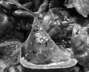
The prevalence of infection of third-stage larvae of D. renale in T. dorbigni was 50%, with one larva per cyst occurring in the body cavity, muscles, mesentery, stomach serous lining and on surfaces of the lung, heart, liver, pancreas, spleen and intestine (fig. 1). A significantly higher prevalence of 87.5% was found in turtles from urban sites, compared with 7.1% in rural sites (P< 0.05). On the other hand, there were no significant differences between values of mean intensity (13.9 in urban compared with 12.1 in rural sites) nor in mean abundance of infection (1.5 in urban compared with 0.1 in rural sites).
Coelomic cavity of Trachemys dorbigni to show cysts (arrowed) of third-stage larvae of Dioctophyme renale in muscles and mesentery.

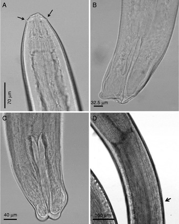
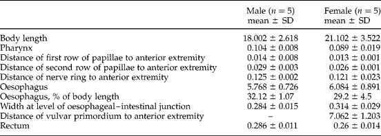
Third-stage larvae of D. renale (fig. 2) recovered from T. dorbigni were larger (table 1) in size than those recovered from oligochaetes, frogs or fish (Mace & Anderson, Reference Mace and Anderson1975; Measures & Anderson, Reference Measures and Anderson1985; Pedrassani et al., Reference Pedrassani, Hoppe, Tebaldi and Nascimento2009a). This finding corroborates that of Measures & Anderson (Reference Measures and Anderson1985), i.e. size differences for third-stage larvae can be related to the host species.
Lateral view of third-stage larvae of Dioctophyme renale to show (A) anterior region with cephalic papillae (arrowed); (B) posterior region of male; (C) posterior region of female; and (D) female with primordium of the vulva (arrowed).

Mean measurements (mm) of third-stage larvae of Dioctophyme renale recovered from naturally infected Trachemys dorbigni in southern Brazil.
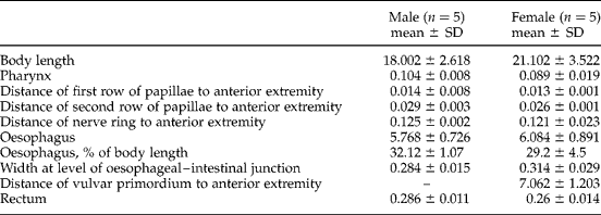
n, Number of specimens examined; SD, standard deviation.
Morphological characteristics of the female third-stage larvae in T. dorbigni confirm that the vulvar primordium is situated in the anterior half of the body, near the oesophageal–intestinal junction (fig. 2D), as described by Mace & Anderson (Reference Mace and Anderson1975), Measures & Anderson (Reference Measures and Anderson1985) and Pedrassani et al. (Reference Pedrassani, Hoppe, Tebaldi and Nascimento2009a). This contrasts to Eustrongylides spp., where the vulvar primordium is located at the posterior end of the body (Measures, Reference Measures1988).
Fish and amphibians act as paratenic hosts when they become infected by ingesting aquatic oligochaetes with third-stage larvae (Mace & Anderson, Reference Mace and Anderson1975; Measures & Anderson, Reference Measures and Anderson1985). In Brazil there is a large gap in the knowledge of D. renale paratenic hosts, despite several reports of the parasitic helminth in domestic dogs and wild animals. Pedrassani et al. (Reference Pedrassani, Hoppe, Tebaldi and Nascimento2009a) identified the frog, Chaunus ictericus (Spix, 1824) in Santa Catarina State (Brazil), as a paratenic D. renale host with third-stage larvae and a prevalence of 5.17% (3/58).
Lumbriculus variegatus (Müller, 1774) (Oligochaeta: Lumbriculida) was the only species identified as an experimental and natural D. renale host (Mace & Anderson, Reference Mace and Anderson1975). The aquatic oligochaete is not registered in South America (Brinkhurst & Jamieson, Reference Brinkhurst and Jamieson1971, cited by Marshall, Reference Marshall1978) and there are no known oligochaete species which act as intermediate hosts for D. renale in South America. This leads us to believe that in this region the nematode's cycle occurs with the participation of other aquatic oligochaete species.
The participation of freshwater turtles in the life cycle of D. renale is not understood. Turtles may have acquired the third-stage larvae by ingesting aquatic oligochaetes, fish or frogs. All have been identified in the diet of T. dorbigni by Lema & Ferreira (Reference Lema and Ferreira1990), who reported that turtles eat snakes, frogs, teleosts, saurians, caecilians, oligochaetes, bovine meat and lettuce. Hahn (Reference Hahn2005) studied the stomach content of 75 turtles, and reported that 85.27% of their diet was composed of plant matter, while 13.23% of the total volume was represented by animal matter (molluscs, crustaceans, arthropods, Hirudinea and vertebrates). With regard to vertebrates as a component of the T. dorbigni diet, the author identified Anura in two turtles and Characiformes in another.
We observed domestic sewage discharge in the channels where turtles were collected in the urban area. This favours a larger population of oligochaetes, which are used as organic pollution indicators. The oligochaetes Tubificidae, for example, are abundant in organically polluted waters, where food is abundant and predators or competitors are non-existent (Alves & Lucca, Reference Alves and Lucca2000; Dornfeld et al., Reference Dornfeld, Alves, Leite and Espíndola2006; Martins et al., Reference Martins, Stephan and Alves2008). In Florida, USA, the epizootiology of the nematode Eustrongylides ignotus Jagerskiold, 1909, in wading birds and fish (as intermediate hosts) was related to nutrient input and human activity. The parasite caused an 80% mortality rate among nestling herons and egrets in one colony alone. The infection enhancement was attributed to eutrophication, which led to increased oligochaete densities in the first intermediate host (Spalding et al., Reference Spalding, Bancroft and Forrester1993; Coyner et al., Reference Coyner, Spalding and Forrester2002). Further research should be conducted to investigate the influence of urban environmental changes on the presence of D. renale in turtles of the region.
The presence of third-stage larvae in freshwater turtles examined in southern Brazil suggested that the reptile could act as a paratenic host. However, turtles are not ideal paratenic hosts for D. renale, because adult turtles are rarely preyed upon by wild carnivores or domestic dogs. To confirm the involvement of T. dorbigni as paratenic hosts for D. renale, experimental infections in definitive hosts (such as dogs) would be required in order to confirm the turtle's potential participation in the cycle.
In relation to the occurrence of D. renale in wild animals in Brazil, there are records for Speothos venaticus (Lund, 1842) (Proença, Reference Proença1935); Chrysocyon brachyurus (Illiger, 1815) (Varzone et al., Reference Varzone, Aquino and Rodovalho2008, Duarte et al., Reference Duarte, Costa, Katagiri, Martins, Oliveira and Ribeiro2013); Nasua nasua (Linnaeus, 1766) (Milanelo et al., Reference Milanelo, Moreira, Fitorra, Petri, Alves and Santos2009); Lontra longicaudis (Olfers, 1818) (Vicente et al., Reference Vicente, Rodrigues, Gomes and Pinto1997); Choloepus didactylus (Linnaeus, 1758) (Rocha et al., Reference Rocha, Serra and Grecm1965); Galictis cuja (Molina, 1782) (Barros et al., Reference Barros, Lorini and Persson1990; Pesenti et al., Reference Pesenti, Mascarenhas, Krüger, Sinkoc, Albano, Coimbra and Müller2012; Zabott et al., Reference Zabott, Pinto, Viott, Tostes, Bittencourt, Konell and Gruchouskei2012); Cerdocyon thous (Linnaeus, 1766) (Ribeiro et al., Reference Ribeiro, Verocai and Tavares2009) and Cebus apella (Linnaeus, 1758) (Ishizaki et al., Reference Ishizaki, Imbeloni, Muniz, Scalercio, Benigno, Pereira and Lacreta2010).
Canine dioctophymosis has been reported in several Brazilian states: Maranhão (Sousa et al., Reference Sousa, Sousa, Coelho, Quessada, Freitas and Moraes2011), Pará (Miranda et al., Reference Miranda, Benigno, Galvão and Oliveira1992), Minas Gerais (Costa & Lima, Reference Costa and Lima1988), Espírito Santo (Pereira et al., Reference Pereira, Girardelli, Trivilin, Lima, Nunes and Martins2006, Reference Pereira, Holanda, Oliveira, Lopes and Costa2008a), Rio de Janeiro (Lemos et al., Reference Lemos, Santos, Rodrigues, Goulart, Almeida and Silveira2010), São Paulo (Costa et al., Reference Costa, Pizzi, Faria, Souza, Silva and Gentil2011), Paraná (Leite et al., Reference Leite, Círio, Diniz, Luz, Navarro-Silva, Silva, Leite, Zadorosnei, Musiat, Veronesi and Pereira2005; Cottar et al., Reference Cottar, Dittrich, Ferreira, Carvalho, Albernaz, Luz and Tasquet2012), Santa Catarina (Neves et al., Reference Neves, Morais, Nogueira and Chquiloff1983; Pedrassani et al., Reference Pedrassani, Hoppe, Acvancini and Nascimento2009b), Rio Grande do Sul (Kommers et al., Reference Kommers, Ilha and Barros1999; Monteiro et al., Reference Monteiro, Sallis and Stainki2002; Colpo et al., Reference Colpo, Silva, Monteiro, Stainki, Camargo and Colpo2007).
The high prevalence of third-stage larvae of D. renale in urban turtles in this geographical region is of veterinary importance. In the urban area of Pelotas there are no data on the prevalence of canine dioctophymosis and only a few case reports (Nunes et al., Reference Nunes, Araújo, Caetano, Sarmento, Rios, Toaldo, Chapon, Schuch and Scopel2008; Pereira et al., Reference Pereira, Silva, Wilhelm, Muller, Campello, Barbosa, Gallina, Silva, Santos and Nobre2008b; Stainki et al., Reference Stainki, Pedrozo, Gaspar, Zanette, Silva and Monteiro2011; Milech et al., Reference Milech, Perera, Ramos, Pedrozo and Braga2012). In the UFPel Veterinary Hospital, studies are being conducted to estimate the prevalence of D. renale in dogs in Pelotas. To date, of 48 dogs examined by urinalysis, 5 were positive (Dr Josaine Rapetti Pedrozo, pers. comm.). According to Kommers et al. (Reference Kommers, Ilha and Barros1999) dioctophymosis occurs more frequently (preferably) in stray dogs, due to their less finicky eating habits.
The dog population in the city of Pelotas is roughly 66,723 animals (13,345 domesticated, 46,706 semi-domesticated and 6672 strays) (Municipal Administration, 2012). At sites where turtles were collected, several dogs were observed wandering in the streets. In this context, we believe that the prevalence of canine dioctophymosis in the region is high due to the likewise high prevalence of third-stage larvae of D. renale in freshwater turtles, T. dorbigni. Still, we do not consider this reptile to be a paratenic host with the ability to transmit the infective form and continue the cycle of D. renale in urban areas.
The public health risk that the presence of D. renale presents to the people of this region should be emphasized since, in other countries, human infections in the skin and kidneys have been reported (Hanjani et al., Reference Hanjani, Sadighan, Mikakhfar and Arfaa1968; Urano et al., Reference Urano, Hasegawa, Katsumata, Toriyama and Aoki2001; Sardjono et al., Reference Sardjono, Purmono, Iskandar and Gunawan2008; Katafigiotis et al., Reference Katafigiotis, Fragkiadis, Pournaras, Nonni and Stravodimos2013).
Acknowledgements
Special thanks to Instituto Chico Mendes de Conservação da Biodiversidade (ICMbio) for permissions. Special thanks to our colleagues: Jéssica Dias Souza, Mariana de Moura Mendes and Marco Antônio Afonso Coimbra for their assistance; to Dr Daniela Pedrassani for bibliographic aid and to Dr Josaine Rapetti Pedrozo for general information.
Financial support
This work was supported by CAPES (Coordenação de Aperfeiçoamento do Pessoal de Nível Superior) – process number 2010/039.
Conflict of interest
None.
Ethical standards
This study was approved by the Research Ethics Committee of the Universidade Federal de Pelotas (CEEA – 3026/2010).