Introduction
The mixed valence Mn phosphate mineral bermanite, Mn2+Mn3+2 (PO4)2(OH)2⋅4H2O, is known from many localities worldwide. It is a late-stage secondary hydrothermal mineral found replacing primary phosphates in granite pegmatites. Bermanite was first described by Hurlbut (Reference Hurlbut1936) as small reddish brown crystals in veins in triplite from a pegmatite on the 7U7 Ranch, 40 km west of Hillside, Bagdad district, Yavapai Co., Arizona. Hurlbut (Reference Hurlbut1936) and Leavens (Reference Leavens1967) proposed orthorhombic symmetry for the mineral. Kampf and Moore (Reference Kampf and Moore1976) solved the crystal structure of bermanite and determined that the true symmetry is monoclinic. The relatively large temperature factor that they reported for the Mn3 site could indicate some significant occupancy by Mg. The bermanite on which Kampf and Moore (Reference Kampf and Moore1976) refined the structure was collected from the Wiperaminga Hill West Quarry, Boolcoomatta Reserve, South Australia in the 1970s and was not chemically analysed (Anthony Kampf, personal communication, 2021). Chemical data from specimens in the collection of the South Australian Museum from the same find have been obtained by scanning electron microscopy energy-dispersive spectroscopy analysis and show very little Mg.
Magnesiobermanite was found on a specimen collected from the White Rock No.2 quarry situated in the Bimbowrie Conservation Park, 24 km N of Olary, South Australia, Australia in the 1960s. The new mineral is the Mg analogue of bermanite in which Mg replaces Mn2+ in the bermanite structure, providing the ideal formula MgMn3+2(PO4)2(OH)2⋅4H2O. The new mineral and the name (symbol Mbrm) have been approved by the Commission on New Minerals, Nomenclature and Classification of the International Mineralogical Association (IMA2018-115, Elliott, Reference Elliott2019). The holotype specimen is deposited in the collection of South Australian Museum, Adelaide, South Australia, Australia, with registration number G34762.
Occurrence
The White Rock pegmatite is one of more than 70 pegmatite bodies in the Olary Province of South Australia that have been intruded into rocks of the Archaean Willyama Complex. The Willyama Supergroup rocks comprise upper greenschist- to amphibolite-grade metamorphosed and strongly deformed sedimentary and minor igneous rocks (Lottermoser and Lu, Reference Lottermoser and Lu1997) that are overlain unconformably by late Proterozoic Adelaidean metasediments. The pegmatites are typically zoned and comprise an inner quartz core of coarse-grained muscovite, quartz, microcline and minor plagioclase surrounded by an intermediate zone of coarse-grained muscovite, quartz, microcline and minor plagioclase and an outer zone of fine- to medium-grained microcline, quartz, plagioclase and muscovite. The contact between the intermediate zone and the quartz core hosts rare-element minerals such as columbite-(Fe), samarskite and Nb-bearing rutile, and pods of a triplite–zwieselite series mineral. The triplite–zwieselite mineral has been formed by metasomatic alteration of magmatic fluorapatite and has been transformed by hydrothermal alteration and weathering in an oxidising, low-temperature, low-pH environment, to give a complex, microcrystalline intergrowth of secondary phosphate minerals. The White Rock pegmatite is 120 m long and up to 16 m wide with poor outcrops (Olliver and Steveson, Reference Olliver and Steveson1982). Over the period 1932–1973, three quarries were excavated to a depth of 10 m which produced 860 tonnes of feldspar (both albite and microcline) and 8.1 tonnes of beryl. At White Rock, triplite and associated secondary phosphate minerals have been exposed in only the No.2 quarry. Magnesiobermanite occurs in cavities in a matrix comprising quartz and hydrothermally altered dark brown to black dufrénite-group minerals (dufrénite–natrodufrénite–gayite–bimbowrieite) that have nearly completely replaced earlier triplite. Associated minerals are fluorapatite, leucophosphite, jahnsite-(NaFeMg), ushkovite, laueite, perloffite and mitridatite.
Physical and optical properties
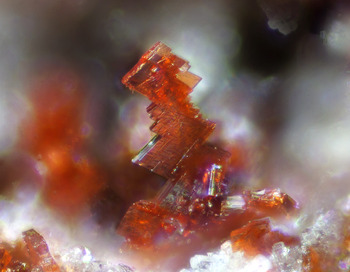
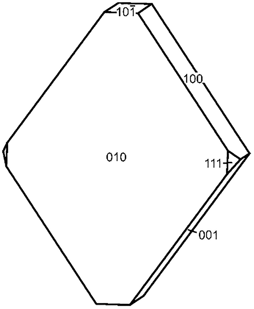
Magnesiobermanite occurs as aggregates of orange–red to brownish red, twinned, bladed to tabular crystals up to 1.2 mm across (Fig. 1). Individual crystals are up to 0.3 mm in length. The observed crystal forms are {010}, {100}, {001}, {10$\bar{1}$ } and {111} (Fig. 2). The mineral is translucent with a salmon-pink streak, vitreous lustre and does not fluoresce under ultraviolet light. It is brittle with a splintery fracture and a perfect cleavage on {001}. The Mohs hardness is 3½. The density, measured with a sodium polytungstate and water mixture, is 2.75(3) g/cm3, in agreement with the calculated value of 2.73 g/cm3 from the empirical formula. Magnesiobermanite is biaxial (–) with indices of refraction; α = 1.690(2), β = 1.719(2), γ = 1. 1.734(2) and 2V(calc.) = 70.4°. Pleochroism is strong; X = pale orange red, Y = pale yellow, Z = dark orange red; Z > X>Y.
Magnesiobermanite crystal group, 0.3 mm in height.

Crystal drawing of magnesiobermanite.

Chemical composition
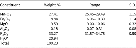
Quantitative chemical analyses were performed using a CAMECA SX50 electron microprobe operating in wavelength-dispersive mode. Operating conditions were: 15 kV accelerating voltage, 20 nA beam current and 5 μm beam diameter. Standards (element, emission line) were: almandine–pyrope (MgKα and FeKα), rhodonite (MnKα), albite (AlKα) and apatite (PKα). The data were reduced and corrected by the PAP method of Pouchou and Pichoir (Reference Pouchou, Pichoir and Armstrong1985). The small amount of available material precluded the determination of H2O by direct measurement, however its presence is inferred from the crystal structure analysis and is confirmed by infrared spectroscopy (see below). The electron microprobe analysis results are given in Table 1. The empirical formula, based on 14 O atoms per formula unit, is Mg1.02(Mn3+1.49Fe3+0.47Al0.02)1.98(PO4)2.01(OH)1.95⋅4.01H2O. The ideal formula of magnesiobermanite is MgMn3+(PO4)2(OH)2⋅4H2O, which requires MgO 9.37, Mn2O3 36.70, P2O5 32.99, H2O 20.94, total 100.00 wt.%.
Compositional data for magnesiobermanite.

*calculated from the ideal formula. S.D. – standard deviation.
Infrared spectroscopy
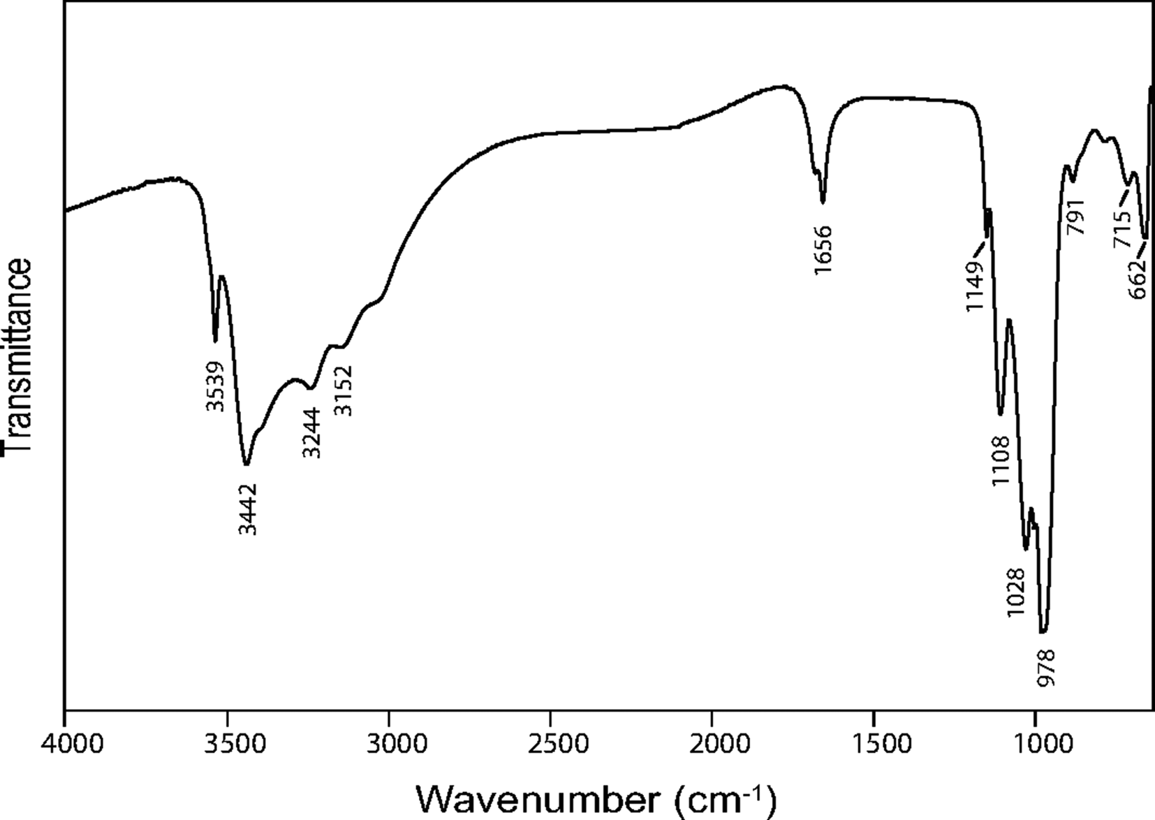
The Fourier transform infrared (FTIR) spectrum (Fig. 3) of powdered magnesiobermanite was recorded using a Nicolet 5700 FTIR spectrometer (range 4000 to 650 cm–1, transmission mode) equipped with a Nicolet Continuum IR microscope and a diamond-anvil cell. In the OH-stretching region, a broad band from 3625 to 2665 cm–1 has peaks and shoulders at 3539, 3442, 3244, 3152 and 3055 cm–1. The peaks correspond to H-bonded water molecules and hydroxyl ions with O⋅⋅⋅O donor–acceptor distances in the range 2.6 to 2.9 Å (Libowitzky, Reference Libowitzky1999); i.e. of strong to intermediate strength, consistent with the observed distances. The band at 1656 cm–1 corresponds to H–O–H bending vibrations of water molecules. Strong bands at 1149, 1108, 1028 and 978 cm–1 are assigned to the ν3 vibrations of PO4 tetrahedra. Weak bands attributed to the PO4 ν1 vibration occur at 883, 791 and 715 cm–1 and a weak band attributed to the PO4 ν4 vibration at 662 cm–1.
The FTIR spectrum of powdered magnesiobermanite.

X-ray diffraction data and refinement of the structure
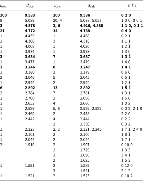
The powder X-ray diffraction pattern of magnesiobermanite (Table 2) was recorded using a Rigaku Hiflux Homelab diffractometer (phi scan, d = 90.0 mm, CuKa X-radiation, λ=1.54187 Å, 40 kV and 30 mA). Calculated intensities were obtained using the program PowderCell (Kraus and Nolze, Reference Kraus and Nolze1996) on the basis of the structural model. The cell parameters, refined on the basis of 26 indexed reflections using the UnitCell software of Holland and Redfern (Reference Holland and Redfern1997), are consistent with the results of single-crystal refinement: a = 5.423(1), b = 19.101(2), c = 5.368(1) Å, β = 110.16(2)° and V = 522.11(7) Å3.

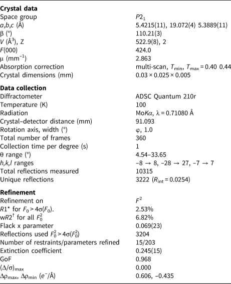
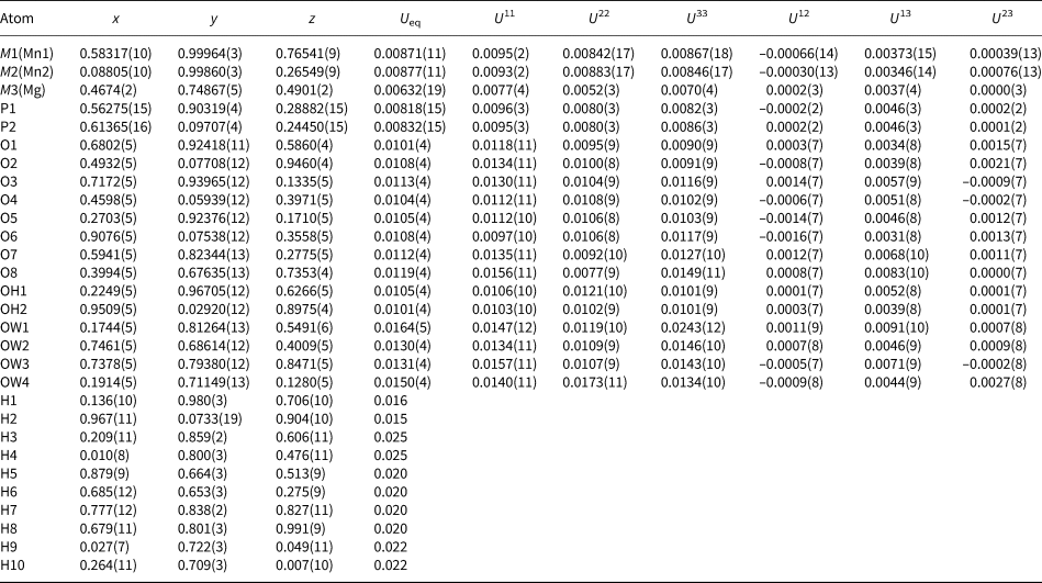
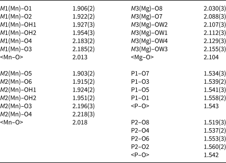
An untwined crystal fragment was mounted on a MiTeGen polymer loop for X-ray data collection on the macromolecular beam line MX2 of the Australian Synchrotron. Data were collected using an ADSC Quantum 315r detector and monochromatic radiation with a wavelength of 0.710800 Å. The detector-to-crystal distance was 91.093 mm. A phi scan was employed and 360 frames were collected with frame widths of 1° and a counting time per frame of 1 s. The intensity data sets were processed using XDS (Kabsch, Reference Kabsch2010) without scaling and with absorption correction and scaling carried out using SADABS (Bruker, 2001). The structure was solved in space group P21 using SHELXT (Sheldrick, Reference Sheldrick2015a) and SHELXL-2018 (Sheldrick, Reference Sheldrick2015b) was used for the refinement of the structure. Following the solution of the structure, the atom coordinates were transformed to correspond to those in the structure of bermanite (Kampf and Moore, Reference Kampf and Moore1976). The final cycle of the refinement converged to R 1 = 0.0243. Difference-Fourier syntheses then located all H atom positions. The H sites were refined with soft restraints on the O–H distances of 0.85(3) Å for the O–H groups and 0.90(3) Å for the water molecules plus H–H restraints of 1.40 Å. Crystal data and experimental details are reported in Table 3. Final atom coordinates and anisotropic-displacement parameters are listed in Table 4, selected interatomic distances and framework angles are given in Table 5, and bond-valence values are given in Table 6. The crystallographic information files have been deposited with the Principal Editor of Mineralogical Magazine and are available as Supplementary material (see below).
Crystal data, data collection and refinement details.

Fractional atomic coordinates, isotropic displacement parameters (in Å2) and site occupancy factors for magnesiobermanite.

Selected interatomic distances (Å) for magnesiobermanite.

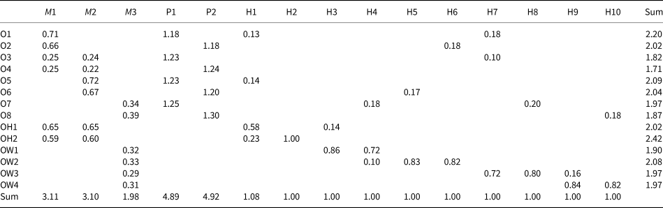
Bond-valence analysis for magnesiobermanite.

Bond-valence parameters are from Gagné and Hawthorne (Reference Gagné and Hawthorne2015).
Structure description
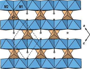
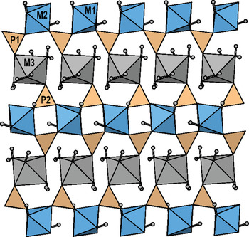
In the structure of magnesiobermanite there are three M sites each coordinated by six O atoms in an octahedral arrangement. The M1 and M2 sites are occupied by Mn3+ and display the (4 + 2)-Jahn–Teller distortion characteristic of Mn3+ in octahedral coordination, with four short M–O distances (1.901–1.954 Å) and two long M–O distances (2.183 and 2.218 Å). The Jahn–Teller distortion is reduced relative to that in bermanite by the presence of significant Fe3+ at the sites as shown by the chemical analysis. Using the relationship of Burns et al. (Reference Burns, Cooper and Hawthorne1994), the calculated distortion Δ is 0.0034 for M1 and 0.0041 for M2 in magnesiobermanite and 0.0040 for M1 and 0.0050 for M2 in bermanite. The M3 site is occupied by Mg and displays a more regular coordination. Occupancy refinement, using the scattering factor for Mg, yielded a value of 12.8 electrons, consistent with full occupancy by Mg as shown by the electron-microprobe data. As expected, the site is smaller in size (<M3–O> = 2.104 Å), compared to that in bermanite (<M3–O> = 2.185 Å). M1 and M2 octahedra share trans edges to form a [Mφ4] chain that extends along [101]. Chains are decorated by corner-connected PO4 tetrahedra to form a [(Mn3+(OH)2(PO4)2] sheet in the (010) plane (Fig. 4). Sheets link through corner-sharing of [M3(H2O)4O2] octahedra and by hydrogen-bonds (Fig. 5).
The crystal structure of magnesiobermanite viewed along [010]. H atoms are grey circles. The unit cell is outlined.

The crystal structure of magnesiobermanite viewed along [101]. H atoms are grey circles.

Hydrogen bonds
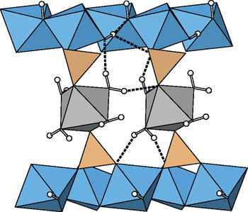
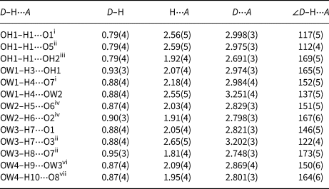
The hydrogen-bonding scheme for magnesiobermanite is reported in Table 7 and is illustrated in Fig. 6. Although H sites were not located in the structure of bermanite by Kampf and Moore (Reference Kampf and Moore1976), a hydrogen bonding scheme was inferred from geometrical and electrostatic valence balance arguments. This scheme is confirmed in the present study, with the addition of four H bonds. OH1 is a donor of two hydrogen bonds to an adjacent sheet, accepted by O1 and OH2. A third hydrogen bond from OH1 is donated to O5, an acceptor within the sheets. Water molecules associated with the intersheet [M3O2(H2O)4] octahedron donate five hydrogen bonds to adjacent octahedral–tetrahedral sheets, OW1–H3⋅⋅⋅OH1, OW2–H5⋅⋅⋅O6, OW2–H6⋅⋅⋅O2, OW3–H7⋅⋅⋅O1 and OW3–H7⋅⋅⋅O3 and also donate five hydrogen bonds to an adjacent [M3O2(H2O)4] octahedron, OW1–H4⋅⋅⋅O7, OW3–H8⋅⋅⋅O7, OW4–H9⋅⋅⋅OW3, OW4–H10⋅⋅⋅O8 and OW1–H4⋅⋅⋅OW2.
Hydrogen bonding in magnesiobermanite.

Details of hydrogen bonding in magnesiobermanite (Å, °).

Symmetry codes: (i) x – 1, y, z; (ii) x, y, z + 1; (iii) x – 1, y + 1, z; (iv) –x + 2, y + ½, –z + 1; (v) –x + 1, y + ½, –z + 1; (vi) x – 1, y, z – 1; (vii) x, y, z – 1.
Related minerals
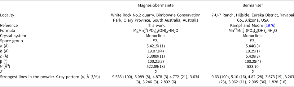
Magnesiobermanite is isostructural with bermanite (Kampf and Moore, Reference Kampf and Moore1976). Comparative data for the two minerals are given in Table 8. As expected, the unit-cell parameters of magnesiobermanite are smaller than those of bermanite, owing to the replacement of Mn2+ (effective ionic radius 0.83 Å; Shannon, Reference Shannon1976) by Mg (effective ionic radius 0.72 Å). The intersheet separation in the two structures is very similar, a difference of ~0.1 Å. Magnesiobermanite is structurally related to ercitite, Na2(H2O)4[Mn3+2(OH)2(PO4)2], (Fransolet et al., Reference Fransolet, Cooper, Cerný, Hawthorne, Chapman and Grice2000; Cooper et al., Reference Cooper, Hawthorne and Černý2009) and coralloite, Mn2+Mn3+2(AsO4)2(OH)2⋅4H2O, (Callegari et al., Reference Callegari, Boiocchi, Ciriotti and Balestra2012). Each of these structures is based on [(Mn3+(OH)2(PO4)2] sheets, which are topologically identical, however, linkage between sheets is different in each structure. In ercitite, sheets link through [Mn2+(H2O)4O2] octahedra that share corners with PO4 groups and through corner-sharing [7]-coordinated Na polyhedra that share edges with [Mn2+(H2O)4O2], the polyhedra of the structural units. In coralloite, adjacent sheets link through [Mn2+(H2O)4O2] octahedra that share corners with PO4 groups. The PO4 groups share cis vertices of the [Mn2+(H2O)4O2] octahedra, whereas in bermanite and magnesiobermanite, PO4 groups share trans vertices of the [Mn2+(H2O)4O2] octahedra.
Comparison of magnesiobermanite and bermanite.

*Powder X-ray diffraction pattern from Anthony et al. (Reference Anthony, Bideaux, Bladh and Nichols2000).
Acknowledgements
The author thanks Ben Wade of Adelaide Microscopy, The University of Adelaide for assistance with the microprobe analysis. The infrared spectrum was acquired with the assistance of the Forensic Science Centre, Adelaide. The single-crystal X-ray diffraction data set was collected at the microfocus beam line MX2 at the Australian Synchrotron. Structures Editor Peter Leverett and reviewers Anthony Kampf and Igor Pekov are thanked for constructive comments that improved the quality of the manuscript.
Supplementary material
To view supplementary material for this article, please visit https://doi.org/10.1180/mgm.2021.87