INTRODUCTION
The gut is characterized by an extensive and much differentiated intrinsic nervous system, the enteric nervous system (ENS). It extends from the oesophagus to the sphincter ani internus and includes the nervous elements within the walls of the gallbladder, the cystic duct, the common bile duct and the pancreas. Because of the high number of neurons (108) and the aforementioned structural characteristics reflecting certain parallels with the central nervous system (CNS), the ENS is called the brain of the gut (Krammer et al., Reference Krammer, Süss, Singer and Wedel2002; von Boyen et al., Reference von Boyen, Reinshagen, Steinkamp, Adler and Kirsch2002a, Reference von Boyen, Reinshagen, Steinkamp, Adler and Kirschb).
Various neuronal networks or plexuses are localized within the individual layers of the intestinal wall. The nerve cells are mainly localized within the ganglia which are extensively interconnected by nerve bundles or strands, giving the plexus the appearance of a meshwork with nodes (ganglia) at the junctions of the mesh. The nerve strands are also referred to as intermodal strands of the plexus. There are up to three ganglionic plexuses, containing nerve cells and four aganglionic plexuses composed of nerve fibres and characterized by a completely absence of nerve cell bodies (Krammer and Kühnel, Reference Krammer and Kühnel1993; Radke and Krammer, Reference Radke and Krammer1996).
All ganglionic and aganglionic plexus are characterized by enteric glia cells (EGCs), a special glia cell form, which outnumbers enteric neurons.
The ENS was mostly characterized by gastroenterologists as the centre of motility. Thus, the research was concentrated on morphological, physiological and biochemical characteristics of enteric neurons. The EGC was only seen as simple supporting cell entity of neurons and did not attract the interest of gastroenterologists nor neuroscientists (Gabella, Reference Gabella1981; Gershon and Rothman, Reference Gershon and Rothman1991; Krammer et al., Reference Krammer and Kühnel1993; Wedel et al., Reference Wedel, Roblick, Gleiss, Schiedeck, Bruch and Kühnel1999; von Boyen et al., Reference von Boyen, Reinshagen, Steinkamp, Adler and Kirsch2002a, Reference von Boyen, Reinshagen, Steinkamp, Adler and Kirschb).
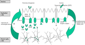
Since 1998 it is clear that EGCs are an essential cell entity for the integrity of the intestines as in animal models destruction of EGCs by genetic ablation led to a fatal haemorrhagic jejuno-ileitis followed by death (Bush et al., Reference Bush, Savidge, Freeman, Cox, Campbell and Mucke1998; Bush, Reference Bush2002) (Fig. 1). In the last decade different groups of scientists have done a lot of interesting research of this cell entity: (i) the cell morphology and characteristics were investigated in detail, (ii) their role in gut inflammation using animal models were described, (iii) different factors released by EGCs modulating the epithelial barrier were proved, (iv) glia-epithelial and neuro-epithelial feedback loops were postulated (Bush et al., Reference Bush, Savidge, Freeman, Cox, Campbell and Mucke1998; Cornet et al., Reference Cornet, Savidge, Cabarrocas, Deng, Colombel and Lassmann2001; von Boyen et al., Reference von Boyen, Steinkamp, Geerling, Reinshagen, Schäfer and Adler2006a, Reference von Boyen, Steinkamp, Reinshagen, Schäfer, Adler and Kirschb; Savidge et al., Reference Savidge, Newman, Pothoulakis, Ruhl, Neunlist and Bourreille2007a, Reference Savidge, Sofroniew and Neunlistb; Abdo et al., Reference Abdo, Derkinderen, Gomes, Chevalier, Aubert and Masson2010).
Model of EGCs in the gut. A leaky barrier of epithelial cells leads to a translocation of bacteria, endotoxins or LPS in the mucosa followed by an increase of proinflammatory cytokines. (i) These cytokines and LPS induce an increase of GFAP-positive EGCs, which secrete high amounts of neurotrophic factors. (ii) High amounts of proinflammatory cytokines, especially TNF-α and interferon γ, over a longer disease period might be possible to start apoptosis in EGCs. The secretion of NGF might be protective for the epithelial barrier and change the content of neurotransmitters in the neurons, which could lead to hypersensitivity of the gut. The secreted GDNF stimulates mitogen-activated-kinase- and protein kinase B pathways, which protects epithelial cells from apoptosis. As the balance of apoptosis and regeneration of epithelial cells is an important component of an intact barrier function of the mucosa, the secreted GDNF might be part of the puzzle in gut integrity. Thus, neurotrophic factors are involved in mucosal healing. After regeneration of the epithelial barrier the secretion of proinflammatory cytokines decrease, which leads to reduced levels of neurotrophic factors in the mucosa.

Although EGCs are part of the peripheral nervous system, EGCs differ markedly from other glia cells like Schwann cells in the peripheral nervous system and resemble CNS astrocytes with respect to S-100 protein and glial fibrillary acidic protein (GFAP) expression (Jessen and Mirsky, Reference Jessen and Mirsky1980; Ferri et al., Reference Ferri, Probert, Cocchia, Michetti, Marangos and Polak1982; Gershon and Rothman, Reference Gershon and Rothman1991; Walz et al., Reference Walz and Lang1998; Walz, Reference Walz2000).
Besides ganglionic plexuses, where EGCs outnumbers the protected neurons, EGCs are the main cell type in aganglionic plexuses.
The internal structure of EGCs, as well as their shape, is different from the Schwann cells. The most striking differences are the high concentration of densely packed intermediate filaments like GFAP (Ferri et al., Reference Ferri, Probert, Cocchia, Michetti, Marangos and Polak1982; Gershon and Rothman, Reference Gershon and Rothman1991). Thus, like in the CNS, a gliosis of EGCs could be shown in the ENS during gut inflammation, although the exact role of gliosis in the nervous system is not understood so far (von Boyen et al., Reference von Boyen, Steinkamp, Reinshagen, Schäfer, Adler and Kirsch2004).
Further investigation described somewhat gliosis of EGCs in a different interesting animal model, which by the way confirmed the data of Bush et al. (Reference Bush, Savidge, Freeman, Cox, Campbell and Mucke1998): A destruction of EGC by auto-immune mechanisms was shown to induce gut inflammation resembling Crohn's disease (CD) (Cornet et al., Reference Cornet, Savidge, Cabarrocas, Deng, Colombel and Lassmann2001). Additionally these authors described for the first time a diminished enteric glia network in patients suffering from CD, although signs of gliosis were seen in the inflamed intestines. These findings are confirmed by a recent study (von Boyen et al., Reference von Boyen, Schulte, Pfluger, Spaniol, Hartmann and Steinkamp2011).
Several mechanisms are investigated how EGCs contribute to the homeostasis of the gut. One of the most interesting is the secretion of neurotrophic factors (Reinshagen et al., Reference Reinshagen, Rohm, Steinkamp, Lieb, Geerling and Von Herbay2000; Steinkamp et al., Reference Steinkamp, Geerling, Seufferlein, von Boyen, Egger and Grossmann2003; von Boyen et al., Reference von Boyen, Steinkamp, Reinshagen, Schäfer, Adler and Kirsch2004, Reference von Boyen, Steinkamp, Geerling, Reinshagen, Schäfer and Adler2006a, Reference von Boyen, Steinkamp, Reinshagen, Schäfer, Adler and Kirschb; Neunlist et al., Reference Neunlist, Aubert, Bonnaud, Van Landeghem, Coron and Wedel2007) and S-nitrosoglutathione (GSNO) (Savidge et al., Reference Savidge, Newman, Pothoulakis, Ruhl, Neunlist and Bourreille2007a, Reference Savidge, Sofroniew and Neunlistb). The release of factors, like interleukins (interleukine-6) (Rühl et al., Reference Rühl, Franzke, Collins and Stremmel2001), endothelins (von Boyen et al., Reference von Boyen, Degenkolb, Hartmann, Adler and Steinkamp2010) or transforming growth factor-β1 (TGF-β1) (Neunlist et al., Reference Neunlist, Aubert, Bonnaud, Van Landeghem, Coron and Wedel2007) by EGCs and the possibility to express HLA-DR complex class II (da Silveira et al., Reference da Silveira, de Oliveira, Neto, Luquetti, Fujiwara and Oliveira2011) are additional mechanisms to stabilize the gut integrity.
EGCs AND INFLAMMATORY BOWEL DISEASE
The ENS is characterized by high specialized demands. As the gut lumen and bacteria are in close proximity to ENS it needs complex mechanisms of protection for the ENS in comparison to the CNS to prevent, for example, invasive infections by luminal antigens (Gabella, Reference Gabella1981; Gershon and Rothman, Reference Gershon and Rothman1991; Krammer and Kühnel, Reference Krammer and Kühnel1993; Wedel et al., Reference Wedel, Roblick, Gleiss, Schiedeck, Bruch and Kühnel1999; von Boyen et al., Reference von Boyen, Reinshagen, Steinkamp, Adler and Kirsch2002a, Reference von Boyen, Reinshagen, Steinkamp, Adler and Kirschb).
Thus, the epithelial mucosa is the crucial barrier. The intestinal mucosa is characterized by epithelial cells, immune cells, nerve fibres and EGCs as part of the ENS. The ‘mucosal plexus’ is aganglionic, which means there are no neuron cell bodies (Gershon and Rothman, Reference Gershon and Rothman1991; Krammer and Kühnel, Reference Krammer and Kühnel1993; von Boyen et al., Reference von Boyen, Steinkamp, Reinshagen, Schäfer, Adler and Kirsch2004). The main cell entity in this area is EGCs directly beneath the epithelium, which are in contact with epithelial cells by their glial fibre strands (Neunlist et al., Reference Neunlist, Aubert, Bonnaud, Van Landeghem, Coron and Wedel2007; Savidge et al., Reference Savidge, Newman, Pothoulakis, Ruhl, Neunlist and Bourreille2007a, Reference Savidge, Sofroniew and Neunlistb). A disruption of the mucosal plexus with loss of EGCs in an animal model induce gut inflammation with histopathological changes closely resembling changes described in human CD (Bush et al., Reference Bush, Savidge, Freeman, Cox, Campbell and Mucke1998; Cornet et al., Reference Cornet, Savidge, Cabarrocas, Deng, Colombel and Lassmann2001; Bush, Reference Bush2002).
This murine model of CD was an unexpected outcome when astroglial cells were targeted for genetic ablation. In this study, transgenic mice were generated, which express the thymidine kinase gene of the herpes simplex virus (HSV-Tk) from the mouse GFAP promoter. Proliferating cells that express the HSV-Tk metabolize the antiviral drug gangciclovir to toxic nucleotides, which interfere with nucleic acid synthesis and induce cell death. Depending on where and when gangciclovir is administered, cell ablation can be controlled regionally and temporally. Astrocytes from the transgenic mice were killed by the gangciclovir without any evidence of toxicity to neighbouring cells. Nevertheless, the continuous administration of gangciclovir to transgenic mice was fatal after 2–3 weeks, with no apparent structural abnormality in major organs other than the intestines. The intestinal changes were characterized by pronounced inflammation, necrosis, surface epithelial injury, crypt hyperplasia, transmigration of neutrophils and intravascular microthrombi. Immunhistochemical staining showed a selective ablation of EGCs in the inflamed intestines. Conventional microbiologic screening found no evidence to suggest that the enteric inflammation was caused by either pathogenic organisms or bacterial overgrowth. Thus, it was concluded that EGCs are essential for the mucosa integrity and that loss or impairment of EGC function may contribute to the pathogenesis of CD. This is underlined that the focal nature of the early aphtoid lesions in CD is reminiscent of lesions that might be produced by a herpes-like neurotropic virus (Bush et al., Reference Bush, Savidge, Freeman, Cox, Campbell and Mucke1998; Bush, Reference Bush2002).
A different animal model confirms the findings of increasing gut inflammation after continuous disruption of EGCs (Cornet et al., Reference Cornet, Savidge, Cabarrocas, Deng, Colombel and Lassmann2001).
In this model, mice expressing a viral neoself antigen in astrocytes and enteric glia were crossed with specific T cell receptor transgenic mice. This resulted in apoptotic depletion of EGCs accompanied by gut inflammation. Furthermore, Cornet et al. (Reference Cornet, Savidge, Cabarrocas, Deng, Colombel and Lassmann2001) showed for the first time that the ENS of patients with CD was characterized by a diminished EGC network to a level comparable to the mouse model.
As intestinal and mesenteric T cell infiltration, vasculitis, T helper 1 cytokine production and fulminant bowel inflammation were characteristic hallmarks of both CD and the double transgenic mice model with CD8+ T cell-mediated autoimmune targeting of EGCs (Cornet et al., Reference Cornet, Savidge, Cabarrocas, Deng, Colombel and Lassmann2001), it was suggested that the disruption of the EGC network in CD patients represents another early pathological feature.
CD is a multifactorial, polygenic disease with genetic heterogeneity. There are a lot of data that EGCs indeed play a role in the multifactorial causes of CD (Bush et al., Reference Bush, Savidge, Freeman, Cox, Campbell and Mucke1998; Cornet et al., Reference Cornet, Savidge, Cabarrocas, Deng, Colombel and Lassmann2001; Bush, Reference Bush2002; Cabarrocas et al., Reference Cabarrocas, Savidge and Liblau2003; Neunlist et al., Reference Neunlist, Van Landeghem, Bourreille and Savidge2008; von Boyen et al., Reference von Boyen, Schulte, Pfluger, Spaniol, Hartmann and Steinkamp2011). The disease is characterized by a reduced glial network of the mucosa (Cornet et al., Reference Cornet, Savidge, Cabarrocas, Deng, Colombel and Lassmann2001; von Boyen et al., Reference von Boyen, Schulte, Pfluger, Spaniol, Hartmann and Steinkamp2011). The cause for the loss of EGCs in CD is not definitely known but the striking decrease in GFAP immunoreactive processes in CD gut samples (Cornet et al., Reference Cornet, Savidge, Cabarrocas, Deng, Colombel and Lassmann2001; von Boyen et al., Reference von Boyen, Schulte, Pfluger, Spaniol, Hartmann and Steinkamp2011) may even suggest that EGCs are specifically targeted by immune or nonimmune (e.g. viral) mechanisms.
Regarding the data of animal models (Bush et al., Reference Bush, Savidge, Freeman, Cox, Campbell and Mucke1998; Cornet et al., Reference Cornet, Savidge, Cabarrocas, Deng, Colombel and Lassmann2001) the reduced mucosal glial structures might lead to a leakage of the epithelial barrier, which allows the translocation of luminal antigens (e.g. components of bacteria) into the mucosa, where inflammatory processes and release of proinflammatory cytokines are initiated. A puzzle of different protecting mechanisms of the mucosal EGCs is discussed. (1) They are able to produce high amounts of neurotrophic factors and neurotrophins in response to inflammatory stimuli, whereas the glial-derived neurotrophic factor (GDNF) is able to protect colonic epithelial cells from receptor-mediated apoptosis (Reinshagen et al., Reference Reinshagen, Rohm, Steinkamp, Lieb, Geerling and Von Herbay2000; von Boyen et al., Reference von Boyen, Reinshagen, Steinkamp, Adler and Kirsch2002a, Reference von Boyen, Reinshagen, Steinkamp, Adler and Kirschb, Reference von Boyen, Steinkamp, Geerling, Reinshagen, Schäfer and Adler2006a, Reference von Boyen, Steinkamp, Reinshagen, Schäfer, Adler and Kirschb; Steinkamp et al., Reference Steinkamp, Geerling, Seufferlein, von Boyen, Egger and Grossmann2003). (2) Additionally, EGCs are a source of interleukin (interleukine-6) (Rühl et al., Reference Rühl, Franzke, Collins and Stremmel2001), endothelins (von Boyen et al., Reference von Boyen, Degenkolb, Hartmann, Adler and Steinkamp2010) and substances like TGF-β1 (Neunlist et al., Reference Neunlist, Aubert, Bonnaud, Van Landeghem, Coron and Wedel2007) or S-nitrosoglutathione (GSNO) (Savidge et al., Reference Savidge, Newman, Pothoulakis, Ruhl, Neunlist and Bourreille2007a, Reference Savidge, Sofroniew and Neunlistb). (3) Otherwise EGCs have the potency of antigen-presenting cells (Geboes et al., Reference Geboes, Rutgeerts, Ectors, Mebis, Penninckx and Vantrappen1992).
The loss of enteric glia structures maybe caused by immune (high levels of tumour necrosis factor-α (TNF-α) or nonimmune (e.g. viral) mechanisms leads to a reduced protection of apoptosis in epithelial cells of the gut mucosa as the secreted neurotrophic factors protect the gut epithelial lining from apoptosis (Reinshagen et al., Reference Reinshagen, Rohm, Steinkamp, Lieb, Geerling and Von Herbay2000; von Boyen et al., Reference von Boyen, Reinshagen, Steinkamp, Adler and Kirsch2002a, Reference von Boyen, Reinshagen, Steinkamp, Adler and Kirschb, Reference von Boyen, Steinkamp, Geerling, Reinshagen, Schäfer and Adler2006a, Reference von Boyen, Steinkamp, Reinshagen, Schäfer, Adler and Kirschb; Steinkamp et al., Reference Steinkamp, Geerling, Seufferlein, von Boyen, Egger and Grossmann2003). Furthermore, a reduction of TGF-β1 and GSNO although increase the leakage of the epithelial lining as TGF-β1 promotes the cell density and surface area of epithelia cells (Neunlist et al., Reference Neunlist, Aubert, Bonnaud, Van Landeghem, Coron and Wedel2007) and the secreted GSNO is a novel potent inducer of intestinal barrier function in human colon (Savidge et al., Reference Savidge, Newman, Pothoulakis, Ruhl, Neunlist and Bourreille2007a, Reference Savidge, Sofroniew and Neunlistb).
Thus, the disturbed epithelial barrier with increased leakage might be the onset or the endpoint of a recurrent chronic inflammation of the gut (Irvine and Marshall, Reference Irvine and Marshall2000; Shanahan, Reference Shanahan2001).
Furthermore, EGCs acquire the ability to process and present antigens efficiently to specific T-cells (Hollenbach et al., Reference Hollenbach, Rühl, Zoller and Stremmel2000). These observations strongly argue that EGCs can act as antigen-presenting cells. In vivo, EGCs constitutively express major histocompatibility complex (MHC) class I molecules and can be directly targeted by autoreactive MHC class I-restricted CD8+ T-cells (Cornet et al., Reference Cornet, Savidge, Cabarrocas, Deng, Colombel and Lassmann2001). Moreover, EGCs have been shown to express MHC class II in inflammatory conditions such as CD (Koretz et al., Reference Koretz, Momburg, Otto and Moller1984; Geboes et al., Reference Geboes, Rutgeerts, Ectors, Mebis, Penninckx and Vantrappen1992), and electron microscopy analysis of CD lesions revealed that infiltrating T-cells could make close contact with MHC class II-expressing EGCs (Geboes et al., Reference Geboes, Rutgeerts, Ectors, Mebis, Penninckx and Vantrappen1992). Altogether, these observations support the hypothesis that EGCs are likely immune mediators in the gastrointestinal tract. T-lymphocyte infiltration of neuronal plexus and enteric axonal abnormality and necrosis are typical in noninflamed CD tissues and are strongly predictive of disease recurrence. This begs the question whether EGCs are indeed involved in the pathology of CD, and studies aimed at examining potential enteric glial autoantigens, antigen presentation and their relative contribution in inducing intestinal and extra-intestinal neuropathy are therefore warranted.
If the loss of EGCs as an antigen-presenting cell leads to a deficient immunological response is only speculation regarding many different forms of antigen-presenting cells in the gut mucosa.
The results of a reduction of endothelins and interleukin by a reduced enteric glia network cannot be correctly interpreted in the knowledge of pathophysiology in CD so far. Immunologic and vasoregulatory dysfunctions in the intestines are imaginable.
Studies of EGCs in correlation with CD are very rare.
A severe inflammation of the ENS like a plexitis with damage of the glial structures is associated with early clinical recurrence of CD and could be taken into account in studies searching for new treatment strategies in the immediate postoperative period (Sokol et al., Reference Sokol, Polin, Lavergne-Slove, Panis, Treton and Dray2009). A diminished EGC network during chronic inflammation was shown for patients with CD in two other papers (Cornet et al., Reference Cornet, Savidge, Cabarrocas, Deng, Colombel and Lassmann2001; von Boyen et al., Reference von Boyen, Schulte, Pfluger, Spaniol, Hartmann and Steinkamp2011). Although a lot of data underline the essential role of EGC in regulating the integrity of the gut, a pathogonomic proof for its rank in CD is still missing (Reinshagen et al., Reference Reinshagen, Rohm, Steinkamp, Lieb, Geerling and Von Herbay2000; Cabarrocas et al., Reference Cabarrocas, Savidge and Liblau2003; Steinkamp et al., Reference Steinkamp, Geerling, Seufferlein, von Boyen, Egger and Grossmann2003; von Boyen et al., Reference von Boyen, Steinkamp, Geerling, Reinshagen, Schäfer and Adler2006a, Reference von Boyen, Steinkamp, Reinshagen, Schäfer, Adler and Kirschb; Neunlist et al., Reference Neunlist, Aubert, Bonnaud, Van Landeghem, Coron and Wedel2007; Savidge et al., Reference Savidge, Newman, Pothoulakis, Ruhl, Neunlist and Bourreille2007a, Reference Savidge, Sofroniew and Neunlistb; Neunlist et al., Reference Neunlist, Van Landeghem, Bourreille and Savidge2008).
Regarding the recent discussions, which suggest ‘mucosal healing’ predictive of reduced disease activity and decreased need for active treatment and surgery in CD it is necessary to study the EGC with its physiological potency of mucosal integrity.
Recently published data underline that ‘mucosal healing’ might be an important aim in the therapy of CD. ‘Mucosal healing’ is defined as an intact epithelium, which is diagnosed by endoscopic view, e.g. by the Simple Endoscopic Score (SES-CD) (Daperno et al., Reference Daperno, D'Haens, Van Assche, Baert, Bulois and Maunoury2004). However, this definition of ‘mucosal healing’ has to be discussed critically as the correlation of endoscopically diagnosed intact epithelium and histologically diagnosed uninflamed mucosa is low.
Nevertheless, Baert et al. (Reference Baert, Moortgat, Van Assche, Caenepeel, Vergauwe and De Vos2010) proved that complete endoscopic ‘mucosal healing’ had superior long-term clinical outcomes in patients with CD.
Furthermore, the SONIC study (Colombel et al., Reference Colombel, Sandborn, Reinisch, Mantzaris, Kornbluth and Rachmilewitz2010) showed a significant improved endoscopic ‘mucosal healing’ after a combination of immunosuppressive therapy with anti-TNF-α antibodies (infliximab) and azathioprine. As high doses of TNF-α induce a dramatic apoptosis in EGC in vitro (von Boyen et al., Reference von Boyen, Steinkamp, Reinshagen, Schäfer, Adler and Kirsch2004, unpublished data) it is imaginable that anti-TNF-α antibody therapies (infliximab) in CD lead to a recovery of the EGC network, which might support mucosal healing by the described mechanisms.
Mucosal healing indeed protects from recurrent flares of CD due to previous data (Rutgeerts et al., Reference Rutgeerts, Geboes, Vantrappen, Kerremans, Coenegrachts and Coremans1984, Reference Rutgeerts, Geboes, Vantrappen, Beyls, Kerremans and Hiele1990; D'Haens et al., Reference D'Haens, Noman, Baert, Vermeire and Van Assche2002; Frøslie et al., Reference Frøslie, Jahnsen, Moum and Vatn2007). In comparison to a clinical remission, proved by a less valid Crohn's disease activity index, ‘mucosal healing’ is a respectable aim of therapy in CD. Thus, therapy should switch over to a differentiated discussion about regulating factors of the epithelial barrier, e.g. neurotrophic factors, TGF-β1 or GSNO under therapeutically aspects. Furthermore, a protective treatment of EGC as source of factors protecting the gut integrity, might be part of future therapy in CD.
EGCs AND CHAGAS' DISEASE
Chagas' disease, caused by infection with the intracellular protozoan parasite Trypanosoma cruzi, is a major public health problem in Latin America.
The disease is characterized by an acute phase and later on by a chronic phase. Although during the chronic phase no circulating parasites are detected there is an ongoing damage of several tissues (Dias, Reference Dias1989; Reed, Reference Reed1998; Oliveira et al., Reference Oliveira, Fujisawa, Hallal Longo, Farias, Contin Moraes and Guariento2009). The gastrointestinal tract is involved in 8–10% of infected patients. The main pathology of the gut is the development of a megaoesophagus and/or megacolon with severe defects of motility (de Souza et al., Reference de Souza, de Carvalho and Barrias2010).
Interestingly, the loss of neurons in the inflamed ENS of Chagas' disease patients is the same in dilated and nondilated intestines (da Silveira et al., Reference da Silveira, Freitas, de Oliveira, Neto, Luquetti and Furness2009). Thus, besides defected neurons further changes are needed for developing megaintestines. da Silveira et al. (Reference da Silveira, Freitas, de Oliveira, Neto, Luquetti and Furness2009) showed in an interesting paper that the dilated intestines are characterized by a diminished enteric glia network, whereas in the nondilated portion there is an increase of GFAP-positive EGC (Nascimento et al., Reference Nascimento, de Souza Lisboa, Fujiwara, de Freitas, Adad and Oliveira2010).
Regarding these results the inflammation caused by Trypanosoma cruzi lead to defects in the enteric glia system, which is followed by a loss of neurons.
The exact mechanisms, how EGCs protect neurons is not definitely known. Nevertheless, recent data show that reduced gluthatione is secreted by EGC and protect neurons from oxidative stress (Abdo et al., Reference Abdo, Derkinderen, Gomes, Chevalier, Aubert and Masson2010). Thus, a protective glia-neuronal loop can be postulated. It would be interesting, if neurotrophic factors like GDNF or neurotrophins like nerve growth factor (NGF) also play a role in this protective system like in inflammatory bowel disease (von Boyen et al., Reference von Boyen, Steinkamp, Geerling, Reinshagen, Schäfer and Adler2006a, Reference von Boyen, Steinkamp, Reinshagen, Schäfer, Adler and Kirschb).
The increase of GFAP in the infected intestines of patients with Chagas' disease (da Silveira et al., Reference da Silveira, Lemos, Adad, Correa-Oliveira, Furness and D'Avila Reis2007, Reference da Silveira, Freitas, de Oliveira, Neto, Luquetti and Furness2009) remember the unspecific increase of GFAP-positive EGC in infectious enteritis or inflammatory bowel disease (von Boyen et al., Reference von Boyen, Schulte, Pfluger, Spaniol, Hartmann and Steinkamp2011), which underline the important role of EGCs in different inflammatory diseases of the gut.
A high interesting finding is the expression of HLA-DR complex class II on EGCs in the megaintestines of Chagas' disease patients (da Silveira et al., Reference da Silveira, de Oliveira, Neto, Luquetti, Fujiwara and Oliveira2011), remembering changes in CD again (Geboes et al., Reference Geboes, Rutgeerts, Ectors, Mebis, Penninckx and Vantrappen1992). This could be a sign that EGCs act as antigen-presenting cells in particular disease.
Therefore, further studies of EGCs in modulating the neuronal survival are of high interest.