Transcatheter stent implantation to treat obstructed cardiovascular lesions is an established approach as an alternative or as an additional procedure to surgery. In structural cardiac disease, branch pulmonary arteries, congenital or post-operative coarctation of the aorta, systemic veins, or right ventricular outflow tract obstructions are frequently affected,Reference O’Laughlin, Perry, lock and Mullins1–Reference Ovaert, Calderone and McCrindle4 and stents with different designs are used, depending on the indication and stent availability.Reference Ing5, Reference Kreutzer and Rome6 An ideal stent design does not exist; therefore, a compromise between the probable strength and weakness of a utilised stent in the context of the patient’s lesion has to be chosen. In addition, the surgical alternative has to be considered in every individual case.
Based on our institutional experience of percutaneous stent placement in more than 400 patients with various forms of cardiovascular obstructions, the aims of this prospective study were to evaluate the feasibility and the current technical aspects of the recently marketed pre-mounted V12 large-diameter graft stent and to report the early results in congenital and acquired structural cardiac diseases.
Patients and methods
This is an interventional and clinical study at a tertiary referral centre from July, 2009 to April, 2010. The primary outcome measures were procedural success and complication rates presented as early results. The study was undertaken under the guidelines of the hospital investigation review board, and with written informed consent.
Patients
All 16 patients received an Advanta V12 large-diameter covered stent (Atrium Medical, New Hampshire, United States of America). The details of the novel designed stent have been described in detail elsewhere.Reference Hussein and Walsh7, Reference Bruckheimer, Birk, Santiago, Dagan, Esteves and Pedra8 The V12 stent has an open-cell design with a polytetrafluoroethylene graft; the stent is pre-mounted on a balloon catheter available in two lengths of 28 and 41 millimetres, and three widths of 12, 14, and 16 millimetres, respectively. Echocardiography and cardiac magnetic resonance imaging were performed to confirm cardiovascular morphology before the transcatheter interventions and angiographies in all but one case during cardiac catheterisation.
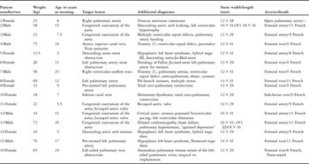
The patient’s characteristics are shown in Table 1, and are listed consecutively as admitted to our paediatric cardiac center. Indications for stenting the target lesions in coarctation with the aorta and pulmonary artery stenosis were invasively measured using a systolic pressure gradient of 20 millimetres of mercury or more. In systemic veins and total cavo-pulmonary connection, the presence of morphological stenosis was taken as a treatment indication even in the absence of a pressure gradient. The decision to use pre-mounted covered V12 stents was based on the following considerations. First were the lesion characteristics: if there was a risk for rupture and bleeding or treatment of an existing aneurysm; in one patient a previous balloon rupture during re-dilation due to a previously placed stent in the junction of the inferior caval vein–total cavo-pulmonary connection tunnel; in three patients to cover a (re)-opened atretic long-segment lesion. Second were the stent characteristics in the context of the patient’s age with regard to the balloon stent size, as re-dilatable widths ranging from 12 to 20 (22) millimetres due to the open-cell design, despite the polytetrafluoroethylene graft.
Summary of patient’s demographics, diagnosis, and procedures performed.

LD = large diameter
Congenital coarctation of the aorta was the target lesion in five patients; and obstruction of the descending aortic arch after hybrid stage three repair in two patients with hypoplastic left cardiac syndrome, both aged 5 years. In one of them (number 5) a Jo-med stent (Abbott Laboratories, Illinois, United States of America) was previously placed to treat a post-operative obstruction, but the stent was already broken when the 12 × 29-millimetre V12 stent was implanted in order to further augment the aortic arch. Five patients had pulmonary artery obstructions. In one patient, with a truncus arteriosus, the obstructed right pulmonary artery was treated with a 12 × 29-millimetre V12 stent as a hybrid approach during open-heart homograft replacement (number 1). In four patients, the left pulmonary artery was stented with a V12 device (n = 1 after an earlier repair of tetralogy of Fallot (n = 2) following total cavo-pulmonary connection completion (n = 1) with congenital multiple pulmonary artery stenoses. Three patients had already received stent placements (n = 2) a Jomed stent (Abbott, Germany), and (n = 1) a Palmaz Genesis stent (Cordis Company, Johnson & Johnson, New Jersey, United States of America), the latter was broken before V12 implantation. One patient had an acquired atretic junction of the anonyma vein to the right superior caval vein. One patient with trisomy 21 and pulmonary atresia with ventricular septal defect had severe cyanosis due to a stenotic shunt and not a grown aortic-pulmonary shunt. The long-segment atretic right ventricular outflow tract was perforated with a high-frequency technique and subsequently underwent gradual balloon dilation before the implantation of a 12 × 41-millimetre covered V12 stent. The last patient in our series suffered from a complete obstruction of the pulmonary veins after surgical re-implantation by using the vertical vein in a 24-year-old woman suffering from partial anomalous pulmonary venous return with drainage of the left-sided pulmonary veins.
Cardiac catheterisation and stent placement
All interventions were carried out under sedation, using local anaesthesia for vascular access without any use of a vessel occlusion system for later haemostasis (Table 1). Based on the origin and shape of the target lesion, vascular access was attempted from the femoral vein (n = 8), femoral artery (n = 7), and in one patient (patient 9) from the left subclavian vein due to bilaterally completely obstructed femoral veins. Open, direct pulmonary artery access was used for intra-operative stent placement in the operation theatre during corrective surgery. Stent positioning was performed when right pulmonary artery was visible and open, the length of the stent was chosen based on the pre-operative angiographic data. Diagnostic catheterisation was performed to determine the exact morphology and the pressure gradient of the stenotic lesions. Once the decision for stenting the target lesion utilising a covered V12 stent angiographically was made, the smallest delivery system possible was chosen. In the two 5-year-old patients with an obstructed descending aorta after Norwood-like aortic arch repair an 8 French sheath (Cook, Copenhagen, Denmark) was placed through the femoral artery, and in the patient with an acquired pulmonary vein obstruction (patient 16) after a trans-septal approach to the Brockenbrough technique. A 9 French sheath as recommended by the stent manufacturer was used in four patients for the placement of a V12 stent pre-mounted on a balloon with a width of 12 millimetres. Two patients received a V12 stenting without a long guiding sheath: in one as a hybrid surgical approach, and in the other as a percutaneous approach for left pulmonary artery stenting. Advanta V12 stents with a balloon width of 14 or 16 millimetres were implanted utilising an 11 French delivery sheath (Cook). After the advancement of the mounted stent to the obstructed target lesion through the sheath, manual injection of the contrast through the sheath or angiographies by means of a separate access confirmed the desired position of the stent immediately before implantation. The extent of the different vessel stenoses was estimated in the context of the corresponding vessel diameters to choose the best stent model currently available. In one patient, the graft stent was used to cover an aneurysm coexisting with a post-surgical descending aortic arch obstruction. Secondary dilation with larger diameter balloons was performed when the residual pressure gradient, the stent–vessel wall relationship, or stent re-coiling due to different reasons did need a re-intervention by ballooning or second stent placement.
Procedural characteristics of stent implantation within the various lesions and immediate follow-up results were analysed to determine the strengths and weaknesses of the novel large Advanta V12 stent was used for various indications. All patients received heparin 50–100 International Units per kilogram body weight before intervention and 300 International Units per kilogram body weight per day for the next 24 hours. Aspirin (2–3 milligrams per kilogram body weight per day) was recommended for 6 months. Patients with a total cavo-pulmonary connection and the patient with a post-stent placement of the obstructed pulmonary vein received warfarin to an international normalised ratio between 2 and 2.5 prior to and post-intervention.
Follow-up
Clinical evaluation, non-invasive blood pressure measurements, echocardiography, or magnetic resonance imaging were done in the post-interventional period. Special attention was directed to the femoral artery in children with coarctation of the aorta and weight of less than 20 kilograms. Repeated duplex sonography of the groins was performed to document the integrity of the vessels at discharge and during follow-up.
Results
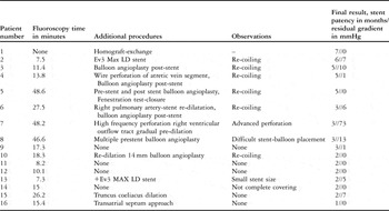
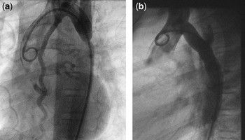
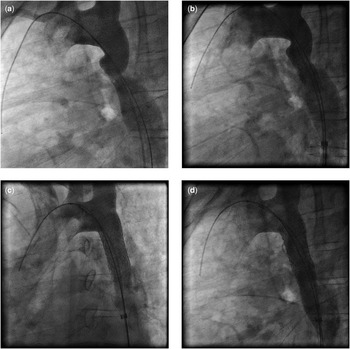
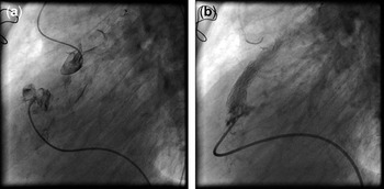
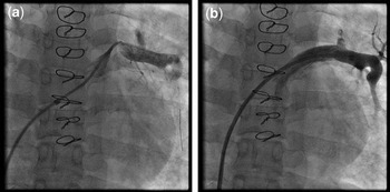
Between July, 2009 and April, 2010, 16 patients underwent implantation of the Advanta V12 large-diameter stent for the treatment of various lesions. Data are described as median, and a range of minimum and maximum values. The median age of the patients was 15 years (5–46 years); the median weight was 26 kilograms (range 14–75 kilograms). The target lesions, additional diagnoses, age, size of the Atrium V12 stents, and vascular access are shown in Table 1. Table 2 summarises the fluoroscopy time of all procedures, the procedures that were performed in addition to the V12 implantation, the facts that complicated the graft stent placement and the final results with early follow-up of stent patency as well as the residual pressure gradients. The variability of the treated lesions and the need for additional interventions were responsible for a wide range of fluoroscopy times between 7.3 and 48.2 minutes (median 17.3 minutes). Considering only the V12 stent implantation in circumscribed lesions enabled stenting with the pre-mounted graft stent with fluoroscopy times of 7 to 10 minutes. Re-coiling of the open-cell-designed stent was observed in seven patients, in whom re-dilation of the V12 stent became necessary, either by ballooning (n = 4) or placement of a second stent (n = 3). The placement of the second stent was performed when the re-coiling of the V12 stent, and/or the stent shortening resulted in an unsatisfactory primary result. The desired final result of V12 stent placement without an additional approach after the procedure was achieved in the patient with the intraoperative stent placement (number 1), in two young patients (number 11, 12, Figs 1a and b) with coarctation of the aorta and in patient 16 with pulmonary vein obstruction. In these patients a re-established descending aortic arch without a pressure gradient could be achieved and a smooth stent–vessel wall relationship with residual compliance within the affected vessel part was angiographically documented. With regard to the additional procedures, as in patient 13, in whom an additional non-covered stent placement was performed (Figs 2a–d); V12 stent placement achieved the desired result in all cases. There were no technical problems in performing V12 stenting, with one exception: in patient 8 with multiple congenital pulmonary artery stenoses, positioning of the pre-mounted V12 stent through a previously placed left pulmonary artery stent was complicated due to the trial of placing the stent without long sheath guiding. Intra-operative stenting of the right pulmonary artery in patient 1, as a hybrid approach, was simple and shortened the open-heart surgery significantly. In the patients with an acquired atretic vein segment as well as the patient with a congenital long atretic right ventricular outflow tract (Fig 3a), V12 stent placement achieved a covered re-opened vein for relief of the caval vein superior syndrome and a reconstructed right ventricular outflow tract as an additional source for improving pulmonary blood flow (Fig 3b). In addition, the complete obstructed pulmonary vein drainage (Fig 4a) of the left lung was resuscitated by placing a V12 (12 × 29 millimetres) graft stent without a residual pressure gradient (Fig 4b). The mean pressure within the left pulmonary vein decreased from 30 millimetres of mercury to 15 millimetres of mercury corresponding to the left atrial pressure. However, exact and sufficient stent placement was performed after pre-dilation of the obstructed area with a high-pressure balloon with a width of 8 and 10 millimetres and a length of 30 millimetres.
Summary of procedure data.

LD = large diameter
Depicted is a congenital coarctation in an 11-year-old patient (number 11) in 30° left anterior oblique projection (a); the final result after placement of a 12 × 29 millimetres Advanta V12 stent projected in 90 degrees lateral view of (b). The V12 covered stent-vessel wall relationship is nearly ideal due to minimal stent re-coiling to the native vessel dimensions surrounding the obstructed part of the descending aorta.

Angiogram performed through a multi-track catheter positioned in the aortic arch shows an aortic coarctation with kinking of the descending arch course together with a small “pseudo”-aneurysm (a); the exactly positioned Advanta V12 (16 × 41 millimetres) stent, shortened to 35 millimetres by balloon inflation, but with an anterior deviation with the upper end within the pseudo-aneurysm meaning that the stent might influence the blood flow due to its coverage (b); the final result in left anterior oblique projection (30 degrees) after placement of an additional eV3 large diameter 36 millimetres bar metal stent inside the V12 dilated to 18 millimetres (c); in lateral projection that both stents are positioned in telescope technique reconstruct the hypoplastic isthmus region, from a morphological point of view (d).

A patient with pulmonary atresia and a ventricular septal defect palliated with an aorto-pulmonary shunt (number 7): Shown is the long segment right ventricular outflow tract obstruction before radiofrequency perforation (a); gradual balloon dilation allowed placement of premounted Atrium V12 (12 × 41 millimetres) to reconstruct the right ventricular outflow tract as an additional source to improve pulmonary blood flow (b).

A patient (number 16) with an acquired left-sided pulmonary venous obstruction: Shown is the short segment obstruction after crossing with a 0.014 inch floppy wire (a); the final result after gradual balloon dilation and placement of an Atrium V12 (12 × 29 millimetres) stent is depicted; the surgically re-connected pulmonary vein is reconstructed without residual pressure gradient (b).

All stents were placed in the target lesions without stent dislodgement, dissection, or aneurysm formation. However, in patient 14 (he received a 12 × 19-millimetre covered stent) with a post-surgical descending aortic arch obstruction accompanied by an aneurysm, a minimal residual flow within the aneurysm was still observed at the end of the intervention despite a complete relief of the obstruction.
At short-term follow-up with a median of 4 months, all patients are alive and well with no evidence of stent failing.
Discussion
The initial results of our study demonstrate that the covered Advanta V12 large-diameter stent can be used safely and effectively for the treatment of various cardiovascular obstructions. Considering the advantages and the weakness of the pre-mounted polytetrafluoroethylene graft stent with an open-cell design, the availability of the Advanta V12 expands the interventional tool to treat high-risk obstructed lesions.
Pulmonary artery stenosis, primarily or postsurgically, can be effectively treated by percutaneous stent placement,Reference O’Laughlin, Perry, lock and Mullins1 or as an intra-operative approach.Reference Bokenkamp, Blom and deWolf9 Stent placement within the pulmonary arteries as a curative or palliative procedure should have the capability of being dilated to a final diameter of at least 15 millimetres.Reference Tomita, Yazaki and Kimura10 The disadvantages of larger stents with the necessity for a large implantation sheath can be overcome by implanting the stent intra-operatively as in the hybrid procedureReference Bacha, Marshall, McElhinney and del Nido11 (patient 1). Vascular tears and aneurysm formation have been reported for pulmonary artery stenting, both at the time of implantation and at re-dilation.Reference Ing, Grifka, Nihill and Mullins12, Reference Carano, Agnetti, Tchana, Squarcia and Saquarcia13 Considering that the predictive risk factors for severe complications are not well defined, we opted for the novel, designed V12 graft stent to re-dilate the pulmonary branch stenosis in one patient with congenital branch pulmonary artery stenosis, and in three patients with pre-stented left pulmonary arteries. However, the potential to cover pulmonary arteries should be considered; therefore, the V12 graft stent should not be used in locations with the risk of side branch occlusions. Since most lesions occur in the great vessels, stents have to be dilated to large diameters and still have sufficient radial force to provide mechanical stability. Moreover, in smaller children it is necessary to use stents, initially dilated to smaller diameters providing the possibility of later adapting the stent diameter by re-dilatation after the somatic growth of the patient. However, carrying the same potential of dilatability to larger diameters the risk for re-dilation has to be taken into account. The novel designed V12 has some advantages, particularly in growing children and might reduce the risk factors for stenting a large vessel with the need for re-dilation. The new large-diameter V12 stent has been designed to be dilatable to a maximal diameter of 22 millimetres with the polytetrafluoroethylene cover remaining intact. The device is pre-mounted on a high pressure balloon with a 12-millimetre balloon recommended for the use with a 9 French introducer sheath but is also implantable through an 8 French sheath without problems. This was useful in two 5-year-old children weighing 14 kilograms, with an obstructed anastomosis after aortic arch repair and acquired coarctation that was pre-treated by bar metal stent implantation. The low profile enables the use of the Advanta V12 for the treatment of coarctation of the aorta in smaller children and in patients with pulmonary vein obstructions if a trans-septal approach is already performed utilising an 8 French sheath with a Brockenbrough needle. The size of the introduced sheath for the V12 stent is comparable to the sheath, which is needed for angioplasty, using only high-pressure balloons. This compares favorably with the Cheatham platinum stent (NuMed, New York, United States of America), which requires a 2 French larger sheath than is necessary for the dilatation balloon and, in the case of a polytetrafluoroethylene-covered Cheatham platinum stent: a 3 French larger sheath.Reference Ewert, Schubert, Peters, Abdul-Khaliq, Nagdyman and Lange14 This study was not designed to compare the V12 graft stent with the covered Cheatham platinum stent, but the weakness of the Cheatham platinum seems to be the strength from the V12 and vice versa. Hence, the lower radial force of the V12, the higher recoil rate has the disadvantage of treating rigid lesions, but might be associated with the advantage of a higher residual compliance after stenting of a native, still-elastic obstruction, which was the case in the two young patients with congenital coarctation of the aorta. Whether the higher compliance of the stented aortic segment will have an immediate or long-term impact on the degree of systemic blood pressure and, therefore, a lower incidence of a residual systemic hypertension, remains to be evaluated in a direct comparative study between the V12 and the stent with more radial strength (e.g. Cheatham platinum stent). However, the availability of the V12 stent in a covered version is useful in extremely stressed vessel areas providing an additional seal to avoid vessel dissection and bleeding, which is crucial when later re-dilation will become necessary because of somatic growth. Furthermore, it offers a treatment solution for the combination of stenosis and vessel aneurysm by stenting the stenosis and covering the aneurysm, as demonstrated in patient 14. The graft design, together with the smallest possible delivery system, was beneficial when treating the atretic systemic and pulmonary veins and the right ventricular outflow tract to improve blood flow with lowering the risk of bleeding. In conclusion, this study emphasises on the preliminary short-term results of the open-cell V12 graft stent implantations utilised in various cardiovascular lesions. However, mid-term and long-term follow-up are necessary to assess the fracture rate, incidence of re-obstruction, and stent behaviour in the long term.