

Introduction
Sperm DNA integrity is essential for embryo development (Fatehi et al., Reference Fatehi, Bevers, Schoever, Roelen, Colenbrander and Gadella2006). Sperm with DNA damage have the ability to fertilize oocytes, however embryo development failures can occur. Infertility and genetic abnormalities in offspring have also been associated with sperm DNA damage (Ahmadi and Ng, Reference Ahmadi and Ng1999; Tamburrino et al., Reference Tamburrino, Marchiani, Montoya, Marino, Natali, Cambi, Forti, Baldi and Muratori2012). Traditionally, conventional sperm analysis does not include the evaluation of sperm DNA. In human, studies demonstrated that injury in sperm DNA is related to human reproduction failures (Aitken et al., Reference Aitken, Gordon, Harkiss, Twigg, Milne, Jennings and Irvine1998; Ahmadi and Ng, Reference Ahmadi and Ng1999; Saleh et al., Reference Saleh, Agarwal, Nada, El-Tonsy, Sharma, Meyer, Nelson and Thomas2003; Tamburrino et al., Reference Tamburrino, Marchiani, Montoya, Marino, Natali, Cambi, Forti, Baldi and Muratori2012; Simon et al., Reference Simon, Murphy, Shamsi, Liu, Emery, Aston, Hotaling and Carrell2014b). Moreover, human sperm DNA damage can affect embryo development, pregnancy rates (Wdowiak et al., Reference Wdowiak, Bakalczuk and Bakalczuk2015) and reduce implantation rates (Tesarik et al., Reference Tesarik, Mendoza and Greco2002).
Sperm DNA
Spermatozoa are formed in the seminiferous tubules, through a process called spermatogenesis. This process occurs after puberty and involves meiotic, mitotic and cellular remodelling. It is divided into three phases: (1) spermatogonia proliferation and differentiation; (2) spermatocytogenesis (meiosis); and (3) spermiogenesis. Type A spermatogonia, which are the stem cells present in the seminiferous tubules, are in constant mitotic division to generate more spermatogonia, including type B spermatogonia. Type B spermatogonia divide and differentiate, giving rise to primary spermatocytes. In spermatocytogenesis, primary spermatocytes undergo the first meiotic division (meiosis I) to form secondary spermatocytes. Secondary spermatocytes undergo the second meiotic division (meiosis II) to give rise to the spermatids (reviewed by Zini and Agarwal, Reference Zini, Agarwal, Sharma and Agarwal2011a).
Spermiogenesis is the final process of spermatozoa formation and is characterized by the differentiation of round spermatids in spermatozoa (Zini and Agarwal, Reference Zini, Agarwal, Sharma and Agarwal2011a). Spermatozoa have two distinct regions: the tail that conducts the sperm to the oocyte, and the head that contains a haploid nucleus with compacted and stable DNA (Ward and Coffey, Reference Ward and Coffey1991). Sperm chromatin has transcriptional activity repressed and condensed DNA. Histones are basic nuclear proteins responsible for DNA packaging in somatic cells. When histones are hyperacetylated and nucleosomes dissociated (Carrell et al., Reference Carrell, Emery and Hammoud2007), DNA undergoes remodelling, as histone and non-histone proteins are replaced, wholly or partially, by transition proteins (TP)1 and 2. TPs are extremely important during normal DNA condensation because they reduce DNA breaks (Balhorn, Reference Balhorn2007). At the end of spermiogenesis, TPs are replaced by specific nucleoproteins called protamines (PRM). PRM or keratinous proteins are half the size of a histone; they have basic characteristics and are rich in arginine and oxidized cysteine, which can form inter-protamine and intra-protamine disulfide bonds that contribute to stabilizing the chromatin (Vilfan et al., Reference Vilfan, Conwell and Hud2004). Protamination is the process that replaces histones by protamines. Protamine types 1 and 2 (PRM1, PRM2) are found in mammalian sperm. PRM1 is expressed in all mammals (Carrell et al., Reference Carrell, Emery and Hammoud2007), whereas PRM2 is described in human, primate, murine and equine sperm (Maier et al., Reference Maier, Nussbaum, Domenjoud, Klemm and Engel1990; Queralt et al., Reference Queralt, Adroer, Oliva, Winkfein, Retief and Dixon1995) and recently in bovine (Hamilton et al., Reference Hamilton, Simões, Mendes, Goissis, Nakajima, Martins, Visintin and Assumpção2019). Protamine type 3 is described as a PRM2 isoform and is reported in rat (Schluter and Engel, Reference Schluter and Engel1995), human, ape and Old World monkeys (reviewed by Steger and Balhorn, Reference Steger and Balhorn2018). PRM compacts the sperm DNA in such a manner that it is six times more compact than somatic cell DNA, and promotes stabilization and protection of sperm nucleus (McLay and Clarke, Reference McLay and Clarke2003). Moreover, DNA compaction decreases cell volume, facilitating passage through the female reproductive tract, penetration and activation of oocyte, which will then initiate embryonic development without DNA damage (Oliva, Reference Oliva2006).
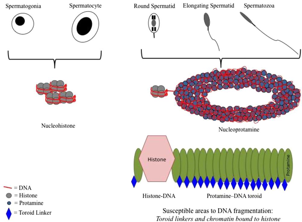
PRMs bind to the DNA minor curvature and form highly compacted complexes called nucleoprotamines (Wouters-Tyrou et al., Reference Wouters-Tyrou, Martinage, Chevaillier and Sautiére1998). After protamine binding, sperm chromatin becomes extremely condensed and inert (Rocha et al., Reference Rocha, Beletti, Marcolini and Amorin2002). The crosslinking between cysteine residues promotes high compaction of protamine toroids and greater DNA stability (Gosálvez et al., Reference Gosálvez, López-Fernández, Fernández, Gouraud and Holt2011). The toroids of the nucleoproteins contain highly compacted DNA located in each DNA curvature, and attached together by linkers. In linker regions, chromatin is more vulnerable to DNA fragmentation (Balhorn et al., Reference Balhorn, Corzett, Mazrimas and Watkins1991; Balhorn, Reference Balhorn1982, Reference Balhorn2007). A schematic representation of sperm chromatin changes is presented in Fig. 1.
Schematic representation of chromatin changes during spermiogenesis and susceptible regions of sperm chromatin to DNA fragmentation.
The enzyme DNA topoisomerase promotes DNA condensation by physiological breaking and linking DNA during spermiogenesis. Sperm DNA physiological breaks that occur during spermiogenesis relieve torsion of the chromatin, supporting the protamination process. Therefore, these endogenous DNA breaks should not persist in mature spermatozoa (McPherson and Longo, Reference McPherson and Longo1993), and must be repaired by the cell itself before the end of the protamination process (Balhorn, Reference Balhorn1982; Andrabi, Reference Andrabi2007). However, cells in advanced stages of differentiation (spermatids and spermatozoa) have no mechanism of DNA repair, therefore cells with DNA damage could be present in the ejaculate (Lewis and Agbaje, Reference Lewis and Agbaje2008). The sperm with DNA damage can fertilize but embryo development can be compromised (Tesarik et al., Reference Tesarik, Mendoza and Greco2002).
The effect of abnormal sperm chromatin depends on the ability of the oocyte to repair the DNA damage (Evenson et al., Reference Evenson, Larson and Jost2002a). Oocytes and embryos at an early stage of development are able to repair sperm DNA damage, but this capacity is limited, and problems such as double-stranded breaks (DSB) and breaks on histone-associated regions are not solved (Menezo et al., Reference Menezo, Dale and Cohen2010).
Therefore, sperm DNA integrity is an important and independent sperm trait that has been associated with fertility potential in reproductive biotechnologies (Larson-Cook et al., Reference Larson-Cook, Brannian, Hansem, Kasperson, Aamold and Evenson2003). In men, a recent review described the beneficial role of sperm DNA fragmentation testing in evaluation of patients with unexplained infertility (Cho and Agarwal, Reference Cho and Agarwal2018).
Sperm DNA fragmentation causes
There are three possible causes for chromatin damage and sperm DNA fragmentation: protamination failures, apoptosis and oxidative stress. Sperm DNA fragmentation can be a consequence of these processes, either combined or not (Berenguer et al., Reference Berenguer, Peregrin, López-Fernández, Fernández, Fernández and Calonge2008).
When protamination occurs incorrectly, chromatin condensation disorders occur (Sotolongo et al., Reference Sotolongo, Huang, Isenberger and Ward2005). Incomplete protamination generates sperm that is more vulnerable to endogenous and exogenous agents, such as nucleases (Sotolongo et al., Reference Sotolongo, Huang, Isenberger and Ward2005), free radicals (Alvarez and Storey, Reference Alvarez and Storey1992) or mutagens. The enzyme DNA topoisomerase II is responsible for repairing DNA breaks during the spermiogenesis process. When this procedure fails, DNA breaks will remain in the sperm. A study with infertile men has correlated translational damage in protamines with sperm protamination injury (Aoki et al., Reference Aoki, Emery, Liu and Carrell2006). A study with knockout mice for TPs, detected protamine alterations and sperm chromatin damage leading to reduced fertility and failures in embryo implantation (Suganuma et al., Reference Suganuma, Yanagimachi and Meistrich2005). In bovine, sperm with low motility presented a decrease in protamine transcripts (Ganguly et al., Reference Ganguly, Gaur, Kumar, Mandal, Kumar, Singh, Kumar and Sharma2013)
The second possible cause of sperm DNA fragmentation is the apoptotic process. Apoptosis is the term used for programmed cell death that normally occurs in many physiological processes. The sperm express apoptosis markers during spermatogenesis and during maturation through the epididymis. In the testis, apoptosis is responsible for maintaining the proportion between germ and Sertoli cells, by removing defective germ cells and controlling sperm production (abortive apoptotic process) (Shukla et al., Reference Shukla, Mahdi and Rajender2012). The presence of molecules involved in apoptosis pathways such as caspases 1, 3, 8 and 9 indicates double-stranded sperm DNA breaks. Double-stranded DNA breaks are attached to the sperm nuclear matrix and are related to a lack of DNA repair during meiosis, inducing miscarriage and implantation failures (Ribas-Maynou and Benet, Reference Ribas-Maynou and Benet2019). In addition, the presence of Fas and p53 in sperm of infertile men indicates that these cells could have escaped from the abortive apoptotic process (Sakkas et al., Reference Sakkas, Moffatt, Manicardi, Mariethoz, Tarozzi and Bizarro2002).
The third possible cause of sperm DNA fragmentation is oxidative stress. Oxidative stress occurs when there is an imbalance between the reactive oxygen species production (ROS) and antioxidants in seminal plasma, including enzymes such as superoxide dismutase and catalase, and blocking reaction of reduction. This type of reaction generates damage at least in one of the DNA strands, inducing sperm DNA fragmentation (Agarwal et al., Reference Agarwal, Saleh and Bendaiwym2003). In fact, it was observed that ram sperm susceptibility to lipid peroxidation might compromise sperm quality and interferes with the oxidative homeostasis generating oxidative stress, which may be the main cause of sperm chromatin damage (Hamilton et al., Reference Hamilton, Castro, Delgado, Assis, Siqueira, Mendes, Goissis, Muiño-Blanco, Cebrián-Pérez, Nichi, Visintin and Ortiz D’Ávila Assumpção2016). The same relationship was verified in human sperm (Agarwal et al., Reference Agarwal, Saleh and Bendaiwym2003; Sakkas and Alvarez, Reference Sakkas and Alvarez2010). Confirming this hypothesis, testicular heat stress generates oxidative stress and increases sperm DNA fragmentation, however without changes in protamination and apoptotic patterns (Hamilton et al., Reference Hamilton, Siqueira, Castro, Mendes, Delgado, Assis, Mesquita, Maiorka, Nichi, Goissis, Visintin and Assumpção2018).
Assessment of sperm DNA integrity
Chromatin stability evaluated by SCSA modified test
Sperm DNA integrity is qualitatively evaluated by flow cytometer after heat- or acid-induced denaturation using the metachromatic fluorophore acridine orange (AO). When the test was first described, the study used heat (100°C, 5 min) to denature DNA at sites of sperm DNA strand breaks, followed by staining with AO. Recently, the protocol changed to denaturation of the sperm DNA by low pH (reviewed by Evenson, Reference Evenson2016). As this protocol uses DNA denaturation, it is considered an indirect assessment of chromatin integrity. This technique measures large numbers of sperm cells rapidly, and therefore provides potent statistical power with precision and repeatability. AO will intercalate with DNA strands and emits a green colour, designating the presence of double-stranded DNA, while red designates the presence of single-stranded DNA, which indicates denaturation of the chromatin structure (fragmented sperm DNA). The spermatozoa go through a flow cytometer, and the relationship obtained between red and total fluorescence is used to determine the DNA fragmentation index (DFI) (Evenson et al., Reference Evenson, Larson and Jost2002b). Sperm DNA with breaks are greatly susceptible to denaturation; therefore spermatozoa with higher DFI have damaged DNA. In regions with DNA fragmentation there is possible alteration of genetic information (Evenson et al., Reference Evenson, Larson and Jost2002b). Many publications have validated the use of this assessment in animals and humans (De Ambrogi et al., Reference De Ambrogi, Ballester, Saravia, Caballero, Johanniston, Wallgren, Anderson and Rodriguez-Martinez2006; Dias et al., Reference Dias, Retamal, Tobella, Arnholdt and Lopez2006; Garcia-Macias et al., Reference Garcia-Macias, Martinez-Pastor, Alvarez, Borragan, Chamorro, Soler, Anel and Paz2006; Hernandez et al., Reference Hernandez, Roca, Ballester, Vazquez, Martinez, Johannisson, Saravia and Rodriguez-Martinez2006; Kasimanickam et al., Reference Kasimanickam, Nebel, Peeler, Silvia, Wolf, McAllister and Cassell2006; Perez et al., Reference Perez, Rocha, Vannuchi, Mendes, Cavalcanti, Nichi, Ambrosio, Miglino, Visintin and Assumpção2014; Hamilton et al., Reference Hamilton, Castro, Delgado, Assis, Siqueira, Mendes, Goissis, Muiño-Blanco, Cebrián-Pérez, Nichi, Visintin and Ortiz D’Ávila Assumpção2016; Castro et al., Reference Castro, Siqueira, Hamilton, Mendes, Visintin and Assumpção2018); and only sperm chromatin integrity assay has an exact standardization (reviewed by Evenson et al., Reference Evenson2016). In our experience, no differences were observed in the percentage of positive sperm for AO between bulls with high and low fertility in IVF (Castro et al., Reference Castro, Siqueira, Hamilton, Mendes, Visintin and Assumpção2018). However, ram sperm with high susceptibility to induced lipid peroxidation presented a high percentage of native positive cells stained with AO, indicating higher fragility of sperm DNA (Hamilton et al., Reference Hamilton, Castro, Delgado, Assis, Siqueira, Mendes, Goissis, Muiño-Blanco, Cebrián-Pérez, Nichi, Visintin and Ortiz D’Ávila Assumpção2016). When rams were submitted to heat stress by testicular insulation, an increase in the percentage of AO-positive sperm cells was observed (Hamilton et al., Reference Hamilton, Siqueira, Castro, Mendes, Delgado, Assis, Mesquita, Maiorka, Nichi, Goissis, Visintin and Assumpção2018). In boar semen, it was verified that the degree of DNA damage evaluated by the percentage of AO-positive sperm cells might be a useful to evaluate boar fertility performance (Broekhuijse et al., Reference Broekhuijse, Sostaric, Feitsma and Gadella2012). In stallion, studies have correlated the damage in sperm DNA (evaluated by using AO) with cooling and cryopreservation effects (Serafini et al., Reference Serafini, Varner, Bisset, Blachard, Teague and Love2017, Reference Serafini, Varner, Blanchard, Teague, LaCaze and Love2018; Ferreira et al., Reference Ferreira, Ferreira-Silva, Rocha, Farras, Calixto, Moura, Alvarenga and Oliveira2018).
Chromatin stability evaluated by sperm chromatin dispersion (SCD) test
The SCD test measures the absence of damage in sperm DNA and was first described in human sperm (Fernández et al., Reference Fernández, Muriel, Rivero, Goyanes, Vazquez and Alvarez2003). The test is based on the principle that sperm with fragmented DNA do not produce a halo of dispersed DNA loops, even after acid denaturation and removal of nuclear proteins. In this test, the sperm suspension is mixed with low-melting-point agarose, sequentially this suspension is pipetted onto a slide pre-coated with standard agarose. After drying, slides are submitted to acid denaturation to generate restricted single-stranded DNA motifs from DNA breaks. Proteins are removed by transferring slides to a tray with neutralizing and lysis solution. Finally, slides are dehydrated, air dried and cells are stained with DAPI (4′,6-diamidino-2-phenylindole) or another staining solution provided with the kit and mixed with antifade solution and then evaluated by fluorescence microscope. The technique may detect single-stranded breaks (SSB) and extensive DSB (Ribas-Maynou and Benet, Reference Ribas-Maynou and Benet2019). The halo size of each cell is evaluated by a relative parameter: surface of halo divided by surface of the whole nucleoid (Fernández et al., Reference Fernández, Muriel, Rivero, Goyanes, Vazquez and Alvarez2003). The SCD test is performed on commercial tests kits and is available in fertility laboratories for in-house use. The protocol is highly standardized, however the analysis is non-standardized, low numbers of sperm cells are analyzed and this method does not detect DSB attached to sperm nuclear matrix (MAR region) (Ribas-Maynou and Benet, Reference Ribas-Maynou and Benet2019). Moreover, less distinctive images can be obtained due to weak low-density nucleoids (Sharif, Reference Sharif2013)
DNA fragmentation evaluated by comet assay
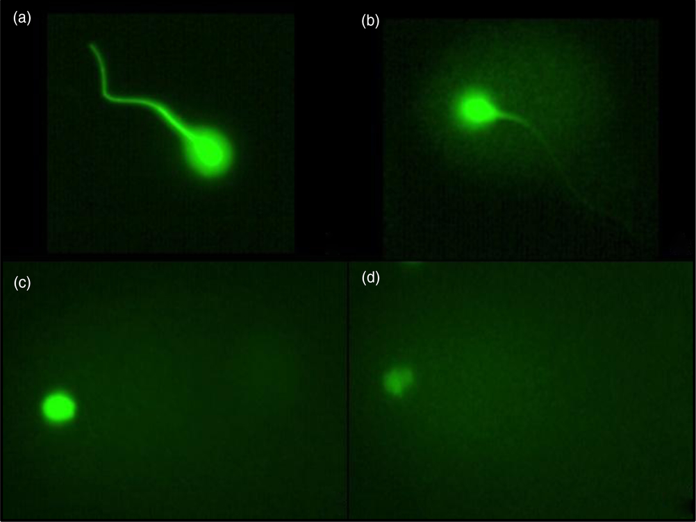
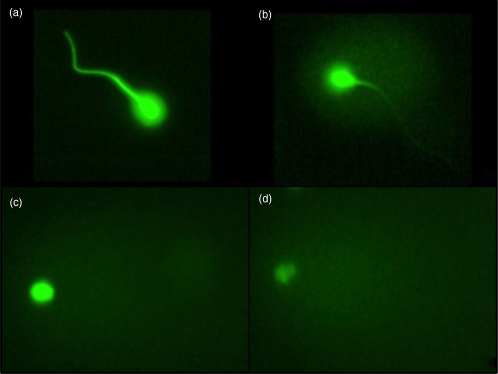
The comet assay is a sensitive and simple method to assess DNA damage using small numbers of spermatozoa (Simon and Carrell, Reference Simon and Carrell2013). The approach consists of single-cell gel electrophoresis that allows the migration of denatured DNA in a thin layer of agarose, which after staining with a fluorescent dye shows a electrophoretic track (tail of comet). Alkaline (pH > 13) or neutral conditions of electrophoresis are used to denature sperm DNA, forming nucleoids. The DNA migration of nucleoids occurs depending on the level of DNA damage and electrophoresis conditions. In the alkaline comet assay the main component of the comet tail is the SSB. The DSB in the comet tail represent high levels of DNA damage when the neutral condition of electrophoresis is used (Afanasieva and Sivolob, Reference Afanasieva and Sivolob2018). Localized DSBs represent very few break points in the genome, and remained attached to the sperm nuclear matrix (Ribas-Maynou et al., Reference Ribas-Maynou, Gawecka, Benet and Ward2014), an important attribute as the nuclear matrix is inherited with the male pronucleus in the zygote (Gawecka et al., Reference Gawecka, Ribas-Maynou, Benet and Ward2015), enabling the embryo to correct DSB. A study comparing five methodologies of analyzing sperm DNA fragmentation verified that the neutral comet assay is the only technique able to detect MAR region DSB, whereas transferase dUTP nick end labelling (TUNEL), SCSA and SCD may detect extensive DSB (Ribas-Maynou et al., Reference Ribas-Maynou, Garcia-Peiro, Fernanez-Encinas, Abad, Amenguai, Prada, Navarro and Benet2013). In humans, SSB may impair fertilization (Ribas-Maynou et al., Reference Ribas-Maynou, Garcia-Peiro, Fernanez-Encinas, Amenguai, Prada, Cortes, Navarro and Benet2012), whereas DSB interfere with embryonic development, implantation (Lewis and Aitken, Reference Lewis and Aitken2005) and cause recurrent miscarriages (Lewis and Simon, Reference Lewis and Simon2010). The comet test consists in applying sperm in a thin layer of agarose on a slide; posteriorly, cells are lysed in high salt conditions. In this process, protamines and histones are removed, with only the supercoiled DNA remaining. Serafini et al. (Reference Serafini, Romano, Varner, Di Palo and Love2015) describe the neutral comet assay as the more appropriate for distinguishing groups with different sperm quality than the alkaline comet assay. Under electrophoresis the broken strands will migrate to the anode pole, forming a comet tail that can be visualized under a fluorescence microscope after staining with SYBR or ethidium bromide. The relative fluorescence of the comet tail, relative to sperm head, provides the grade of sperm DNA damage. The images are randomly selected and analyzed by visual grading in degrees from I to IV, according to the intensity of DNA damage (Hamilton et al., Reference Hamilton, Castro, Delgado, Assis, Siqueira, Mendes, Goissis, Muiño-Blanco, Cebrián-Pérez, Nichi, Visintin and Ortiz D’Ávila Assumpção2016). Grade I consists of sperm with a surrounding halo and stained nuclei, without an evident comet tail, indicating little fragmentation. Grade II consists of sperm with comet tail formation and a pale core light, indicating DNA damage. Grade III consists of sperm with fully formed comet tails, yet with the core partially stained, indicating obvious DNA damage and Grade IV consists of sperm with a fully formed comet tail and with a completely stained core, indicating intense DNA fragmentation. Figure 2 shows an example of these comet grades. In one of our studies, no differences between any comet grades were found when comparing sperm from bulls with high and low in vitro fertility (Castro et al., Reference Castro, Siqueira, Hamilton, Mendes, Visintin and Assumpção2018). When we worked with rams, we observed an increase in Grade III DNA fragmentation in sperm with high susceptibility to lipid peroxidation (Hamilton et al., Reference Hamilton, Castro, Delgado, Assis, Siqueira, Mendes, Goissis, Muiño-Blanco, Cebrián-Pérez, Nichi, Visintin and Ortiz D’Ávila Assumpção2016) and submitted to heat stress (Hamilton et al., Reference Hamilton, Siqueira, Castro, Mendes, Delgado, Assis, Mesquita, Maiorka, Nichi, Goissis, Visintin and Assumpção2018). In men, less SSB and DSB were observed in sperm from fertile men compared with subfertile men (Ribas-Maynou et al., Reference Ribas-Maynou, Garcia-Peiro, Fernanez-Encinas, Amenguai, Prada, Cortes, Navarro and Benet2012). Since 2010, this test has been used for diagnosis of male infertility and prediction of successful IVF in humans (Sharif, Reference Sharif2013).
Fluorescence microscopy of sperm DNA damage as revealed by comet test in ram sperm. Slides stained by Syber Green (SYBR green I, Sigma), diluted 1:1000, for 1 h. (a) Grade I sperm DNA damage. (b) Grade II sperm DNA damage. (c) Grade III sperm DNA damage. (d) Grade IV sperm DNA damage. ×200 magnification.
DNA fragmentation evaluated by TUNEL assay
TUNEL (terminal deoxynucleotidyl transferase dUTP nick end labelling) assay is one of the most commonly used tests to assess sperm DNA damage. This test quantifies the incorporation of fluoresceinated dUTP into SSB or DSB in DNA that contain free 3′OH extremities by labelling only the 3′OH terminal with TdT (Ribeiro et al., Reference Ribeiro, Sharma, Gupta, Cakar, De Geyter and Agarwal2017). The fluorescence will be proportional to the number of strand breaks (Zini and Agarwal Reference Zini, Agarwal, Sharma and Agarwal2011b). TUNEL test consists of respectively fixation and permeabilization of sperm, marking DNA fragments, staining and evaluation of sperm. It is possible to perform this assay on a slide using fluorescence microscopy or in a flow cytometer. Some studies standardized the TUNEL assay for healthy and infertile men (Sharma et al., Reference Sharma, Sabanegh, Mahfouz, Gupta, Thiyagarajan and Agarwal2010, Reference Sharma, Ahmad, Esteves and Agarwal2016). Therefore, this assay identifies actual sperm DNA damage, in opposition to the others that evaluate DNA damage caused by denaturing conditions. However, as in other assays, TUNEL is unable to differentiate clinically important DNA damage from clinically insignificant damage.
Tests used to assess sperm nuclear proteins
Assessment of the lack of protamination by toluidine blue (TB) stain
The assay is based on the low ability of the free phosphate groups of DNA and protamines to bind to basic dyes (Shamsi et al., Reference Shamsi, Imam and Dada2011). Toluidine blue is a dye used for metachromatic and orthochromatic staining of chromatin. Semen smears are air dried, fixed with 96% ethanol:acetone (1:1) and submitted to hydrolysis with 0.1 N HCl. A 0.05% TB solution (50% McIlvain’s citrate phosphate buffer, pH 3.5) is applied to evaluate sperm chromatin. Sperm heads without DNA damage will stain light blue, and those with DNA damage will stain purple. Sperm nuclei that stain purple in the TB test fluoresce red in the AO test, and the sperm nuclei that stain in light blue, fluoresce green in the AO test. This technique is used for sperm DNA analysis in men (Erenpreisa et al., Reference Erenpreisa, Erenpreiss, Freivalds, Slaidina, Krampe, Butikova, Ivanov and Pjanova2003) as it is fast, low-cost and efficient. Actually, TB is standardized in several domestic species such as canine (Flores et al., Reference Flores, Angrimani, Rui, Brito, Abreu and Vannucchi2016; Rui et al., Reference Rui, Angrimani, Bicudo, Losano, Nichi and Pereira2018), feline (Rui et al., Reference Rui, Angrimani, Bicudo, Losano, Nichi and Pereira2018), ruminants (Kamimura et al., Reference Kamimura, Jacomini and Beletti2010; Quintero-Moreno et al., Reference Quintero-Moreno, Ramirez, Nava-Trujillo, Osorio, Rodríguez-Gil and Hidalgo2010; Rui et al., Reference Rui, Angrimani, Bicudo, Losano, Nichi and Pereira2018), equine (Sardoy et al., Reference Sardoy, Carretero and Neild2008; Rui et al., Reference Rui, Angrimani, Bicudo, Losano, Nichi and Pereira2018) and avian (Rui et al., Reference Rui, Shibuya, Kawaoku, Losano, Angrimani, Dalmazzo, Nichi and Pereira2017), as well as laboratory animals such as leporine (Beletti and Mello, Reference Beletti and Mello2004) and murine (Krzanowska, Reference Krzanowska1982).
Protamine deficiency assessment in sperm chromatin
Chromomycin A3 (CMA3) is a fluorochrome that competes with protamine binding sites (Bianchi et al., Reference Bianchi, Manicardi, Urner, Campana and Sakkas1996). It is a robust tool to estimate sperm chromatin packaging and allows indirect visualization of protamine deficiency. CMA3 analysis can be assessed in microscopy analysis or flow cytometry (Evenson, Reference Evenson2016; Hamilton et al., Reference Hamilton, Castro, Delgado, Assis, Siqueira, Mendes, Goissis, Muiño-Blanco, Cebrián-Pérez, Nichi, Visintin and Ortiz D’Ávila Assumpção2016, Reference Hamilton, Siqueira, Castro, Mendes, Delgado, Assis, Mesquita, Maiorka, Nichi, Goissis, Visintin and Assumpção2018; Castro et al., Reference Castro, Siqueira, Hamilton, Mendes, Visintin and Assumpção2018). Protamine deficiency is described as one of the causes of human male infertility (Agarwal et al., Reference Agarwal, Shyam and Allamaneni2005) In cattle, stress does not appear to be sufficient to alter the protamine pattern (Simões et al., Reference Simões, Feitosa, Mendes, Marques, Nicacio, Barros, Visintin and Assumpção2009). A recent study comparing sperm from bulls with high and low in vitro fertility verified greater protamine deficiency on bulls with high in vitro fertility (0.19% ± 0.03) compared with low (0.04% ± 0.04). However, authors suggested that protamine deficiency in bovine sperm may not have a strong biological effect to explain the difference of in vitro fertility (Castro et al., Reference Castro, Siqueira, Hamilton, Mendes, Visintin and Assumpção2018) In agreement with this hypothesis, a study comparing young, adult and senile bulls verified that differences in protamination did not interfere in embryo in vitro production (Kipper et al., Reference Kipper, Trevizan, Carreira, Carvalho, Mingoti, Beletti, Perri, Franciscato, Pierucci and Koivisto2016). In rams, no differences were observed between animals submitted to testicular heat stress in the percentage of sperm with protamine deficiency assessed by flow cytometer CMA3 test (Hamilton et al., Reference Hamilton, Siqueira, Castro, Mendes, Delgado, Assis, Mesquita, Maiorka, Nichi, Goissis, Visintin and Assumpção2018).
Protamine expression in sperm nuclei
The study of protamines is reported as an important predictor of human male fertility (Sharif, Reference Sharif2013), it is believe that the relation 1:1 is ideal for PRM1:PRM2. In mice, DNA integrity is altered when the ratio between protamines 1 and 2 is disrupted (Suganuma et al., Reference Suganuma, Yanagimachi and Meistrich2005). In bovine, a recent study from our research group identified, for the first time, PRM2 in bovine epididymal spermatozoa and in testis (Hamilton et al., Reference Hamilton, Simões, Mendes, Goissis, Nakajima, Martins, Visintin and Assumpção2019). This finding could suggest that the ratio PRM1:PRM2 could be important in bovine fertility also. The knowledge of the amount of these proteins in the sperm nucleus is fundamental to establish the ratio between protamines, as the ratio between protamine types (PRM1:PRM2) differs among the species. To extract and analyze protamines it is important to break disulfide bridges with DTT (Yebra and Oliva, Reference Yebra and Oliva1993; Dogan et al., Reference Dogan, Vargovic, Oliveira, Belser, Kaya, Moura, Sutovsky, Parrish and Topper2015). After that, cysteines must be reduced and protamines separated by electrophoresis. SDS polyacrylamide acetic acid–urea gel electrophorese is indicated (Seligman et al., Reference Seligman, Kosower, Weissenberg and Shalgi1994; Hamilton et al., Reference Hamilton, Simões, Mendes, Goissis, Nakajima, Martins, Visintin and Assumpção2019). Scanning the gel previously stained with Coomassie blue allows the identification of protamines. Relative amounts of PRM1 and PRM2 can be assessed using imaging software. Gels are transferred to a polyvinylidene fluoride membrane and incubated with primary antibodies for PRM1 and PRM2. After application of secondary antibody, positive bands are detected by chemiluminescence or fluorescence, then the protamines are quantified and the ratio between these is estimated.
Assessment of the sperm nucleoprotein structure by determination of free cysteine radicals
Disruption of the disulfide bonds between cysteine radicals from protamines alters the sperm nucleoprotein structure and generate DNA fragmentation. The correct degree of sperm thiol oxidation is important for sperm maturation, motility and activation, and for the events during fertilization (Seligman et al., Reference Seligman, Kosower, Weissenberg and Shalgi1994). The alteration of sperm nucleoprotein structure by analysis of sperm thiols and disulfides is verified in cryopreserved boar sperm (Yeste et al., Reference Yeste, Estrada, Casas, Bonet and Rodríguez-Gil2013), asthenozoospermia (Seligman et al., Reference Seligman, Kosower, Weissenberg and Shalgi1994), old rats (Zubkova et al., Reference Zubkova, Wade and Robaire2005), and rams with diminished fertility (Rodrigues et al., Reference Rodrigues, Ohanian and Bustos Obregon1985). The technique verifies the presence of non-oxidized thiols groups in the cysteine radicals of the protamines. Therefore, the sperm nuclear proteins are previously isolated and then incubated with fluorescent thiol labelling agent monobromobimane (mBBr). mBBr-labelled spermatozoa could be examined using fluorescence microscope or flow cytometer.
Conclusion
Sperm DNA fragmentation can be the consequence of protamination failures, apoptosis and oxidative stress. Therefore, the cause of sperm DNA fragmentation should be considered when choosing the optimum test for assessment of sperm DNA fragmentation. The numerous variations of sperm DNA fragmentation tests make it very difficult to compare data to assess male infertility. Moreover, different DNA fragmentation tests produce different threshold estimations. For humans, there is hesitation to incorporate sperm DNA fragmentation tests into clinical routine, as there is a lack of reliable thresholds. However, some publications have supported the benefits of human sperm DNA testing and showed the effect of the paternal genome on fertilization and early embryo quality (Simon et al., Reference Simon, Lui, Murphy, Ge, Hotaling, Aston, Emery and Carrell2014a, Reference Simon, Murphy, Shamsi, Liu, Emery, Aston, Hotaling and Carrell2014b). Therefore, according to Lewis (Reference Lewis2015), its possible continue to use traditional tests to evaluate male infertility, or include innovation in male fertility testing, such as DNA testing, and verify a possible improvement in fertility rates. For animal production (such as for bulls and rams), pressure of selection and fertility-oriented breeding programmes have allowed the removal of unacceptable sperm traits, including sperm DNA damage. Therefore, it is difficult to standardize tests and measure sperm DNA damage and correlate this damage with fertility rates in livestock. However, use of DNA fragmentation tests could be important for livestock selection, as the males will be used several times for breeding. For other species, such as companion animals, the use of DNA fragmentation tests in routine semen analysis will depend on breeding performance.
Acknowledgements
We thank Marcelo Goissis for English review (School of Veterinary and Animal Science, USP).
Ethics statements
Authors declare that they have no conflict of interest.