Introduction
Echinococcus granulosus and Echinococcus multilocularis are important zoonotic pathogens that cause serious disease in humans. The former has the greatest geographic distribution worldwide whereas the latter is restricted to transmission in the northern hemisphere. It is prevalent in western China (Craig et al., Reference Craig, Rogan and Campos-Ponce2003). Both pathogens have become a severe public health problem in China (Chai, Reference Chai1995; Schantz et al., Reference Schantz, Wang, Qiu, Liu, Saito, Emshoff, Ito, Roberts and Delker2003), especially in the Provinces of Sichuan, Tibet, Xinjiang, Gansu and Ningxia. In 2006 the Chinese government launched a national hydatid disease control programme in the above north-western provinces and also Inner Mongolia. Dog management and treatment of dogs with praziquantel were two of the key control measures. An important part of the programme was diagnosing Echinococcus spp. infection in dogs. The prevalence in dogs is an indicator of the effectiveness of control measures.
For the diagnosis of intestinal Echinococcus spp. infections in carnivores, classical diagnostic methods for direct parasite detection include sedimentation and counting eggs in faeces, arecoline purging or necropsy of the carnivore definitive hosts (Ziadinov et al., Reference Ziadinov, Mathis, Trachsel, Rysmukhambetova, Abdyjaparov, Kuttubaev, Deplazes and Torgerson2008). In addition, immunological and molecular techniques (detection of coproantigen by enzyme-linked immunosorbent assay (ELISA) and of copro-DNA by polymerase chain reaction (PCR)) are available (Lahmar et al., Reference Lahmar, Lahmar, Boufana, Bradshaw and Craig2007). All these tests have been validated in many parts of the world (Carmena et al., Reference Carmena, Benito and Eraso2006). Depending on the aims of the investigation, different methods and diagnostic strategies may be applied (Torgerson & Deplazes, Reference Torgerson and Deplazes2009).
Coproantigen testing is now the main surveillance tool of choice for dog infections, once it has been developed and validated in each country. Serum antibodies have been examined as a diagnostic tool (Benito et al., Reference Benito, Carmena, Martínez and Guisantes2006) and may be useful as a second test to prove validity of coproantigen results (Torgerson & Deplazes, Reference Torgerson and Deplazes2009). Copro-PCR can also provide a validation for coproantigen results (Nonaka et al., Reference Nonaka, Sano, Inoue, Teresa Armua, Fukui, Katakura and Oku2009). There are now three commercial coproantigen kits being sold in China (Shenzhen Combined Biotech Co. Ltd, Guang Dong (‘Combined kit’); Zhuhai Special Economic Zone Haitai Biological Pharmaceuticals Co. Ltd, Guang Dong (‘Haitai kit’); Xinjiang Tiankang Animal Husbandry Biotech Co. Ltd, Xinjiang (‘Tiankang kit’)). Details of validation have not been supplied by the manufacturers for any of the kits. We have conducted a small evaluation of the usefulness of these kits, and compared them to the results obtained with an experimental kit (‘Laboratory kit’) that had been partially developed and validated in Sichuan Province, China (Huang et al., Reference Huang, Yang, Qiu, Chen, Yang, Qiu, Xiao, Xiao and Heath2007).
Materials and methods
Collection and examination of faecal samples
A set of faecal samples from four dogs experimentally infected with E. granulosus were collected prior to infection and after infection (the animal trial was approved by the Qinghai Institute of Endemic Disease Prevention and Control Animal Ethics Committee). Each dog was individually housed and sequential individual faecal samples were collected.
Echinococcus granulosus cysts were obtained from sheep from a local slaughter house in Qinghai province. Brood capsules were collected from fertile cysts. The number of viable protoscoleces in a well-mixed aliquot of brood capsules was determined by dye uptake (0.1% eosin) and flame cell activity. Four dogs, before being infected with protoscoleces, were orally treated with praziquantel (5 mg/kg) and albendazole (10 mg/kg) to eliminate any existing intestinal parasites. Ten days later, each of the four dogs was infected orally with brood capsules containing 20,000 viable protoscoleces (dog 1, male, 2 years old; dog 2, male, 1.2 years old; dog 3, female, 4 years old and dog 4, female, 2 years old). At the time of infection and thereafter, 23 (19 positive and 4 control) sequential individual faecal samples were obtained. Forty-five days after infection, cestode eggs were detected around the perianal region of the four dogs, using the sellotape strip method, whereon the dogs were necropsied. All faeces collected from the rectum were frozen at − 70°C, as suggested in all three kits, for at least 3 days before faecal extracts were made, in order to kill the viable eggs.
From stray dogs in Shiqu (18 samples) and Seda (17 samples) a total of 35 faecal samples were collected at necropsy. In brief: the small intestine was removed and opened longitudinally. After visual inspection of the surface of the intestine, the areas containing worms were placed in closed bottles of 0.85% NaCl (saline) and shaken vigorously. Released worms were fixed in 4% of 40% formaldehyde in saline, either before or after relaxing overnight in tap water (Heath & Lawrence, Reference Heath and Lawrence1976). All parasites were observed visually (for big worms) and were identified by morphology using a microscope for E. granulosus and E. multilocularis worms (Eckert et al., Reference Eckert, Gemmell, Meslin and Pawlowski2001). An estimate was made of the worm burden of each species. Necropsy findings for each dog were recorded. Faecal samples from the rectum were obtained and treated as described above. All dog necropsies were carried out with the permission of Sichuan Centers for Disease Control and Prevention Animal Ethics Committee.
Coproantigen test kits
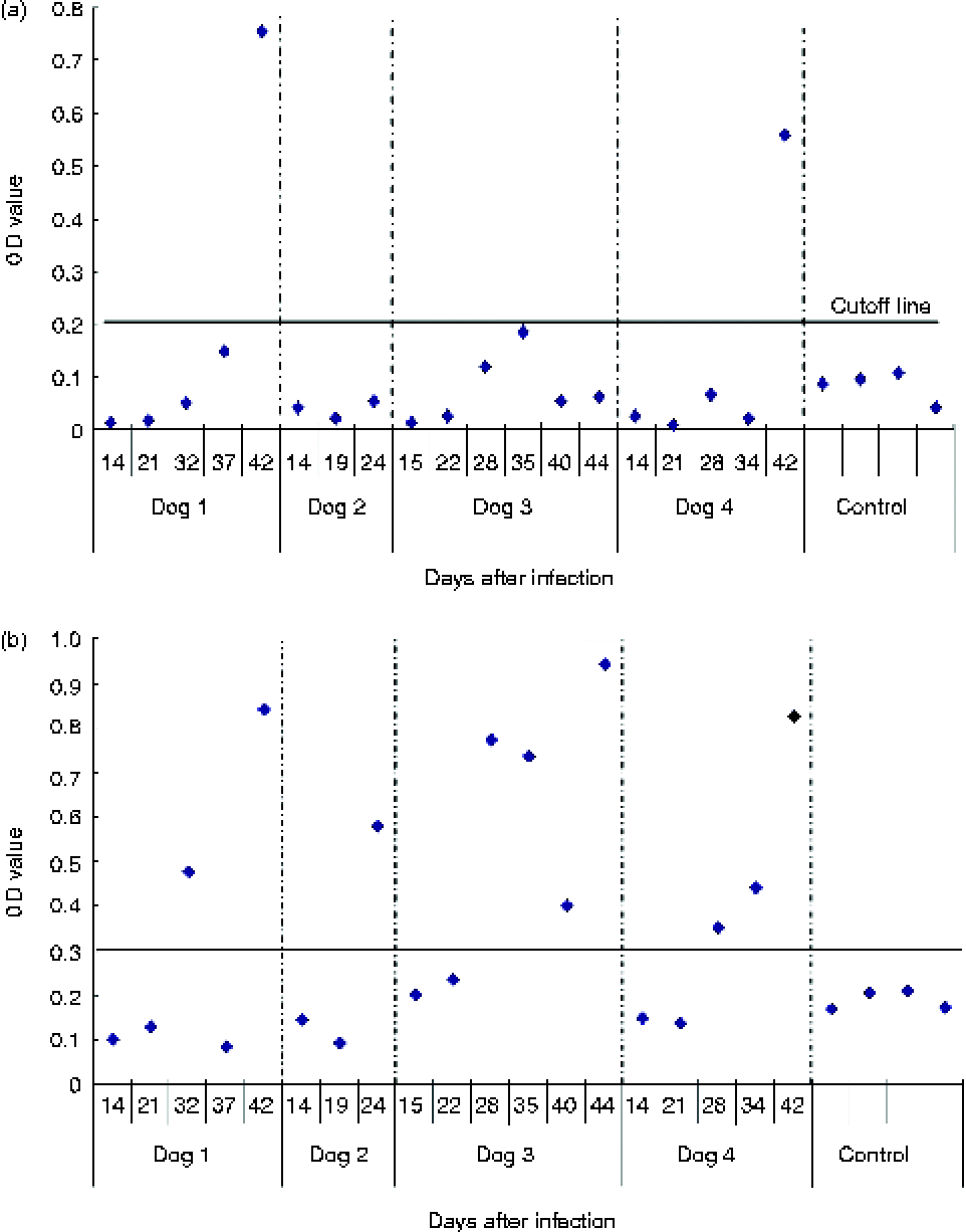
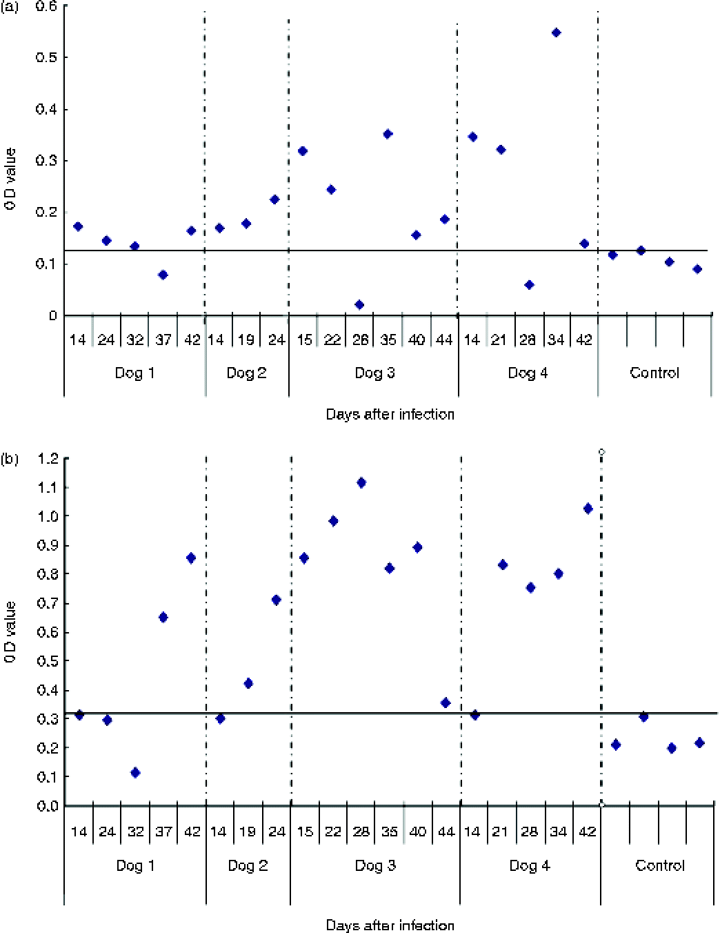
The procedure for each coproantigen test was conducted according to the manufacturer's instructions, given in brief below. Cut-off values for each kit are shown in figs 1 and 2.
(colour online) Detection of coproantigen by ELISA to show OD values from sequential faecal samples of four infected dogs (numbers 1– 4) and control using (a) Haitai and (b) Combined kits.

(colour online) Detection of coproantigen by ELISA to show OD values from sequential faecal samples of four infected dogs (numbers 1–4) and control using (a) Tiankang and (b) Laboratory kits.
Combined kit
The faecal samples were taken from the freezer and thawed at room temperature. About 1 g of faeces from each sample was put into a centrifuge tube with 1.5 ml of sample buffer (components not given), shaken vigorously to mix thoroughly, stood for 10 min, then centrifuged at 5000g for 30 min. The supernatant was used for coproantigen detection. For this test it was stipulated that all reagents and the ELISA plate should be warmed to room temperature (stored at 2–8°C) before using. The ELISA plate was designed to have duplicate wells of positive control and negative control, 100 μl/well. A sample buffer well (100 μl) was included as a blank. The test wells were filled with 50 μl/well of sample buffer first, then faecal supernatant of 50 μl/well for each sample was added. The plate was incubated at 37°C for 30 min, then washed four times with washing buffer (components not given), with 1 min intervals between each wash. Except for the blank, to each well was added two drops of horseradish peroxidase (HRP) conjugate (components not given). The plate was left at 37°C for 30 min, washed three times, one drop of substrate and another drop of developer were added to each well, and all wells were mixed within the plate using a vortex plate mixer. The plate was incubated in the dark at 37°C for 10 min, and the reaction was stopped by the addition of one drop of stop buffer to each well. The plate was then read at 450 nm using an automated ELISA plate reader (Anthos, Austria). The cut-off point for the test was calculated as 3.1 × the mean of the two negative wells. Samples with an OD (optical density) value above or equal to the cut-off point were determined as positives. (If the OD value of the negative mean was less than 0.10, it was recommended to use 0.10 as the negative mean.)
Haitai kit
The procedures conducted were almost the same as those for the Combined kit except: there was only one positive control, one negative control and three cut-off controls. These were all set up at 100 μl/well. One well was left as a blank without adding anything. The cut-off value for this test was determined by the mean of the three cut-off controls. A sample OD value above or equal to the cut-off was considered positive. This test required a quality control for each plate. The OD value of the negative control should be less than 0.15, the OD value of the positive control should be above 0.30 and the cut-off mean should be above 0.15. Only if all conditions satisfied these criteria should the test results be adopted.
Tiankang kit
The method for this kit was the same as above, except: the plate was designed to have a positive control (one well), four negative controls (four wells) and two quality controls (two wells), all at 100 μl/well. An additional well for a blank control contained 100 μl of sample buffer. To the rest of the wells was added sample buffer, 80 μl/well, first, then 20 μl/well of dog faecal extract. The cut-off was taken as the value of 1.5 × the mean of the four negative control samples.
Laboratory kit
This followed the procedure described by Huang et al. (Reference Huang, Yang, Qiu, Chen, Yang, Qiu, Xiao, Xiao and Heath2007). Briefly, the plate was set up with two positive controls, four negative controls and one blank. All incubations were at 37°C for 1 h. The cut-off value was determined as the mean of the four negative controls +3SD (standard deviations) of the negative control results.
Results and discussion
Each dog contained more than 10,000 E. granulosus worms when the small intestine of experimentally infected dogs was observed at necropsy. The OD value for the same faecal sample varied greatly when tested with different reagents and each kit had some false negatives. The Tiankang kit showed similar results to the Laboratory kit in that 16 out of 19 positives were recognized (84%). Only 2 positives were detected by the Haitai kit (11%). The results are shown in figs 1 and 2.
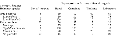
Of the 35 stray dogs, five were found to have Echinococcus spp. worms at necropsy (E. granulosus worms were found in four dogs, one dog contained one E. multilocularis worm (true positives, prevalence 14%)). The Combined kit recognized the five positives (100%) but it also detected ten false positives (33%). The Tiankang, Haitai and Laboratory kits all detected three of the five positives (60%).The Combined kit and Haitai kit detected the single E. multilocularis worm in one dog.The three commercial kits and the Laboratory kit shared six false positives. In three of these, no parasites were found, one had Toxocara canis, another had Dipylidium caninum and the third had Taenia multiceps. The results for all samples differed between tests. Necropsy findings and copro-ELISA results are shown in table 1.
The proportion of coproantigen positives (%) detected by ELISA in faecal samples of stray dogs using the Haitai, Combined, Tiankang and Laboratory kits.
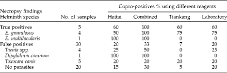
With the samples from experimentally infected dogs and stray dogs, the three commercial coproantigen test kits and one laboratory kit were evaluated for specificity and sensitivity of each kit. Tiankang detected 84% of experimental and 60% of field true infections and had 7% false positives. Haitai detected 10% of experimental and 60% of field and had 20% false positives. Combined detected 53% of experimental and 100% of field true infections but had 33% false positives. Lab detected 84% of experimental and 60% of field true infections and had 20% false positives.
The ELISA test for the detection of antigens in the faeces of definitive hosts was initially developed for the diagnosis of E. granulosus (Allan et al., Reference Allan, Craig, Garcia, Mencos, Liu, Wang, Wen, Zhou, Stringer and Rogan1992). These tests subsequently were shown to have cross-reactivity with E. multilocularis. However, they lacked sensitivity for this species (Allan et al., Reference Allan, Craig, Garcia, Mencos, Liu, Wang, Wen, Zhou, Stringer and Rogan1992; Deplazes et al., Reference Deplazes, Gottstein, Eckert, Ewald, Jenkins and Jimenez-Palacios1992). There is a need to test the kits being evaluated here for their ability to detect E. multilocularis infections because this parasite is very common in western China (Craig et al., Reference Craig, Rogan and Campos-Ponce2003). The detection of one E. multilocularis worm by two of the kits may possibly have been false positives. The sensitivity of coproantigen detection is generally good with moderate to high worm burdens (>100 worms), but less in animals with low burdens (Eckert, Reference Eckert2003). Coproantigens can also detect prepatent infections (Huang et al., Reference Huang, Yang, Qiu, Chen, Yang, Qiu, Xiao, Xiao and Heath2007). The positive predictive value can be poor if used where the expected prevalence of Echinococcus is low (Varcasia et al., Reference Varcasia, Tanda, Giobbe, Solinas, Pipia, Malgor, Carmona, Garippa and Scala2011). Coproantigen ELISA may have high cross-reactions with other parasites, especially reacting with patent infections of Taenia hydatigena (57%) and T. multiceps (33%) (Nonaka et al., Reference Nonaka, Nakamura, Inoue, Oku, Katakura, Matsumoto, Mathis, Chembesofu and Phiri2011). Another study showed the same problem (Casaravilla et al., Reference Casaravilla, Malgor, Rossi, Sakai, Nonaka, Kamiya and Carmona2005). They raised two monoclonal antibodies (MAbs), EgC1 and EgC3, to detect Echinococcus infections in experimentally infected dogs. EgC3 showed a higher sensitivity than EgC1 in detecting E. granulosus infections, but EgC3 also detected false positives caused by T. hydatigena.
The study reported here showed that the reliable diagnosis of Echinococcus spp. in living dogs is still unsatisfactory. The four detection kits examined in this paper often gave different results for the same samples. This kind of problem also occurred in a study that was carried out in Sardinia, Italy (Varcasia et al., Reference Varcasia, Tanda, Giobbe, Solinas, Pipia, Malgor, Carmona, Garippa and Scala2011). They used three ELISA test systems, including the widely used Bommeli kit, to detect dog infections. The results showed different prevalence levels for each of the three kits. It may be that the quality of antibodies is one of the main factors affecting the detections. For example, the study in Sardinia adopted: (a) Chekit Echinotest (Bommeli, Bern, Switzerland) based on polyclonal antibodies against adult excretory/secretory (E/S) antigens, which gave a sensitivity of 83% and a specificity that ranged from 80 to 98% (Eckert et al., Reference Eckert, Gemmell, Meslin and Pawlowski2001); (b) a sandwich ELISA that used rabbit polyclonal antibody EmA9 produced against adult E. multilocularis somatic extract, with a sensitivity of 100% and a specificity of 96% (Malgor et al., Reference Malgor, Nonaka, Basmadjian, Sakai, Carámbula, Oku, Carmona and Kamiya1997); (c) a sandwich assay that used monoclonal antibody EgC3 produced by immunization with adult E. granulosus E/S products, having a sensitivity of 100% and specificity of 100% (Casaravilla et al., Reference Casaravilla, Malgor, Rossi, Sakai, Nonaka, Kamiya and Carmona2005). The results revealed that the MAbs seemed to be more effective in increasing the sensitivity and specificity of the test, but they still reacted with T. hydatigena. Pierangeli et al. (Reference Pierangeli, Soriano, Roccia, Bergagna, Lazzarini, Celescinco, Kossman, Saiz and Basualdo2010) used samples from known uninfected dogs, sequentially collected samples from dogs experimentally infected with E. granulosus, and samples from rural dogs tested by arecoline purging. They validated the coproantigen test with samples from the experimentally infected dogs (92% positive). They also evaluated the effect on the coproantigen test of the presence of Taeniids (3.6% positive) or nematodes (9.9% positive). In the study reported here, the Laboratory kit used polyclonal antibodies against adult E. granulosus worm E/S in rabbits, with a sensitivity of 92% and a specificity of 80% (Huang et al., Reference Huang, Heath, Christine, Yang, Wang, Huang, Yi, Xiao, Qiu and Yang2008). False positives and cross-reaction with other parasites in the small intestine, including Toxocara, T. hydatigena, T. pisiformis, T. multiceps and D. caninum were observed, so further study is necessary to assess specificity and repeatability of these kits. The composition of faecal extract solution might also affect the test (Benito et al., Reference Benito, Carmena, Martínez and Guisantes2006) although Huang et al. (Reference Huang, Yang, Qiu, Chen, Yang, Qiu, Xiao, Xiao and Heath2007) found no significant differences between results where formalin was used and those where it was not used. Unfortunately, details of faecal extract solutions of the three commercial kits in this study are not available.
From a comprehensive evaluation, it seems that the Tiankang kit may identify more positive samples and have fewer false positives than the other two kits. However, its shelf life is only 6 months (the others are at least 12 months) and the Tiankang kit should be stored at − 20°C (the others can be kept at 4°C). These shortcomings limit its use in the field and rural areas.
However, coproantigen tests remain a useful procedure for population studies, provided that the potential pitfalls are fully understood when utilizing such tests. It has become clear that coproantigen tests should be developed specifically for the environment being studied, and should be validated with necropsy and possibly purging data from a statistically valid sample of the population to be tested.
Provided the features of the diagnostic tests are well understood, the absence of a perfect gold standard, or even of a suitable reference test, need not be an insurmountable problem when undertaking population studies. Multiple parallel testing or the use of additional confirmatory test(s) in a diagnostic strategy will also yield valuable information at a population and individual level, provided the study is well designed and any shortcomings of the tests are accounted for in the analysis (Torgerson & Deplazes, Reference Torgerson and Deplazes2009).
The validation of the Haitai kit, Combined kit and Tiankang kit have not been published. Users will need to validate the chosen kit if these kits are used in different areas of China or other places in the world. Based on the current results, the Tiankang kit might be preferred, but it has shelf-life and storage problems for field use.
Cross-reactions to other parasites (Taenia spp., D. caninum, T. canis) were observed in some or all tests, but numbers were too few to draw statistically valid conclusions. The tests differed in their ability to detect parasites other than Echinococcus spp., as well as in their detection of dogs with no parasites.
Acknowledgements
The authors would like to acknowledge the financial support of the Science and Technology Department of Sichuan Province (grant no. 2008SZ0197) and the Health Department of Sichuan Province (grant no. 090401). We also thank Renqing Pengcuo in Shiqu County Centre for Disease Control and Zhen Yuntai in Seda County Centre for Disease Control for their help with the field work.