Introduction
Schizotypy refers to a set of personality traits related to schizophrenia spectrum pathology, including magical ideation, odd behaviour, perceptual aberration as well as anhedonia (Nelson et al. Reference Nelson, Seal, Pantelis and Phillips2013). Anhedonia, the reduced capacity to experience pleasure, is one of the core symptoms of major depressive disorder and also an important negative symptom of schizophrenia (Andreasen, Reference Andreasen1982; APA, 2013). Social anhedonia refers to the phenomenon of reduced capacity to experience pleasure in the social context, and has been considered a component of negative schizotypy and indicates an increased risk for psychosis (Meehl, Reference Meehl1990; Clardige & Beech, Reference Clardige, Beech, Raine, Lencz and Mednick1995). Previous studies have suggested that a high level of social anhedonia was observed not only in patients with schizophrenia and their unaffected first-degree relatives (Katsanis et al. Reference Katsanis, Iacono and Beiser1990), but also in individuals prone to psychosis (Chan et al. Reference Chan, Shi, Lai, Wang, Wang and Kring2012a ). In addition, previous studies have also indicated that the level of social anhedonia reported by individuals with psychometrically defined schizotypy was not different from that reported by patients with schizophrenia (Wang et al. Reference Wang, Lui, Zou, Zhang, Zhao, Yan, Hong, Tan, Cheung and Chan2014). Longitudinal studies have shown that individuals with a high level of social anhedonia developed full-blown psychosis or schizophrenia with a higher probability in 5 and 10 years, whereas no difference was found on the probability of developing depression or bipolar disorder (Kwapil, Reference Kwapil1998; Gooding et al. Reference Gooding, Tallent and Matts2005). All these suggest that the level of social anhedonia could potentially be a predictor of schizophrenia spectrum disorders. Therefore, investigation of the neural correlates of social anhedonia may contribute to a better understanding of the development of psychosis.
The striatum has been identified as one of the key brain structures involved in reward processing. Recent studies have further suggested that the ventral and dorsal parts of the striatum play distinct roles; the former being related to reward expectation and the latter related to the maintenance of the meaning of the reward outcome (O'Doherty et al. Reference O'Doherty, Dayan, Schultz, Deichmann, Friston and Dolan2004). These circuits have also been linked to schizophrenia and other disorders (Pantelis & Brewer, Reference Pantelis and Brewer1995, Reference Pantelis, Brewer, Pantelis, Nelson and Barnes1996; Gabbay et al. Reference Gabbay, Ely, Li, Bangaru, Panzer, Alonso, Castellanos and Milham2013). Di Martino et al. (Reference Di Martino, Scheres, Margulies, Kelly, Uddin, Shehzad, Biswal, Walters, Castellanos and Milham2008) examined the resting-state functional connectivity of the striatum in healthy participants by dividing it into six subdivisions [including the nucleus accumbens (NAc), the ventral caudate, the dorsal caudate, the dorsal caudal putamen, the dorsal rostral putamen (DRP) and the ventral rostral putamen]. Their results described the different functional connectivity patterns of the subdivisions of the striatum. For example, the dorsal caudate is highly connected to the frontal lobe, while the ventral caudate is connected to the limbic areas, which correspond to the cognitive/affective divisions. Taken together, the existing findings suggest that examining corticostriatal connectivity may be an optimal approach.
Altered functional connectivity of the striatum has been found in individuals with psychosis. Fornito et al. (Reference Fornito, Harrison, Goodby, Dean, Ooi, Nathan, Lennox, Jones, Suckling and Bullmore2013) adopted the seed-based approach and found reduced functional connectivity between the dorsal caudate and the prefrontal regions, and increased functional connectivity between the ventral caudate and the prefrontal regions in patients with first-episode psychosis and their unaffected first-degree relatives. Furthermore, the fact that this pattern was also observed in unaffected first-degree relatives suggests that it may be an endophenotype for psychosis (Fornito et al. Reference Fornito, Harrison, Goodby, Dean, Ooi, Nathan, Lennox, Jones, Suckling and Bullmore2013). Further work in individuals at ultra-high risk (UHR) for psychosis found reduced functional connectivity between the caudal caudate and the prefrontal cortex, and increased connectivity between the ventral putamen, and the frontal and the temporal lobes (Dandash et al. Reference Dandash, Fornito, Lee, Keefe, Chee, Adcock, Pantelis, Wood and Harrison2014), suggesting that these changes are apparent before psychosis onset. However, it is still not fully known whether functional connectivity between the striatum and the frontal lobe are disrupted in the whole spectrum of schizophrenic disorders, including individuals with schizotypal traits.
In addition, problems in emotional processing, especially reduced pleasure experience and expression, have been found in patients with schizophrenia (Kring & Elis, Reference Kring and Elis2013) and also in individuals at high-risk for psychosis (Phillips & Seidman, Reference Phillips and Seidman2008). Although corticostriatal circuits are important in emotional processing, few studies have examined the association between altered corticostriatal connectivity and reduced pleasure experience and expression in patients with schizophrenia and individuals at-risk for psychosis. In the two studies mentioned above, reduced functional connectivity was associated with both positive and negative symptoms in patients with first-episode schizophrenia and UHR individuals (Fornito et al. Reference Fornito, Harrison, Goodby, Dean, Ooi, Nathan, Lennox, Jones, Suckling and Bullmore2013; Dandash et al. Reference Dandash, Fornito, Lee, Keefe, Chee, Adcock, Pantelis, Wood and Harrison2014), but the association between reduced emotional experience/expression and corticostriatal circuit in schizophrenia spectrum remains unclear.
The aims of the present study were two-fold. First, we examined corticostriatal functional connectivity in individuals with high level of negative schizotypy (measured by the Social Anhedonia Scale), taking into account both the ventral and dorsal part of the striatum. Second, we examined the associations between emotional processing (including anticipatory/consummatory pleasure experiences and emotional expression) and corticostriatal functional connectivity. We hypothesized that individuals with high level of negative schizotypy would show reduced functional connectivity between the dorsal striatum and the frontal lobe and increased connectivity of the ventral part of striatum, a ‘dorsal-to-ventral gradient of hypo- to hyper-connectivity with the prefrontal cortex’. In addition, the increased functional connectivity of the ventral striatum and the prefrontal cortex would be related to reduced anticipatory pleasure experience.
Method
Participants
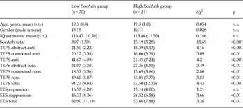
There were two groups of participants in this study: individuals with high social anhedonia score (High SocAnh group) and those with low social anhedonia score (low SocAnh group). All participants were selected from a large sample pool from the Guangzhou Medical University based on their scores on the Chinese version of the Chapman Social Anhedonia Scale (CSAS; Chan et al. Reference Chan, Shi, Lai, Wang, Wang and Kring2012a ). The means and standard deviations (s.d.) from a previous study (Chan et al. Reference Chan, Shi, Lai, Wang, Wang and Kring2012a ) in Chinese college students (n = 887) using the same scale (males: mean = 9.40, s.d. = 6.32; females: mean = 7.61, s.d. = 5.34) were used as reference in this study. Since previous meta-analysis has shown significant gender difference for the Chapman scales (Miettunen & Jaaskelainen, Reference Miettunen and Jaaskelainen2010), we calculated the cut-offs for males and females separately. Participants whose scores were 0.5 s.d. above the mean of their own gender were recruited into the High SocAnh group (n = 21). Participants whose scores were 0.5 s.d. below the mean scores of their own gender were recruited into the low SocAnh group (n = 30). All participants were right-handed as assessed by the Annett Handedness Scale (Annett, Reference Annett1970). They had no history of substance abuse, brain injury, neurological disorders and no personal and family history of mental disorders. The high SocAnh group had a mean age of 19.3 years (s.d. = 1.0) and consisted of 10 males. The low SocAnh group had a mean age of 19.3 years (s.d. = 0.9) and consisted of 15 males. Their IQ scores were estimated using the commonsense, arithmetic, similarity and digit span subtests of the Chinese Version of the Wechsler Adult Intelligence Scale – Revised (WAIS-R; Gong & Dai, Reference Gong and Dai1984). The mean IQ estimates for the high SocAnh and the low SocAnh groups were 115.86 (s.d. = 11.55) and 116.43 (s.d. = 10.39), respectively. No significant difference was found between the two groups on age, gender and IQ estimates, as shown in Table 1. The present study was approved by the Ethics Committee of the Institute of Psychology, Chinese Academy of Sciences. Written informed consents were obtained from each participant prior to the study.
Demographic information and group comparison on scales

SocAnh, The Chinese version of the Chapman Social Anhedonia Scale; TEPS, The Temporal Experiences and Pleasure Scales; anti, anticipatory factor of the TEPS; cons, consummatory factor of the TEPS; EES, Emotional Expressivity Scale.
Measures
Chapman Social Anhedonia Scale (CSAS)
The Chinese version of CSAS was adopted from the original English version (Eckblad et al. Reference Eckblad, Chapman, Chapman and Mishlove1982) after undergoing a series of standard validation procedures (Chan et al. Reference Chan, Shi, Lai, Wang, Wang and Kring2012a ). The CSAS is a 40-item questionnaire assessing the reduced pleasure experiences induced by social interaction. For each item, participants were asked to report their own experience with a ‘true’ or ‘false’ answer. The total CSAS score was calculated for each participant. A higher total score indicates more severe anhedonia in social interactions.
Temporal Experiences and Pleasure Scales (TEPS)
The TEPS is a self-report questionnaire designed to measure individual trait dispositions in both anticipatory and consummatory experiences of pleasure. The original TEPS consists of a 10-item anticipatory and an 8-item consummatory pleasure scale (Gard et al. Reference Gard, Gard, Kring and John2006). In this study, we adopted the validated Chinese version of the TEPS (Chan et al. Reference Chan, Wang, Yan, Zhao, McGrath, Hsi and Stone2012b ), which includes 19 items and has a four-factor construct in the Chinese context, including abstract anticipatory, contextual anticipatory, abstract consummatory, and contextual consummatory factors.
Emotional Expressivity Scale (EES)
The EES, consisting of 17 items, was developed to capture individuals’ differences on the outward display of their emotion regardless of valence (Kring et al. Reference Kring, Smith and Neale1994). Individuals rate themselves on a 6-point Likert scale from 1 (never) to 6 (always) on how often they express their emotions. The Chinese version of the EES has a two-factor construct with good internal consistency: whole scale (Cronbach's α = 0.82), ‘suppression’ factor (Cronbach's α = 0.82; e.g. ‘I keep my feelings to myself’) and ‘expression’ factor (Cronbach's α = 0.78; e.g. ‘I display my emotions to other people’) (Chan et al. Reference Chan, Wang, Li, Shi, Wang, Liu and Huang2010).
Image acquisition and preprocessing
All MRI scans were acquired on a Siemens Verio 3 T MR scanner (Siemens, Germany) at the Guangzhou First People's Hospital, Guangzhou, China. Resting-state fMRI data were acquired using a T2-weighted echo planar imaging (EPI) sequence; 200 whole-brain volumes were collected with slice thickness = 3.5 mm, echo time (TE) = 30 ms, repetition time (TR) = 2500 ms, flip angle = 90°, matrix size = 64 × 64, 42 slices in the coronal plane, field of view (FOV) = 200 mm, voxel size = 3.1 × 3.1 × 3.5 mm3, bandwidth = 2520 Hz/Px. Scans were screened by a radiologist to exclude any incidental clinical abnormalities before further analysis.
Preprocessing was performed using the Data Processing Assistant for Resting-State fMRI (DPARSF) software (Yan & Zang, Reference Yan and Zang2010). The first 10 volumes were removed. Time delay in image acquisition and head motion were corrected. Two participants were excluded due to excessive head motion (>3 mm or 3°) prior to further analysis. The fMRI images were further spatially normalized to the Montreal Neurological Institute (MNI) EPI template, re-sliced to 3 mm cubic voxels and then smoothed using a 4 mm full-width at half maximum Gaussian kernel. Temporal band-pass filtering (0.01 < f < 0.10 Hz) was performed. The Nuisance covariates, including head motion parameters, global mean signal, white-matter signal and cerebrospinal fluid signal were regressed out. To exclude artifacts caused by head motion, we took the Friston 24-parameter model (Friston et al. Reference Friston, Williams, Howard, Frackowiak and Turner1996) as a regressor for the individuals’ first-level analysis, which has been proved to be superior to the six-parameter model (Yan et al. Reference Yan, Cheung, Kelly, Colcombe, Craddock, Di Martino, Li, Zuo, Castellanos and Milham2013). We also calculated the mean framewise displacement (FD) (Power et al. Reference Power, Barnes, Snyder, Schlaggar and Petersen2012) of each participant. Although the group comparison did not show significant difference between the low SocAnh group (mean FD = 0.12, s.d. = 0.06) and high SocAnh group (mean FD = 0.10, s.d. = 0.04) (t = 0.78, p > 0.1), we still took the individuals’ mean FD as a covariate in the second-level analysis as suggested by Yan et al. (Reference Yan, Cheung, Kelly, Colcombe, Craddock, Di Martino, Li, Zuo, Castellanos and Milham2013).
Functional connectivity analyses
To examine the functional connectivity between seeds of the striatum and whole brain voxels, we first defined six seeds of the striatum in each hemisphere as described in a previous study (Di Martino et al. Reference Di Martino, Scheres, Margulies, Kelly, Uddin, Shehzad, Biswal, Walters, Castellanos and Milham2008), including the NAc (MNI coordinates: ±9, 9, −8), the ventral caudate (±10, 15, 0), the dorsal caudate (±13, 15, 9), the dorsal caudal putamen (±28, 1, 3), the DRP (±25, 8, 6) and the ventral rostral putamen (±20, 12, −3). The radius of each seed was set at 4 mm. Voxel-wise functional connectivity analyses were conducted between brain activity of each seed and the whole brain voxels using a toolkit for resting-state functional MRI analysis (REST; Song et al. Reference Song, Dong, Long, Li, Zuo, Zhu, He, Yan and Zang2011).
Statistical analysis
Subjects were compared on age, gender, IQ estimates and scores on the CSAS, TEPS, and EES. One-sample t tests were conducted in both groups for functional connectivity analyses for all six pairs of seeds in the striatum. The results are illustrated in Supplementary Fig. S1. To examine the group effect in functional connectivity, correlation r maps were transformed to Fisher z maps and were analysed with repeated-measures ANOVA (two hemispheres by two groups) using the SPM8 software implemented in Matlab, with mean FD as a covariate in the general linear model. The clusters were considered significant if they reached a threshold of p < 0.001 with a cluster size of >50 voxels. We also extracted the transformed Fisher z values of the clusters that showed significant group effect and correlated these with the scores on the various scales to examine the associations between altered corticostriatal functional connectivity and emotion processing. Significance level was set as two-tailed p < 0.05.
Results
Demographics description and group differences in emotion processing
The two groups were matched in terms of age, gender and IQ estimates (see Table 1). The high SocAnh group reported lower pleasure experience in both anticipatory (t = 4.20, p < 0.001) and consummatory (t = 3.53, p < 0.01) components of the TEPS and lower EES suppression (t = 3.66, p < 0.001) and total scores (t = 3.26, p < 0.01).
Group effects in corticostriatal functional connectivity
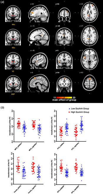
A main effect of group was observed in the functional connectivity between the NAc and the right medial frontal gyrus [extending to the anterior cingulate cortex (ACC)] [BA (Brodmann area) 9/10/32], between the NAc and the left posterior cingulate gyrus (BA 31), between the ventral caudate and the left insula (BA 13), as well as between the DRP and the right superior frontal gyrus (supplementary motor area, BA 6). There was no interaction between group and the two hemispheres.
We then extracted the transformed z values of four significant clusters and compared them between the two groups. The results showed that compared to the low SocAnh group, reduced functional connectivity was observed in the high SocAnh group between the posterior cingulate cortex (PCC) and the bilateral NAc (p = 0.001); while increased connectivity was found between the medial frontal gyrus and the bilateral NAc, between the insula and the ventral caudate, as well as between the superior frontal gyrus and the DRP (all p's < 0.001) (see Fig. 1 for details).
Significant group effect of high and low social anhedonia on the functional connectivity (FC) of the striatum. Repeated-measures ANOVA was conducted. Results are displayed at p < 0.001, cluster size >50 voxels, uncorrected. See Table 2 for details. NAc, Nucleus accumbens; VC, ventral caudate; DRP, dorsal rostral putamen; MFG, medial frontal gyrus; PCC, posterior cingulate gyrus; SFG, superior frontal gyrus.

Effect of the high and low social anhedonia groups on corticostriatal functional connectivity

Hemi, Hemisphere; BA, Brodmann area; R, right; L, left; PCC, posterior cingulate cortex; dmPFC, dorsomedial prefrontal cortex; ACC, anterior cingulate cortex; SMA, supplementary motor area.
Threshold at p < 0.001, cluster size >50 voxels.
Correlations between functional connectivity and self-reported scores
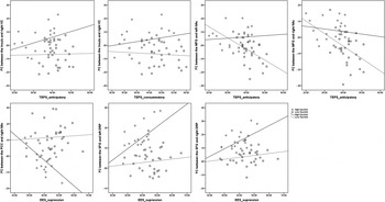
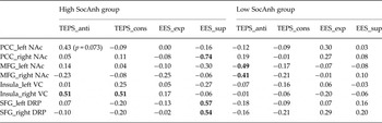
Taking age, gender and IQ estimates as covariates, the altered functional connectivity between the striatum subdivisions and cortical activity were correlated with the self-reported scores of scales capturing emotional processing. In the high SocAnh group, we found that both the TEPS anticipatory and consummatory scores were positively correlated with functional connectivity between the insula and the right ventral caudate (r s = 0.51 for both, p's < 0.05). EES suppression scores were negatively correlated with functional connectivity between the PCC and the left NAc (r = −0.74, p < 0.001) and positively correlated with functional connectivity between the superior frontal gyrus and the bilateral DRP (left DRP: r = 0.57, right DRP: r = 0.54; p's < 0.05). In the low SocAnh group, significant negative correlations were found between the TEPS anticipatory subscale scores and functional connectivity between the medial frontal gyrus and the bilateral NAc (left NAc: r = −0.49; right NAc r = −0.41; p's < 0.05) (see Fig. 2 and Table 3 for details).
Partial correlations between functional connectivity (FC) of the striatal seeds and the self-reported scale scores. SocAnh, Chinese version of the Chapman Social Anhedonia Scale; TEPS, Temporal Experiences and Pleasure Scale; EES, Emotional Expressivity Scale. PCC, posterior cingulate cortex; VC, ventral caudate; SFG, superior frontal gyrus; DRP, dorsal rostral putamen; MFG, medial frontal gyrus; NAc, nucleus accumbens.

Partial correlations between functional connectivity of regions of interest and self-reported scales

TEPS, Temporal Experiences and Pleasure Scales; EES, Emotional Expressivity Scale; anti, anticipatory factor; cons, consummatory factor; exp, expression factor; sup, suppression factor; PCC, posterior cingulate cortex; NAc, nucleus accumbens; MFG, medial frontal gyrus; VC, ventral caudate; SFG, superior frontal gyrus; DRP, dorsal rostral putamen.
Age, gender and estimated IQ were taken as covariates; numbers in bold indicate the significant correlations with p < 0.05 as threshold, two tailed.
Discussion
In this study, we examined resting-state functional connectivity in individuals with high levels of negative schizotypy, which may be a vulnerability trait marker for psychosis. Adopting the seed-based approach, we used six subdivisions of the striatum and calculated functional connectivity between each seed of the striatum and the rest of the brain. Compared to individuals with low level of social anhedonia, the high SocAnh group showed reduced connectivity between the PCC and NAc. Increased connectivity was observed between the dorsomedial prefrontal cortex and NAc, between the insula and ventral caudate, and between the superior frontal gyrus (supplementary motor area) and the DRP.
The idea of taking into account the subdivisions of the striatum is based on empirical findings that the ventral and dorsal striatum play different roles in reward processing. The ventral part is a key brain structure in the reward circuit, and has been suggested to be specifically related to reward expectation. It receives inputs from the prefrontal cortex, including the orbitofrontal cortex, the medial prefrontal cortex and the dorsal ACC, and projects to the ventral pallidum, then back to the prefrontal cortex through the thalamus, constituting the frontal-basal ganglia circuit in reward processing. Through this circuit, sensory information, personal expectation, beliefs and memory could be integrated and goal-directed plans would then be generated (Haber & Knutson, Reference Haber and Knutson2010). Our finding of increased functional connectivity between the ventral striatum and the medial prefrontal cortex, the ACC and the insula in individuals with high level of social anhedonia suggests that decreased pleasure experience in social interaction may be related to dysfunction of this frontal-basal ganglia circuit. Reduced pleasure experience has been observed in schizophrenia patients, their unaffected relatives and high-risk populations (Katsanis et al. Reference Katsanis, Iacono and Beiser1990; Burbridge & Barch, Reference Burbridge and Barch2007; Wang et al. Reference Wang, Lui, Zou, Zhang, Zhao, Yan, Hong, Tan, Cheung and Chan2014). Patients with schizophrenia have been shown to have deficits in anticipatory pleasure, but not consummatory pleasure. Strauss et al. (Reference Strauss2013) proposed that this specific pattern may be due to the low-pleasure beliefs and reduced estimation of past and future pleasure. Similar to previous findings, individuals with social anhedonia in the present study also showed lower anticipatory pleasure than controls (Shi et al. Reference Shi, Wang, Cao, Wang, Wang, Zong, Xu, Tse, Hsi, Stone, Lui, Cheung and Chan2012). Using partial correlation analysis, we found significant associations between TEPS anticipatory scores and functional connectivity between the ventral striatum and the dorsomedial prefrontal cortex/ACC in the low SocAnh group. At the same time, functional connectivity between the ventral caudate and the insula was associated with both TEPS anticipatory and consummatory subscale scores in the high SocAnh group. Hence, our study showed that reduced anticipatory pleasure, which has been found in patients with schizophrenia, is also observed in individuals with high level of social anhedonia. Furthermore, this reduced pleasure experience may be related to dysfunction of the reward system, especially functional connectivity between the ventral striatum and the medial prefrontal cortex and the insula.
We also found reduced functional connectivity between the NAc and PCC. Di Martino et al. (Reference Di Martino, Scheres, Margulies, Kelly, Uddin, Shehzad, Biswal, Walters, Castellanos and Milham2008) reported a connection between the NAc and PCC, both of which are regions involved in emotion processing. A previous study has shown that abnormalities of the PCC may be related to novelty seeking and may be linked to the behavioural activation system (Lei et al. Reference Lei, Chen, Xue, He, Chen, Liu, Moyzis, Xue, Cao, Li, Li, Zhu, Liu, Hsu, Li and Dong2014). Although the PCC is an important midline cortical structure in the default mode network and its dysconnectivity has been reported in patients with schizophrenia and individuals with familial risk (Peeters et al. Reference Peeters, van de Ven, Gronenschild, Patel, Habets, Goebel, van Os and Marcelis2015), little is known whether this is also the case in individuals with schizotypy. In the present study we also found a strong correlation between EES suppression and functional connectivity between the NAc and PCC in the high SocAnh group. This finding suggests that the PCC and its connection with the ventral striatum may be related to emotion regulation and needs to be carefully examined in the future.
We also found increased functional connectivity between the dorsal striatum and the supplementary motor cortex (BA 6). The dorsal striatum is involved in motor control, which is the basic function of the basal ganglia. Both human and non-human studies have shown that the dorsal striatum and its connections to the sensorimotor or associative cortex are important in the learning of action-reward association (Balleine et al. Reference Balleine, Delgado and Hikosaka2007). The reduced pleasure experience and greater suppression of emotion observed in individuals with high social anhedonia in the present study is consistent with this. We also found an association between emotion suppression and functional connectivity between the dorsal striatum and supplementary motor area, suggesting that dysconnectivity of the dorsal striatum may be a neural correlate underlying abnormal emotion processing in individuals with high risk for psychosis. Individuals with high levels of social anhedonia may have problems in the initiation of behaviour that induces pleasure experience, which further affects their beliefs and estimation of pleasure, as well as action-reward association.
In Fornito et al.'s (2013) study, the authors examined the resting-state functional connectivity of the striatum in patients with schizophrenia and their unaffected first-degree relatives. They found a reduced functional connectivity between the dorsal striatum and the prefrontal cortex, and increased functional connectivity between the ventral striatum and the orbitofrontal cortex/dorsolateral prefrontal cortex and the insula in schizophrenia patients, i.e. a ‘dorsal-to-ventral gradient of hypo- to hyper-connectivity with the prefrontal cortex’ (Fornito et al. Reference Fornito, Harrison, Goodby, Dean, Ooi, Nathan, Lennox, Jones, Suckling and Bullmore2013). Similar patterns were also observed in unaffected first-degree relatives. Furthermore, in a study of UHR individuals, Dandash et al. (Reference Dandash, Fornito, Lee, Keefe, Chee, Adcock, Pantelis, Wood and Harrison2014) also found reduced functional connectivity between the dorsal striatum, the prefrontal cortex and the thalamus; as well as increased functional connectivity between the ventral putamen and the superior temporal gyrus and the insula. Our results are consistent with these findings in terms of the hyper-connectivity between the ventral striatum and the prefrontal cortex and the insula in the high SocAnh group. However, the high SocAnh group in our study also exhibited increased connectivity at the dorsal striatum and reduced connectivity at the ventral striatum. This may be related to the fact that our sample was recruited from well-functioning college students screened by self-reported Social Anhedonia Scale, who are likely to be different from the unaffected relatives of schizophrenia patients and UHR individuals with psychotic-like symptoms in previous studies. For example, a previous study has shown that higher levels of positive symptoms predicted lower connectivity at the dorsal striatum and stronger connectivity at the ventral striatum (Dandash et al. Reference Dandash, Fornito, Lee, Keefe, Chee, Adcock, Pantelis, Wood and Harrison2014). It is not clear if this reversed connectivity pattern of the dorsal striatum is related to the more severe positive symptoms in high-risk individuals. Future research is needed to clarify this issue.
In the present study, we recruited college students, whose age ranged from 17 to 21 years. This period is important in examining the development of psychosis, since psychosis usually emerges in late adolescence or early adulthood with a peak age between 18 and 25 years. Existing findings have shown that the human brain continues to develop until young adulthood, especially the prefrontal cortex (Gogtay et al. Reference Gogtay, Giedd, Lusk, Hayashi, Greenstein, Vaituzis, Nugent, Herman, Clasen, Toga, Rapoport and Thompson2004). A very recent study examined the age-related changes of intrinsic functional connectivity to middle adulthood and found decreasing connection strength between the ventral striatum and the ACC and the insula (Porter et al. Reference Porter, Roy, Benson, Carlisi, Collins, Leibenluft, Pine, Luciana and Ernst2015). In particular, there is a quick drop in connection strength from young to middle adulthood between the ventral striatum and the ACC. In the context of this finding, individuals with high negative schizotypy in our study showed hyper-connectivity between the ventral striatum and the ACC, suggesting an association between abnormal cortico-striatum connectivity and negative schizotypy.
Anhedonia or reward processing is important since it is a key symptom not only for schizophrenia spectrum disorders, but also for some other mental disorders, such as depression. A prior study examined the striatum-based circuitry in adolescents with major depressive disorder and found that the anhedonia severity in patients with major depression showed positive correlations with functional connectivity between the caudate and supplementary motor area, the precuneus, the middle frontal gyrus, and the subgeneual ACC, as well as negative correlations with functional connectivity between the NAc and the subgeneual ACC (Gabbay et al. Reference Gabbay, Ely, Li, Bangaru, Panzer, Alonso, Castellanos and Milham2013). Similar findings have also been found in other psychiatric disorders. Gabbay et al. (Reference Gabbay, Ely, Li, Bangaru, Panzer, Alonso, Castellanos and Milham2013) suggested that abnormal functional connectivity of the striatum related to anhedonia may be independent of the major depressive disorder diagnosis. In the present study, however, we adopted the psychologically defined ‘schizotypy’ approach, and found a link between higher levels of social anhedonia and hyper- or hypo-connectivity of the striatum. All these findings support the Research Domain Criteria (RDoC) proposed by the National Institute of Mental Health (NIMH) (Cuthbert & Insel, Reference Cuthbert and Insel2013) that examining anhedonia and its underlying circuits across different disorders might be a helpful step in understanding mental disorders.
There are several limitations in this study. First, we recruited a convenience sample from college students and the categorization of the two comparison groups was based on scores from a self-reported scale. A more rigorous approach adopting an interview-based measure and behavioural tasks to capture emotion processing should be incorporated in future studies. Second, because we focused on anhedonia in the present study, the potential effect of positive symptoms was not examined. The possibility that the reverse pattern of ventral striatum connectivity may be related to positive symptoms needs to be further clarified. Last, since the present study was conducted in relatively well-functioning college students and was exploratory in nature, we did not correct for multiple comparisons in the functional connectivity analyses.
In conclusion, we examined resting-state functional connectivity of the striatum by examining its ventral and dorsal subdivisions, in individuals with high levels of social anhedonia. We observed dysconnectivity between the ventral striatum and ACC, the insula and PCC, and between the dorsal striatum and motor cortex. These dysconnectivities were also associated with self-reported anticipatory pleasure experience and emotion suppression in individuals with high social anhedonia. These may be early changes in brain functional connectivity in the reward system associated with negative schizotypy (especially social anhedonia). Future studies on social anhedonia in UHR populations as well as first-episode schizophrenia patients could provide new evidence in the better understanding of the development of psychosis.
Supplementary material
For supplementary material accompanying this paper visit http://dx.doi.org/10.1017/S0033291715001592.
Acknowledgements
This study was supported by a grant from the ‘Strategic Priority Research Program (B)’ of the Chinese Academy of Sciences (XDB02030002), the National Science Fund China (81088001, 91132701), and a grant from Beijing Training Project For The Leading Talents in S&T (Z151100000315020) to Raymond Chan. Yi Wang was supported by a National Science Fund China (31400884). Christos Pantelis was supported by an Australian National Health and Medical Research Council (NHMRC) Senior Principal Research Fellowship (ID: 628386). The authors thank the student helpers for their help with the data collection in Guangzhou Medial School.
Declaration of Interest
None.