Introduction
The species-rich and morphologically variable genus Arthonia (Ach.) Ach. has been known for a long time to be heterogeneous and polyphyletic (e.g., Santesson Reference Santesson1952; Sundin & Tehler Reference Sundin and Tehler1998). The genus is currently being split into more natural groups based on morphological, chemical and molecular characters (Grube Reference Grube2001; Frisch & Thor Reference Frisch and Thor2010; Frisch et al. Reference Frisch, Thor and Sheil2014a, Reference Frisch, Thor, Ertz and Grubeb). In this contribution, we report on a small group of species with distinct, elevated, white pruinose pycnidia or sporodochia and, as so far known, immersed to adnate, white pruinose apothecia with a well-developed hypothecium, only weakly gelatinized hymenium, transversely pluriseptate ascospores with±enlarged apical cell, and a distinctive chemistry including compounds related to lepraric and confluentic acids. The species are presently classified in Arthonia and Tylophoron in Arthoniaceae, Lecanactis and Schismatomma in Roccellaceae, or else they are new to science. The generic name Inoderma (Ach.) Gray is available for the pycnidiate species after lectotypification of Inoderma with Inoderma byssaceum (Weigel) Gray [=Arthonia byssacea (Weigel) Almq.], while the new genera Sporodophoron and Glomerulophoron are introduced for the species with sporodochia. Using molecular data, several additional species with sporodochia have recently been found to belong in the family Arthoniaceae, namely Reichlingia leopoldii Diederich & Scheid. (Ertz & Tehler Reference Ertz and Tehler2011) and Tylophoron spp. (including Sporodochiolichen; Ertz et al. Reference Ertz, Bungartz, Diederich and Tibell2011, Reference Ertz, Fischer, Killmann, Razafindrahaja and Sérusiaux2013). The differences between all these genera are discussed.
Material and Methods
Collections and morphological investigations
Fieldwork was carried out in France, Japan, Mauritius, Russia, Sweden, Tenerife and Uganda. Additional specimens for this study were investigated at BR, NY, O, TNS, TRH, UPS and the private collections of the authors. Holotype specimens of the new species are placed in BR, TNS or UPS, as indicated. Morphology was studied on hand-cut sections and on squashed preparations mounted in water and lactic cotton blue (LCB). Ascospore and conidium sizes are presented as (minimum value—) $$\bar x$$
–SD–
 $$\bar x$$
+SD(—maximum value) whenever possible (
 $$\bar x$$
= mean; SD = standard deviation; n = number of measurements. Exsiccate specimens are abbreviated as in IndExs (indexs.botanischestaatssammlung.de).
Chemical investigations
Secondary lichen compounds were identified by TLC (Orange et al. Reference Orange, James and White2010) and HPTLC (Arup et al. Reference Arup, Ekman, Lindblom and Mattsson1993) using solvents A, B' and C. Standard compounds for determining Rf values of lichen acids on the TLC and HPTLC plates included usnic acid, atranorin, sphaerophorin, norstictic acid, protocetraric acid and salazinic acid. Roccella fuciformis was used to identify lepraric acid, while 2'-O-methylperlatolic acid was identified by means of Myriostigma candidum and Pertusaria conglomerata.
The amyloidity of thallus and apothecia was examined using 1% aqueous iodine solution (I), without and with pretreatment with 10% aqueous potassium hydroxide (KI). The colour reaction of the thallus was tested using 10% aqueous potassium hydroxide (K), potassium hypochlorite as applied in common household bleach (C), 10% aqueous potassium hydroxide followed by potassium hypochlorite (KC), and 1,4-phenylendiamine in 96% ethanol (Pd). Brown pigments in the pycnidial walls were tested with K and 20% nitric acid. Calcium oxalate crystals were identified by applying 10% sulphuric acid to squashed preparations of thallus samples.
DNA extraction
DNA extraction followed a modified CTAB protocol as described in Hosaka (Reference Hosaka2009). The extracts were kept in a refrigerator for at least 1 hour and then used undiluted or in 1:10 dilutions depending on the DNA concentration in the extracts. For small samples or where contamination with parasitic fungi or moulds was a problem, thin sections of pycnidia or sporodochia of c. 20–50×50–100 µm were used for a direct PCR approach (Grube Reference Grube2005). Carbonized portions were removed from the sections as far as possible and secondary lichen products removed with acetone. These lichen preparations were then placed directly in 0·2 ml microtubes for DNA amplification.
PCR and sequencing
10 µl (DNA extractions) and 20 µl (direct PCR) PCR reactions were used. Each 10 µl of PCR mix contained 1 µl genomic DNA extraction (or the lichen sample), 0·25 µl (mtSSU, nLSU) or 0·35 µl (RPB2) of each primer (10 pmol/ µl), and 5 µl EmeraldAmp PCR Master Mix (TaKaRa Bio Inc.). The following primers were used for PCR amplification: mtSSU (mtSSU1, mtSSU3R and MSU7; Zoller et al. Reference Zoller, Scheidegger and Sperisen1999; Zhou & Stanosz Reference Zhou and Stanosz2001), nLSU (LIC24R and LR7; Vilgalys & Hester Reference Vilgalys and Hester1990; Miadlikowska & Lutzoni Reference Miądlikowska and Lutzoni2000) and RPB2 (RPB2-7cF and RPB2-11aR; Liu et al. Reference Liu, Whelen and Hall1999). PCR cycling conditions for mtSSU were 94°C (3 min), followed by 11 cycles of 95°C (30 s), 62°C to 52°C (30 s) with annealing temperatures lowered by 1°C between cycles, and 72°C (1 min), followed by 30 cycles at 52°C annealing temperature and a final extension at 72°C (7 min). For RPB2, annealing temperatures started at 61°C and were lowered to 51°C; for the nLSU, annealing temperatures started at 64°C and were lowered to 54°C. Sequencing of the PCR products was performed on a 3130xl Genetic Analyzer (Applied Biosystems).
Alignment
Sequences were aligned in MAFFT as implemented in the MEGA5 package (Tamura et al. Reference Tamura, Peterson, Peterson, Stecher, Nei and Kumar2011). The single gene alignments were checked for obvious aligning errors. All ambiguously aligned regions and parsimony-uninformative insertions were removed prior to the analysis. The final concatenated alignment comprised 880 (mtSSU), 876 (RPB2), and 1202 (nLSU) columns, resulting in a combined alignment of 2958 nucleotide positions. Of these, 1269 were variable and 971 were parsimony-informative. A partitioned dataset was used for the phylogenetic analyses to enable independent parameter estimation for the three gene loci. The RPB2 dataset was further partitioned according to codon positions to allow for the higher evolutionary rates of the 3rd codon position.
ML and Bayesian analysis
A general-time-reversible model with a proportion of invariable sites (GTR-I-Γ) was found to best explain the sequence evolution for the mtSSU, RPB2 and nLSU dataset using the Akaike Information Criterion (AIC; Akaike Reference Akaike1973) implemented in MEGA5 (Tamura et al. Reference Tamura, Peterson, Peterson, Stecher, Nei and Kumar2011). Bayesian inference (Huelsenbeck et al. Reference Huelsenbeck, Ronquist, Nielsen and Bollback2001; Holder & Lewis Reference Holder and Lewis2003) and Maximum Likelihood (ML) were used for inferring phylogenetic hypotheses. Prior to concatenation, the single-gene alignments were tested for conflicting tree topologies. Serious conflict was assumed when deviant tree topologies were supported by ≥70% bootstrap values (BS) and ≥0·95 posterior probabilities (PP).
Bayesian analysis was performed with MrBayes 3.2.1 (Ronquist & Huelsenbeck Reference Ronquist and Huelsenbeck2003) implemented in the CIPRES Science Gateway (Miller et al. Reference Miller, Pfeiffer and Schwartz2010). A GTR-I-Γ model of sequence evolution was applied to the partitioned dataset, and the model parameters were estimated during the run for each gene partition separately, starting from a default flat Dirichlet distribution. The analysis was run for 10 000 000 generations in 8 chains and every 500th generation was sampled. The first 30% of trees were discarded as burn-in and a 50% majority-rule consensus tree was calculated.
Maximum likelihood was performed with the RAxML-HPC black box implemented in the CIPRES Science Gateway (Miller et al. Reference Miller, Pfeiffer and Schwartz2010) using rapid bootstrapping and full ML analysis under the GTR+GAMMA approximation allowing for a proportion of invariable sites. The analysis was stopped automatically after 650 bootstrap replicates using the bootstopping option implemented in RAxML 3.2.7 (Pattengale et al. Reference Pattengale, Alipour, Bininda-Emonds, Moret and Stamatakis2009).
Results
Phylogenetic analysis
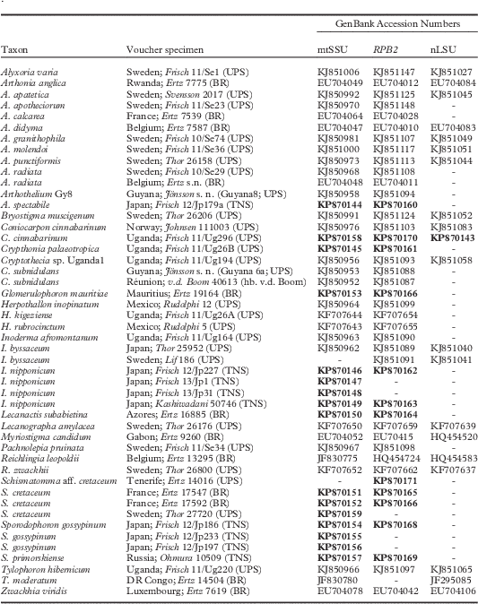
We generated new sequences of the mtSSU (16 specimens), RPB2 (12 specimens) and nLSU (1 specimen) gene loci. An additional 79 sequences were obtained from GenBank. The nLSU gene, which at present is difficult to obtain from species of the cryptothecioid subclade, was added to the analysis to obtain a better backbone support (Table 1). Due to the age of the specimens available for this study, sequence data could not be obtained from Sporodophoron americanum. The final alignment contained 48 specimens and 36 species of Arthoniales, including 3 outgroup taxa: Alyxoria varia (Pers.) Ertz & Tehler, Lecanographa amylacea (Ehrh. ex Pers.) Egea & Torrente and Zwackhia viridis (Ach.) Poetsch & Schied. Seventeen sequenced specimens belong to the genera Inoderma, Sporodophoron and Glomerulophoron, representing all currently recognized species in these genera except Sporodophoron americanum.
Specimen information and GenBank accession numbers for taxa used in this study. New sequences are indicated by accession numbers in bold

Our new analyses show the same general results regarding the phylogenetic relationships within Arthoniaceae as presented in Frisch et al. (Reference Frisch, Thor, Ertz and Grube2014b). Inoderma (clade 1, Fig. 1) and Sporodophoron (clade 2, Fig. 1) are supported as sister clades in the cryptothecioid subclade of Arthoniaceae, both by the ML and the Bayesian analysis. These genera form a well-supported clade with Glomerulophoron (clade 3, Fig. 1) and Cryptothecia, but the exact position of Glomerulophoron in relation to these genera cannot be demonstrated due to lacking branch support. Our analysis shows that the presence of sporodochia is a homoplasious character in Arthoniaceae, having evolved four times in the family in the genera Glomerulophoron, Reichlingia, Sporodophoron and Tylophoron (Fig. 1). In contrast, all species with elevated, white pruinose pycnidia included in the analysis are placed in Inoderma. While Glomerulophoron, Sporodophoron and Tylophoron belong to the cryptothecioid subclade (clade A, Fig. 1), Reichlingia leopoldii is placed in the arthonioid subclade (clade B, Fig. 1). To date, species with sporodochia are not known from the Bryostigma subclade (clade C, Fig. 1).
Phylogenetic relationships of the species used in this study based on Maximum Likelihood and Bayesian analyses using three loci (mtSSU, nLSU, RPB2). Thick black lines indicate support from both Bayesian and ML analyses, the fragmented line indicates support from Bayesian analysis alone and the double line indicates support from the ML analysis alone. 1, 2, 3=branches supporting Inoderma, Sporodophoron and Glomerulophoron, respectively; A, B, C=branches supporting the cryptothecioid-, the arthonioid- and the Bryostigma subclades respectively (Frisch et al. Reference Frisch, Thor, Ertz and Grube2014b). Grey vertical bar indicates species with sporodochia. Black vertical bars indicate species with elevated white pruinose pycnidia. The outgroup taxa in Lecanographaceae and Opegraphaceae are at the base of the tree.
Arthothelium spectabile, the type of the large heterogeneous genus Arthothelium, is sister to Pachnolepia pruinata in the phylogenetic tree and forms a well-supported clade with the two analyzed species of Tylophoron, both known to produce sporodochia. Arthothelium was tentatively excluded from Arthoniales by Tehler (Reference Tehler1990) and the phylogenetic position of the genus has remained controversial since then. Arthothelium is confirmed here for the cryptothecioid subclade in Arthoniaceae.
Thallus chemistry of Inoderma, Sporodophoron and Glomerulophoron
With the exception of Inoderma byssaceum and I. afromontanum, species of Inoderma and Sporodophoron are characterized by a distinct K+ yellow spot reaction of the thallus, sporodochia, and the pruina on conidiomata and apothecia. This reaction is caused in the case of Inoderma by lepraric acid, while in Sporodophoron a compound seemingly related to lepraric acid is found in all species. This ‘lepraric high unknown’ compound runs slightly higher on the TLC plates in solvent systems A, B' and C (Rf values 38, 9, 32, respectively), but otherwise it shows the same spot characteristics as lepraric acid. Confluentic acid is known from Inoderma subabietinum, while 2'-O-methylperlatolic acid occurs in Sporodophoron cretaceum, S. gossypinum and Glomerulophoron mauritiae. The identity of the upper spot of the two ‘byssaceum unknowns’ in I. byssaceum (one pale yellow spot in solvents A and C, or two spots in B; Rf values 52, 46+48, 53 in A, B', C, respectively) with 2'-O-methylperlatolic acid needs confirmation. A series of up to four trace compounds below, and probably related to, lepraric acid, occurs in S. americanum and S. cretaceum. These compounds could not be identified. An unknown fatty acid is found in S. gossypinum.
The Genera
Inoderma (Ach.) Gray
A Natural Arrangement of British Plants 1: 498 (1821); Verrucaria (unranked) Inoderma Ach., Lichenographia Universalis: 294 (1810); non Inoderma Kütz., Algarum Aquae Dulcis Germanicarum 4: 2 (1833), nom. illegit.; non Inoderma P. Karst., Meddeland. Soc. Fauna Fl. Fenn. 5: 39 [1879 (reprint) (‘1880’)], nom. illegit.; non Inoderma Berk., J. Linn. Soc., Bot. 18: 386 (1881), nom. illegit.
Lectotype: Inoderma byssaceum (Weigel) Gray, selected here.
Thallus extensive, whitish to pale olive-grey, continuous, rimose to fissured-areolate but sometimes evanescent, with an ecorticate, weakly felty, scurfy or (sub-)granular to powdery-mealy surface, ±endoperidermal or epilithic; photobiont trentepohlioid (Printzina lagenifera in I. nipponicum; Handa et al. Reference Handa, Frisch and Ohmura2014).
Apothecia present or absent, immersed to adnate, rounded to indistinctly lobed,±convex, thin to thick white pruinose; proper exciple not well differentiated, densely inspersed by pale granular crystals, of paraphysoidal hyphae; hymenium hyaline to pale yellowish brown, only weakly gelatinized, with a dense inspersion by pale granular crystals in the epithecium; hypothecium hyaline to pale yellowish brown or dark brown, well-developed, of intertwined branched and netted prosoplectenchymatous hyphae embedded in a gelatinous matrix; paraphysoidal hyphae a loose three-dimensional mesh in between the asci, wavy, only slightly widened and not pigmented in the tips; asci Arthonia-type, 8-spored, without KI+ blue tholus structures; ascospores narrow obovate to sole-shaped, hyaline, transversely (1–)2–4-septate, without or with slightly enlarged apical cells, not constricted at the septa, with rather thick (0·5–1·0 µm) walls and septa; septation microcephalic, starting in or slightly above the middle of the spore and extending downwards.
Conidiomata elevated pycnidia of the Lecanactis-type (Vobis Reference Vobis1980) with paraplectenchymatous, dark brown to black wall covered by a thick whitish pruina, phialidic conidiogenous cells of type II (Vobis Reference Vobis1980) and bacilliform to long bacilliform conidia.
Chemistry
Lepraric acid, confluentic acid, the byssaceum unknowns. The dark brown pigment in the apothecia and pycnidia turns greenish black in K and slowly changes to orange-brown in nitric acid.
Ecology and distribution
Inoderma is distributed across the Northern Hemisphere, being known from temperate Europe, North America, Eastern Siberia, Japan, and eastern Africa (Uganda).
Notes
When Acharius (Reference Acharius1810) described Verrucaria [unranked] Inoderma, he included five species described as having an arachnoid, flocculose to subspongiose subiculum, which only doubtfully fitted into his concept of Verrucaria: Verrucaria byssacea (Weigel) Ach. (=Inoderma byssaceum), including β. V. stictica (Ach.) Ach. (=Arthonia cinereopruinosa Schaer., fide Zahlbruckner, Cat. Lich. Univ. 2: 20, Reference Zahlbruckner1922) and γ. V. minutissima (Ach.) Ach. (=Arthonia minutissima Nyl., fide Zahlbruckner, Cat. Lich. Univ. 2: 68, Reference Zahlbruckner1922, an unclear name), V. velutina (Bernh.) Ach. (unclear name fide Orange Reference Orange1991), V. epigaea (Pers.) Ach. (=Thrombium epigaeum (Pers.) Wallr.), V. spongiosa (Bernh.) Ach. (=Leptogium subtile (Schrad.) Torss., fide Zahlbruckner, Cat. Lich. Univ. 3: 173, Reference Zahlbruckner1925) and V. rubens (Hoffm.) Flörke (=Inoderma byssaceum fide Zahlbruckner, Cat. Lich. Univ. 2: 15, Reference Zahlbruckner1922). Gray (Reference Gray1821) listed only two species, Inoderma epigeum (Pers.) Gray and I. byssaceum (Weigel) Gray (incorrectly spelled I. epigaea and I. byssacea in the protologue), when he raised Inoderma to generic level. Inoderma is often listed as a synonym of Thrombium Wallr. (Zahlbruckner Reference Zahlbruckner1921; Kirk et al. Reference Kirk, Cannon, Minter and Stalpers2008), however, Inoderma was published earlier than Thrombium Wallr. (Wallroth 1831). To our knowledge, however, Inoderma (Ach.) Gray has never been lectotypified. By lectotypification with Inoderma byssaceum, as proposed in this publication, Inoderma becomes available for A. byssacea and related species in Arthoniaceae, and at the same time Thrombium is saved for T. epigaeum in Verrucariaceae. This decision would be in accordance with Acharius’ concept of Verrucaria [unranked] Inoderma, as the weakly felty, scurfy or (sub-)granular to powdery-mealy thallus of Inoderma can be interpreted as subiculum.
Inoderma is characterized within Arthoniaceae by immersed to adnate, white pruinose apothecia with a well-developed hypothecium and only a weakly gelatinized hymenium; hyaline transversely pluriseptate ascospores with±enlarged apical cell; conspicuous elevated, white pruinose pycnidia; and a secondary chemistry including lepraric acid, confluentic acid and the byssaceum unknowns. The genus is placed in the cryptothecioid subclade by the phylogenetic analyses (Fig. 1). Few genera in this subclade have distinct, well-defined apothecia: Coniarthonia Grube (Reference Grube2001), hypothesized as belonging to the cryptothecioid subclade in Frisch et al. (Reference Frisch, Thor, Ertz and Grube2014b), and Crypthonia Frisch & G. Thor (Reference Frisch and Thor2010) have even less gelatinized apothecia lacking a well-developed hypothecium, and the ascospores are thinner-walled and lack enlarged apical cells. Coniarthonia additionally differs in the poorly delimited maculate, red-pruinose apothecia (anthraquinones) and lack of elevated pycnidia, Crypthonia in loosely adnate byssoid thalli with a well-developed and typically pigmented hypothallus, and a different structure of the pycnidia which are immersed in hemispherical to conical or pseudoisidia-like thallus warts and lack carbonized walls as well as an outer crystal layer. The two genera further lack lepraric acid and the lepraric high unknown which, besides in the closely related genus Sporodophoron (see below), are otherwise not known within Arthoniaceae.
Reichlingia Diederich & Scheid. (Reference Diederich and Scheidegger1996) and Coniocarpon DC. superficially resemble Inoderma in their rather thick-walled, transversely pluriseptate ascospores with enlarged apical cell, the often somewhat felty thallus and, in the case of Reichlingia, presence of perlatolic and 2'-O-methylperlatolic acids, two compounds related to confluentic acid. These genera, however, are shown by molecular data to belong to the arthonioid subclade and are not closely related to Inoderma (Frisch et al. Reference Frisch, Thor, Ertz and Grube2014b; Fig. 1).
Inoderma afromontanum Frisch & G. Thor sp. nov.
MycoBank No.: MB 811890
Species of Inoderma characterized within the genus by 2'-O-methylperlatolic acid as the only thallus compound; weakly convex, pale brown to brown, rounded to broadly lobed apothecia and a pale hypothecium; small 3-septate ascospores, 8–10×3·5–4·0 µm; I+ red and KI+ blue ascomatal gels; and its occurrence in tropical montane rainforests.
Type: Uganda, Kabale District, Bwindi Impenetrable N. P., Western sector, Buhoma, Waterfall trail, 01°01'00''S, 29°37'30''E, on rough bark of an old tree, 1600–1700 m, 14 May 2011, A. Frisch 11/Ug164 (UPS—holotype).
A & B, Inoderma afromontanum; A, fertile thallus with white pruinose apothecia (Frisch 11/Ug1906, UPS); B, pycnidia (Frisch 11/Ug164, UPS). C, Inoderma nipponicum; transverse section of the pycnidial wall showing the outer (right) crystal layer, the dark brown paraplectenchymatous wall layer and conidiogenic cells with conidia (Frisch 12/Jp227—holotype, TNS). D, Inoderma byssaceum; thallus with pycnidia (Thor 25952, UPS). Scales: A, B & D=1 mm; C=20 µm.
Thallus extensive, pale olivaceous grey, continuous to weakly fissured, in section up to 0·1 mm tall, partly endoperidermal; thallus surface ecorticate, matt, scurfy to minutely granular; prothallus a thin dark brown to black line in contact with other lichens; photobiont trentepohlioid; cells elliptical to globose, rarely short cylindrical, 7–18×5–12 µm; calcium oxalate crystals absent.
Apothecia maculate with weakly convex surface, rounded to broadly lobed in outline, pale brown to brown with a thin white pruina, 0·5–0·8 mm diam., in section 75–100 µm tall; proper exciple not well differentiated, of paraphysoidal hyphae; epithecium 5–12 µm tall, inspersed with pale granular crystals c. 1 µm diam.; hymenium hyaline, 35–45 µm tall, only moderately gelatinized; hypothecium hyaline to pale brown in patches, 40–55 µm tall, strongly conglutinated, of densely intertwined hyphae, with lumina 1·5–2·0 µm wide; paraphysoidal hyphae 1·0–1·5 µm wide; tips slightly widened to 2 µm wide, hyaline or with patches of a pale brown pigment in the walls; asci broadly clavate, 25–30×12–15 µm; ascospores narrow obovate, 8–10×3·5–4·0 µm, 3-septate, with slightly enlarged apical cell, few well-developed seen.
Pycnidia numerous, emergent, 0·30–0·45 mm diam., often slightly undulating to irregular in outline, dark brown to black but covered by a whitish pruina; pore usually rugged and gaping, 0·1–0·3 mm wide, the pycnidial wall recurved apically but typically eroded; central cavity conical to broad bowl-shaped, 80–350 µm wide, often with undulating outline in larger pycnidia, typically divided by thin septa formed from the inner pycnidial wall; wall medium to dark reddish brown, 8–15 µm wide, paraplectenchymatous; crystal layer (pruina) up to 70 µm thick, with pale granules embedded between hyaline, frequently branched, 1·5–2·0 µm wide hyphae with 3–6 µm long cylindrical cells; conidiogenous cells 6–8×1·0–1·5 µm, phialidic; conidia bacilliform to long bacilliform, (3·0–)3·7–5·1(–6·0)×(1·0–)1·1–1·7(–2·0) µm (n=50; mean length = 4·4 ± 0·67; mean width = 1·4 ± 0·29).
Chemistry
2'-O-methylperlatolic acid; thallus and pycnidial pruina K−, C−, KC−, Pd−, thallus hyphae I+ and KI+ pale blue. Ascomatal gels I+ red, KI+ blue; asci without KI+ blue tholus structures. The granular crystals in the epithecium and exciple dissolve in K and LCB, but not in sulphuric acid.
Etymology
This species is named after its habitat, montane tropical rainforests in eastern Africa.
Ecology and distribution
Inoderma afromontanum is rather common in the montane rainforests of Bwindi Impenetrable N. P. in south-western Uganda at 1600–2400 m elevation. The species was collected from rather dry, smooth to deeply fissured bark on large trunks of various tree species, including among others, Chrysophyllum albidum, Drypetes ugandensis and Leptonichia mildbraedii.
Notes
Sterile specimens of I. afromontanum are similar to I. byssaceum but differ in containing 2'-O-methylperlatolic acid instead of the byssaceum unknowns (which may include 2'-O-methylperlatolic acid as one of the compounds; see ‘Chemistry’ in the results section and under I. byssaceum) and its habitat in tropical montane forests. Fertile specimens are easily separated by the immersed and only weakly convex apothecia with often lobed margins, smaller ascospores, and a pale instead of dark brown hypothecium. Inoderma afromontanum is presently the only Inoderma species known to occur in tropical latitudes.
Additional specimens examined
Uganda:Kabale Distr.: Bwindi Impenetrable N. P., Eastern sector, Ruhija, Kasone, 01°02'59''S, 29°45'36''E, on smooth bark of Chrysophyllum albidum in mixed montane forest of Chrysophyllum albidum, Symphonia globulifera and Strombosia scheffleri, 2090 m, 2011, A. Frisch 11/Ug1908 (UPS); ibid., Katonve, 01°04'15''S, 29°46'15''E, on bark of tree in mixed montane forest, 2300 m, 2011, A. Frisch 11/Ug212 (UPS, hb. Frisch); ibid., Rushe, 01°05'40''N, 29°42'16''E, on fissured bark of a small tree in mixed montane forest south of summit, 2400 m, 2011, A. Frisch 11/Ug621 (UPS). Kanungu Distr.: Bwindi Impenetrable N. P., Western sector, Buhoma, 00°59'39''S, 29°37'23''E, on smooth bark of Leptonichia mildbraedii in mixed montane forest of Strombosia scheffleri, Entandrophragma cylindricum, Sapium ellipticum and Ficus capensis (Pont Svamp Trail), 1580 m, 2011, A. Frisch 11/Ug1906 (UPS); ibid., 00°59'34''S, 29°37'55''E, on smooth bark of Drypetes ugandensis in mixed montane forest of Newtonia buchananii and Milletia dura (Pont Svamp Trail), 1890 m, 2011, A. Frisch 11/Ug1909 (hb. Frisch).
Inoderma byssaceum (Weigel) Gray
A Natural Arrangement of British Plants 1: 498 (1821); Sphaeria byssacea Weigel, Observationes Botanici: 42 (1772); Verrucaria byssacea (Weigel) Ach., Methodus qua omnes detectos lichenum: 116 (1803); Arthonia byssacea (Weigel) Almq., K. svenska Vetensk-Akad. Handl., ser. 2, 17(6): 25 (1880); type: habitat in corticibus arborum, praesertim Quercus frequens (Herbarium Hausknecht, JE—not found).
(Fig. 2D)
Thallus extensive, whitish to pale fawn, continuous, rimose or evanescent, in section up to 0·05 mm tall, largely endoperidermal; thallus surface ecorticate, matt, scurfy to weakly felty, sometimes patchily granulose, the c. 0·02 mm diam. granules being large photobiont cells; prothallus not observed; photobiont trentepohlioid; cells elliptical to globose, 12–25×5–22 µm; calcium oxalate ‘crystals’ absent.
Apothecia adnate, rounded to slightly undulate in outline, weakly to strongly convex, densely white pruinose, 0·4–1·0 mm diam., in section 110–200 µm tall; proper exciple not well differentiated, of paraphysoidal hyphae; epithecium 10–25 µm tall, greyish to brown, inspersed with pale granular crystals 1–3 µm diam.; hymenium hyaline to pale yellowish brown, 45–65 µm tall, only moderately gelatinized; hypothecium dark brown, 50–120 µm tall, of densely intertwined hyphae to subparaplectenchymatous, with lumina 2–3 µm wide; paraphysoidal hyphae 1·0–1·5 µm wide; tips slightly widened to 2 µm wide, with sparse dark brown pigment attached to the outer wall; asci clavate to broadly clavate, 37–46×15–19 µm; ascospores narrow obovate, (11·0–)13·5–16·7(–19·0)×(4·0–)4·5–5·5(–6·0) µm (n=45; mean length = 15·1 ± 1·58; mean width = 5·0 ± 0·54), (2–)3–4(–5)-septate, with slightly enlarged apical cell.
Pycnidia emergent, 0·15–0·40 mm diam., dark brown to black but covered by a thick whitish pruina, with 0·10–0·35 mm wide entire or rugged pore, the pycnidial wall often slightly recurved apically; pale fawn conidial masses commonly protruding from the pore; central cavity in vertical section bowl-shaped to weakly cerebriform, 100–250 µm wide, divided by thin septa formed from the inner pycnidial wall; wall carbonized, 10–20 µm wide, paraplectenchymatic; crystal layer (pruina) up to 40 µm thick, with pale, 1–5 µm large crystals embedded between hyaline, branched, 1·0–1·5 µm wide hyphae with 3–8 µm long cylindrical cells; conidiogenous cells 8–11×1–2 µm; conidia bacilliform, (4·0–)4·5–5·7(–6·0)×(1·0–)1·1–1·5(–1·5) µm (n=50; mean length = 5·1 ± 0·56; mean width = 1·3 ± 0·17).
Chemistry
Byssaceum unknowns detected by HPTLC, probably 2'-O-methylperlatolic acid and one accessory compound. Thallus, apothecia and pycnidial pruina K−, C−, KC−, Pd−, thallus hyphae I+ pale blue, KI+ pale blue. Ascomatal gels I+ and KI+ deep blue (Zahlbruckner, Krypt. Exs. 2051) or I+ red±mottled with blue, KI+deep blue (Lendemer, Lich. East. N. Amer. Exs. 315; Wetmore, Lich. Exs. 151); asci without KI+blue tholus structures. The granular crystals in the epithecium and exciple dissolve in K and LCB, but not in sulphuric acid.
Ecology and distribution
Inoderma byssaceum in temperate Europe is largely restricted to the trunks of old oaks and shows a preference for old-growth forests or old trees in locally humid climatic conditions. The species has experienced a sharp decline in population size in the past decades and is red-listed in many European countries. Inoderma byssaceum is collected in north-eastern North America from a variety of deciduous and coniferous trees with deeply fissured or flaky bark including, for example, Quercus spp., Acer saccharum, Chamaecyparis thyroides, and Thuja spp. Specimens have been seen from northern hardwoods, white cedar-red maple woods and a bog along a stream. The specimen from Japan was collected in a wooded park along a stream, from a deciduous tree with deeply fissured bark. Inoderma byssaceum is widely distributed in the Northern Hemisphere (e.g. Foucard Reference Foucard2001; Wirth et al. Reference Wirth, Hauck, Schultz, de Bruyn, Bültmann, John, Litterski and Otte2013) and North America (e.g. Fryday et al. Reference Fryday, Fair, Googe, Johnson, Bunting and Prather2001; Thomson Reference Thomson2003; Harris Reference Harris2004). The species is reported here for the first time for Japan and the Russian Far East (Primorsky Krai). The collection from Primorsky Krai (UPS) connects the western Eurasian areal of I. byssaceum with nearby Hokkaido.
Notes
The type of Sphaeria byssacea was searched for but could not be found in the herbarium Hausknecht, Jena (H.-J. Zündorf, pers. comm.). Specimens of I. byssaceum lacking apothecia are similar to I. nipponicum and I. afromontanum, but can be separated by the different thallus chemistry. The thallus substances in I. byssaceum occur only in low concentration. Only one spot showing the same characteristics as 2'-O-methylperlatolic acid on the TLC plates is found in solvents A and C, but a double spot is present in solvent B'. The identification of the upper spot with 2'-O-methylperlatolic acid and the lower spot needs further investigation.
Selected specimens examined
Estonia:Viljandi: Paistu par., N of Loodi, 9 km S of Viljandi, at the crossroad to Ramsi, 58°15'N, 25°35'E, 100 m, on Quercus and Larix in stand of old Larix and in forsaken “Germany Park”, 1993, Hermansson 3491 (UPS); Pärsti par., E of Weimtali, NE exposed slope in a ravine, 58°19'N, 25°30'E, on Acer in mixed deciduous forest, forsaken park, 1993, Hermansson 3557 (UPS).—Russia:Pskov: Pushkinskie Gory, Trigorskoe, 57°04'N, 28°57'E, 100 m, on old-growth Tilia in the park, 1995, Hermansson 4790 (UPS). Primor’e: Laso Reserve, Nogejevskaya Pad, riverjoint Nogejevskaya and Left Nogejevskaya rivers, 43°08'N, 134°01'E, 500 m, 1991, Santesson 33230 (UPS).—Sweden:Nerike: Gothlunda, 1872, Blomberg s. n. (O). Uppland: Lohärad parish, 1 km S of Kristineholm, W of the road, 59°50·779'N, 18°28·084'E, on bark at the base of an old oak, 20 m, 2010, Frisch 10/Se30 (hb. Frisch).—Germany:Baden-Württemberg: ex sylvis prope Karlsruhe, Braun (UPS); Lörrach, Baden, an Eichen, 400 m, 1910, Lettau s. n. (UPS). Bayern: Eichstätt, on Quercus, 1857, Arnold s. n. (UPS); Eichstätt, auf einer alten Eiche im Walde zwischen Wasserzell u. Breitenfurt, 1858, Arnold 825 (UPS).—Japan:Hokkaido: along the road from Kushiro to Teshikaga, Teshikaga-cho, 43°26'48·77''N, 144°30'10·09''E, on the small road to the hotel Mashu-en, on deciduous tree in park with deciduous trees and a small stream, c. 200 m, 2010, Thor 25952 (TNS, UPS).—USA:Maine: Trout Brook Township, Baxter State Park, Boody Brook, 46°10'N, 68°55'W, 250 m, in mixed Acer-Tsuga-Thuja-Carya forest, on trunk of Thuja occidentalis in swampy area, 1997, Tibell 21523 (UPS). Michigan: Cheboygan Co., N of Burt Lake, Reece’s Bog, in Thuja swamp with Betula and Picea glauca, on bark of Thuja occidentalis, 1977, Tibell 7372 (UPS). Wisconsin: Bayfield Co., Apostle Island Nat. Lakeshore, Mainland at Sand Point on Shore of Lake Superior, Grid: Sec. 35, T52N, R5W, on Thuja, low area along shore with Thuja, white birch and balsam fir, 1987, Wetmore 61192 (UPS).
Exsiccate specimens examined. Arnold, Lich. Monac. Exs. 125 (O); Brodo, Lich. Canad. Exs. 227 (TNS); Hepp, Flechten Eur. 229 (TNS); Lendemer, Lich. East. N. Amer. Exs. 315 (TNS); Wetmore, Lich. Exs. Min. 151 (TNS); Zahlbruckner, Krypt. Exs. 2051 (TNS).
Inoderma nipponicum Frisch, Y. Ohmura & G. Thor sp. nov.
MycoBank No.: MB 811891
Species of the genus Inoderma similar to I. subabietinum, but distinguished by the absence of confluentic acid and pycnidial characters: pycnidial wall not outwardly reflexed when old, with mealy instead of coarsely granular pruina; conidia often slightly curved.
Type: Japan, Honshu, Hikone Castle, Hikone-city, 35°16'N, 136°15'E, on the trunk of a huge Machilus thunbergii, 120 m, 12 July 2012, A. Frisch 12/Jp227 (TNS—holotype).
(Fig. 2C)
Thallus extensive, pale olive grey, fissured to fissured-areolate with the substratum or evanescent, the areoles angular, 0·1–0·4 mm diam., in section up to 0·12 mm tall, partly endoperidermal; thallus surface ecorticate, matt, weakly felty, scurfy to irregularly fine-warted or subgranular, with often an up to 15 µm tall, loosely structured necrotic layer above the photobionts; prothallus not observed; photobiont trentepohlioid (Printzina lagenifera); cells elliptical to globose, 9–19×5–14 µm, in short chains or single celled; calcium oxalate crystals absent.
Apothecia sparse and depauperate in the type collection, densely white pruinose, with slightly convex disc immersed in the thallus, emarginate, rounded, 0·4–0·6 mm diam., up to 0·15 mm tall; proper exciple not clearly differentiated, of paraphysoidal hyphae; epithecium 15–25 µm tall, greyish, inspersed with pale, 1–3 µm large crystals; hymenium hyaline, 50–70 µm tall, only moderately gelatinized; hypothecium hyaline to pale yellowish brown, 50–70 µm tall, of intertwined branched and netted prosoplectenchymatous hyphae, 1·0–1·5 µm wide, embedded in gelatinous matrix; paraphysoidal hyphae 1·0–1·5 µm wide; tips not clearly widened, loosely intertwined, often horizontally extending above the asci; asci narrowly clavate, c. 40×12 µm (all asci young and without spores, or poorly developed); spores narrow obovate without enlarged apical cell, 13–16×3–4 µm, hyaline, (2–)3–4-septate.
Pycnidia numerous, strongly emergent or with constricted base adnate, 0·25–0·50 mm diam., dark brown to black but covered by thick whitish pruina, with 0·05–0·40 mm wide entire or rugged pore (probably due to erosion of the apex); pale fawn conidial masses commonly protruding from the pore; central cavity in vertical section narrow to broad bowl-shaped, 100–350 µm wide, often divided by thin septa formed from the inner pycnidial wall; wall strongly carbonized laterally, indistinctly carbonized in the basal portions, 10–25 µm wide, paraplectenchymatous; crystal layer (pruina) up to 50 µm thick, with pale granules and 1–4 µm large crystals embedded between hyaline, frequently branched, 1·5–2·0 µm wide hyphae with 3–8 µm long cylindrical cells; conidiogenous cells 9–11×1·5–3·0 µm; conidia bacilliform to long bacilliform, often slightly curved, (3·0–)3·9–5·9(–8·0)×(1·0–)1·1–1·5(–1·7) µm (n=91; mean length = 4·9 ± 1·04; mean width = 1·3 ± 0·21).
Chemistry
Lepraric acid; thallus and pycnidial pruina K+ lemon yellow, C−, KC−, Pd−, thallus hyphae I− and KI+ pale blue (the I/ KI reaction of the hymenium and asci was not tested due to the poorly developed apothecia in the type specimen). The crystals in the epithecium and the crystal layer of the pycnidia dissolve in K with a yellowish solution, and in LCB, but not in sulphuric acid.
Etymology
The specific epithet refers to the country of origin of the new species.
Ecology and distribution
Inoderma nipponicum was collected in shady to semi-shady conditions, from deeply fissured or flaky bark of various deciduous and coniferous trees including Acer buergerianum, Machilus thunbergii, Picea sp., Pinus densiflora and Torreya nucifera. An additional small collection was made from rain-shadowed rock faces of the basement walls of Hikone Castle, Shiga Prefecture. All specimens were collected below 500 m elevation. Inoderma nipponicum appears to be rather tolerant of air pollution and dry habitat conditions and was found in urban parkland as well as in old-growth forest. The species is known from Japan (Hokkaido and Honshu).
Notes
Only two poorly developed apothecia have been found in the type collection. Both were destroyed for the species description, which needs to be improved when additional fertile collections become available. The specimens from the Imperial Palace Grounds and Hokkaido lack apothecia. Inoderma nipponicum bears a resemblance to I. subabietinum with respect to the distinct elevated pycnidia with a thick whitish pruina and lepraric acid as the thallus compound, but lacks confluentic acid. In addition, older pycnidia of I. nipponicum lack the outwardly reflexed margins and the very coarse, granular pruina covering the expanded ‘disc’, which gives the pycnidia of I. subabietinum the appearance of maculate soralia, for example, on isotype specimens of Lecanactis subabietina (Vězda, Lich. Sel. Exs. 977, TNS!, UPS!). Sterile specimens of I. byssaceum can be similar to the new species, but are easily distinguished by the K− thallus and pycnidia and the slightly shorter, never weakly curved conidia. Inoderma nipponicum was previously reported from Japan as lichen sp. #3 (Kashiwadani & Thor Reference Kashiwadani and Thor2000) and Arthonia sp. 1 (Handa et al. Reference Handa, Frisch and Ohmura2014; Ohmura et al. Reference Ohmura, Thor, Frisch, Kashiwadani and Moon2014).
Selected specimens examined
Japan:Hokkaido: Mt O-Akan along the trail from Lake Akan to the summit, Kushiro-city, 43°26'N, 144°08'E, on very large Picea sp. in old-growth montane forest, 420–500 m, 2010, Thor 26001 (UPS). Honshu: Tokyo, Fukiage-gyoen, Imperial Palace Grounds, 35°41'15''N, 139°44'55''E, on bark of Torreya nucifera, 20 m, 2013, Frisch et al. 13/Jp31 (TNS); ibid., Chikurin, 35°41'00''N, 139°44'49''E, on bark of Acer buergerianum, 30 m, 2013, Frisch et al. 13/Jp1 (TNS); ibid., Kamidokanbori Moat, 35°40'49''N, 139°45'06''E, on bark of Pinus densiflora, 20 m, 2013, Frisch et al. 13/Jp10 (TNS); Prov. Ohmi (Prefecture Shiga), Hikone Castle, Hikone-city, 35°16'N, 136°15'E, on semi-shaded rock wall, 120 m, 2012, Frisch et al. 12/Jp231 (TNS); Prov. Ohmi (Prefecture Shiga), Ohshimizu, Maibara City, on bark of Zelkova serrata, 200 m, 2012, Kashiwadani 50746 (TNS).
Inoderma subabietinum (Coppins & P. James) Ertz & Frisch comb. nov.
MycoBank No.: MB 811892
Lecanactis subabietina Coppins & P. James, Lichenologist 11: 141 (1979); type: Great Britain, England, S. Devon, Kingsbridge, Slapton, Slapton Ley, ad corticem Quercus sp., 4 December 1970, P. W. James & D. L. Hawksworth s. n. (BM—holotype; Vězda, Lich. Sel. Exs. 977, TNS!, UPS!—isotypes).
(Fig. 3B)
A, Inoderma nipponicum; thallus and pycnidia (Frisch 13/Jp1, TNS). B, Inoderma subabietinum; thallus and pycnidia – note the coarse pruina on the pycnidia (Ertz 16885, BR). C–F, Sporodophoron gossypinum (Frisch 12/Jp186—holotype, TNS); C, transverse section through sporodochium showing chains of conidia and photobiont cells; the greyish crystals in the outer parts are dissolved in LCB; D, ascus with spores, paraphysoids; E, thallus with apothecia and sporodochia; F, sporodochial conidia after crystals dissolved. Note the crystal needles formed in the LCB preparations. Scales: A=0·5 mm; B=1 mm; C=200 µm; D=10 µm; E=1·5 mm; F=20 µm.
Thallus extensive, whitish to pale olivaceous grey, continuous to fissured-areolate, the areoles angular, 0·2–0·4 mm diam., in section up to 0·12 mm thick, partly endoperidermal; thallus surface ecorticate, matt, scurfy to minutely granular; prothallus not observed; photobiont trentepohlioid; cells elliptical to globose, 7–15×4–13 µm; calcium oxalate crystals absent.
Apothecia unknown.
Pycnidia numerous, emergent, 0·20–0·45 mm diam., dark brown to black but covered by thick whitish coarsely granular pruina, which may give the pycnidia the appearance of maculate soralia; pore narrow to usually wide open, 0·1–0·3 mm wide, entire, the pycnidial wall often strongly recurved apically; central cavity in vertical section bowl- to wedge-shaped, 100–300 µm wide, often divided by thin septa formed from the inner pycnidial wall; wall carbonized, 10–15 µm thick, paraplectenchymatous; crystal layer (pruina) up to 60 µm thick, with pale granules and 1–3 µm large crystals embedded between hyaline, frequently branched, 1·5–2·0 µm wide hyphae with 3–5 µm long cylindrical cells; conidiogenous cells 6–9×1·0–1·5 µm; conidia bacilliform to long bacilliform, (3·0–)4·2–5·8(–7·0)×(1·0–)1·1–1·5(–1·7) µm (n=50; mean length = 5·0 ± 1·83; mean width = 1·3 ± 0·19).
Chemistry
Confluentic and lepraric acids; thallus and pycnidial pruina K+ lemon yellow, C−, KC−, Pd−, thallus hyphae I+ and KI+ pale blue. The crystals in the crystal layer of the pycnidia dissolve in K, with a yellowish solution, and in LCB but not in sulphuric acid.
Ecology and distribution
Inoderma subabietinum is a highly oceanic species of dry and usually (semi-)shaded acidic bark. It is most often reported from old oak trees, but also occurs on Betula, Calluna, Hedera, Pinus and decaying fern fronds. The species is reported from the Azores, Canary Islands, France, Great Britain, Ireland, and Madeira (Coppins & James Reference Coppins and James1979; Hafellner Reference Hafellner1995; Wolseley et al. Reference Wolseley, Purvis and Rose2009).
Notes
Inoderma subabietinum shows the difficulties involved in placing sterile species without molecular data. The original description in Lecanactis (Roccellaceae) was based on a similar structure of the adnate white pruinose pycnidia with those of L. abietina, the type species of Lecanactis. This superficial similarity was further corroborated by the presence of lepraric acid, a compound that, within Arthoniales, was at the time known only from Roccellaceae. Lecanactis abietina, however, contains lecanoric and schizopeltic acids, compounds that are not known from Inoderma, it has longer conidia [(7–)12–17 µm; Foucard Reference Foucard2001], and its ascomatal characters agree with Roccellaceae, not Arthoniaceae. Our molecular data place L. subabietina in Arthoniaceae related to Inoderma byssaceum, which is corroborated by its corresponding thallus chemistry and conidia size; ascomatal characters remain unknown. Inoderma subabietinum is most similar to I. nipponicum and the distinction of both taxa is discussed under that species.
Additional specimens examined
Ireland: V. C. H1,Kerry: Killarney Lake, Ross Island, Ross Castle, on Pinus, 1982, Jørgensen 9135 (UPS).— Portugal:Azores: Terceira, NE of Serrata, Ponta do Queimado, 38°45'59''N, 27°22'23''W, top of a marine cliff, base of a big trunk, 75 m, 2011, Ertz 16885 (BR; dupl. in UPS).
Sporodophoron Frisch, Y. Ohmura, Ertz & G. Thor gen. nov.
MycoBank No.: MB 811893
Genus of Arthoniaceae with the same overall characteristics as Inoderma but forming sporodochia instead of pycnidia. Sporodochia whitish, convex, discrete or confluent in the thallus centre. Sporodochial conidia formed apically in zigzag-shaped and occasionally branched chains, rounded angular to elliptical to short cylindrical, 0–2-septate, constricted at the septa, with unevenly thickened walls, c. 5–15×3–9 µm depending on the species; in S. cretaceum up to 6-septate, often appearing±submuriform, 10–20×6–14 µm. Apothecia known only from I. gossypinum, densely white pruinose, adnate, 0·6–1·7 mm diam., with poorly differentiated proper exciple of paraphysoidal hyphae, a greyish epithecium inspersed with granular crystals, a hyaline to pale yellowish brown and only moderately gelatinized hymenium, a pale yellowish brown hypothecium up to 65 µm tall, and 1·0–1·5 µm wide paraphysoidal hyphae with only slightly widened, unpigmented, horizontally extending tips. Asci of Arthonia-type, without KI+ blue tholus structures. Ascospores 1–2(–3)-septate, with slightly enlarged apical cell, 11–15×4–6 µm. The lepraric high unknown, 2'-O-methylperlatolic acid, fatty acids and trace compounds probably related to the lepraric high unknown present.
Type species: Sporodophoron gossypinum Frisch, Y. Ohmura & G. Thor.
Etymology
The name of the new genus refers to the distinctive sporodochia, which superficially resemble the anamorph of the only distantly related genus Tylophoron.
Notes
Sporodophoron is morphologically and chemically similar to Inoderma and forms the sister clade to that genus in our phylogenetic analyses (Fig. 1). Sporodophoron is most easily distinguished by the distinctive sporodochia, while pycnidia are unknown. The lepraric high unknown is found only in this genus, while 2'-O-methylperlatolic acid occurs in both Sporodophoron and Inoderma.
Species with superficially similar sporodochia are found in Tylophoron (including Sporodochiolichen Aptroot & Sipman). The sporodochia of Tylophoron hibernicum (D. Hawksw. et al.) Ertz et al. and T. galapagoense Bungartz et al. (Ertz et al. Reference Ertz, Bungartz, Diederich and Tibell2011) resemble those of I. gossypinum, but the 0–1-septate sporodochial conidia are more regularly elliptical or oblong, have evenly thickened walls, and are not formed in zigzag-shaped and occasionally branched chains. The thallus chemistry consists of lecanoric acid, while compounds related to lepraric and confluentic acids are lacking. Sporodochiolichen flavus Aptroot & Sipman was excluded from Tylophoron by Diederich (Reference Diederich2013) due to differences in conidiogenesis, particularly the conidia being formed by the conidiophores breaking into fragments of variable length. The species is thought to belong in Arthoniaceae due to the trentepohlioid photobiont, but cannot be placed with certainty as molecular data are not available. Besides its distinctive type of conidiogenesis, S. flavus differs from Sporodophoron in its 1–pluriseptate conidia not being constricted at the septa, with cylindrical to oblong cells and evenly thickened walls. The species contains an unidentified yellow pigment (Aptroot & Sipman Reference Aptroot and Sipman2011; Diederich Reference Diederich2013), while compounds related to lepraric and confluentic acids are absent.
Sporodophoron americanum (Lendemer, E. Tripp & R. C. Harris) Ertz & Frisch comb. nov.
MycoBank No.: MB 811894
Tylophoron americanum Lendemer et al., Mem. New York Bot. Gdn. 104: 43 (2013); type: USA, Tennessee, Blount County, Great Smoky Mountains N. P., Rich Mountain gap at Ace Gap Trail, 0–0·25 mi of Old Cades Cove Road at N. P. boundary, 35°40'13''N, 83°49'50''W, 1660 ft, limestone outcrops in upland forest of Pinus and mixed hardwoods (Nyssa, Quercus, Cornus and Carya), on bark of grooves of large Quercus, 30 June 2010, J. C. Lendemer 23590 (NY!—holotype).
Thallus extensive, whitish to pale olivaceous grey with often a weak brownish tinge, continuous to fissured, in section up to 0·1 mm tall, partly endoperidermal in corticolous and epilithic in saxicolous specimens; thallus surface ecorticate, matt to weakly glossy, compact to locally scurfy or minutely granular; prothallus thin, whitish, fibrous, of loosely woven irregular hyphae, up to 3 mm wide; photobiont trentepohlioid; cells elliptical to globose, 9–20×4–11 µm; calcium oxalate crystals absent.
Apothecia unknown.
Sporodochia whitish, 0·2–0·5 mm diam., up to 0·4 mm tall, strongly convex; sporodochial conidia formed from c. 2 µm wide hyaline hyphae in long zigzag-shaped and occasionally branched chains constricted at the septa, that disintegrate into 0–2-septate fragments of irregular shape: rounded angular to elliptical to short cylindrical,±constricted at the septa, and often wavy in outline, bent or indistinctly branched, (6·0–)8·3–12·5(–14·0)×(4·0–)5·1–6·9(–9·0) µm (n=90; mean length = 10·4 ± 2·06; mean width = 6·0 ± 0·91); walls hyaline, irregularly thickened, 0·5–1·0 µm wide with a thick gelatinous coating and densely adspersed with greyish granules 0·5–1·5 µm diam.
Chemistry
Lepraric high unknown, up to four trace compounds below the lepraric high unknown as found in S. cretaceum; thallus and sporodochia K+ lemon yellow, C−, KC−, Pd−, thallus hyphae I+ and KI+ pale blue. The crystals in the sporodochia dissolve in K with a yellowish solution, and in LCB without precipitation of hyaline crystals. They do not dissolve in sulphuric acid.
Ecology and distribution
Sporodophoron americanum is presently known only from a small area of North America including North Carolina and easternmost Tennessee. The specimens examined here were collected in mixed hardwoods and oak-juniper forest, from various trees including Quercus and Acer spp., but also from dead roots and rock below an overhang. The elevation of these collections ranges from c. 700–2000 m a.s.l.
Notes
In the absence of molecular data, this species is placed in Sporodophoron as it agrees well with other species of the genus in terms of morphology and thallus secondary chemistry. Sporodophoron americanum is identical to S. cretaceum regarding its content of lichen acids, except that it lacks 2'-O-methylperlatolic acid. It differs morphologically in the much thinner, not cretaceous thallus and the discrete sporodochia not getting confluent in the thallus centre. The conidia are smaller (6–14×4–9 µm vs. 10–20×6–14 µm) and do not partly appear±submurifom.
Additional specimens examined
USA:North Carolina: Swain County, Great Smoky Mountains N. P., Lakeshore trail, 0–2·5 mi E of jct. with Eagle Creek Trail at Horseshoe Bend in Eagle Creek, S slopes of Pinnacle Ridge, 35°28'47''N, 83°44'51''W to 35°29'04''N, 83°46'29''W, 1700–2100 m, on large Acer saccharum in acid forest with mixed hardwoods (Acer, Quercus, Liriodendron, Carya) and Pinus strobus, 2012, Lendemer 33141-A (NY); ibid., on rock in overhang, 2012, Lendemer 33130 (NY); Wake County, William B. Umstead State Park, vicinity of Lower Sycamore Lake, 1 mi SW of Ebenezer Church, Cary Quad., 35°51'49''N, 78°45'06''W, ridges and rocky ravines with small creeks and forest varying from disturbed (Pinus, Betula, Acer) to mature (Fraxinus, Acer, Quercus), open soil banks and large rock outcrops along spillway, abundant on the bark of a large maple (Acer), 2007, Lendemer 8413 (NY); Transylvania County, Gorges State Park, west banks of Auger Fork, north of confluence with Maple Spring Branch, c. 1600 ft, 35°05'33''N, 82°53'46''E, very steep east-facing slope and stream bed below waterfalls (forest with Tsuga canadensis dominant, Fraxinus americana, Fagus grandifolia and Acer), on dead roots, 2005, Lendemer 5648 (NY); Jackson County, Ceda Cliff Mountain, c. 3·5 mi E of NC 107 at Tuckasegee along NC 281, 35°15'N, 83°05'W, 710–1010 m, oak–juniper forest over gneiss and schist, 1998, Buck 35113 (NY).
Sporodophoron cretaceum (Hue) Ertz & Frisch comb. nov.
MycoBank No.: MB 811895
Crocynia cretacea Hue, Bull. Soc. Bot. Fr. 71: 347 (1924); Schismatomma cretaceum (Hue) J. R. Laundon, Lichenologist 16: 56 (1984); type: exact locality unknown, possibly in France, on bark, hb. J. L. Thuillier ex hb. Richard (PC—holotype, not seen).
= Schismatomma virgineum D. Hawksw. & P. James, Lichenologist 6: 194 (1974); type: Great Britain, England, South Somerset, Tarr Steps, on bark of Quercus, 19 October 1972, D. L. Hawksworth 3183, P. W. James, F. Rose & S. R. Davey (BM—holotype, not seen; IMI 170232—isotype, not seen).
A & B, Sporodophoron cretaceum; A, thallus with confluent sporodochia in the thallus centre; B, sporodochial conidia, unevenly thickened cell walls are indicated by arrows (all Ertz 17547, BR). C, Sporodophoron primorskiense; thallus with sporodochia (Ohmura 10509, TNS). D–F, Glomerulophoron mauritiae; D, thallus with irregular sporodochia; E, section through sporodochium; F, sporodochial conidia (all Ertz 19164—holotype, BR). Scales: A=2 mm; B=10 µm; C=1 mm; D=0·5 mm; E=20 µm; F=10 µm.
Thallus often forming extensive patches, whitish grey at first, becoming pure chalk white due to the confluence of sporodochia, often thick and tartareous, in section up to 0·9 mm tall, continuous to rimose, rarely areolate with the areoles flat, angular, 0·5–2·0(–3·0) mm wide; thallus surface ecorticate, powdery-mealy; medulla whitish; prothallus not observed; hyphae covered by numerous tiny hyaline crystals that dissolve in K (polarized light); calcium oxalate crystals absent; photobiont trentepohlioid; cells subglobose or ellipsoid, 10–18×6–15 µm, in short chains or single celled.
Apothecia unknown.
Sporodochia whitish, at first delimited, becoming irregular and erose, then covering most of the thallus surface; sporodochial conidia formed from c. 2 µm wide hyaline hyphae disintegrating into 1–6-septate fragments of irregular shape, often with a ±submuriform appearance: rounded angular to elliptical,±constricted at the septa, and often wavy in outline, bent or indistinctly branched, (10·0–)11·3–16·7(–20·0)×(6·0–)7·7–11·2(–14·0) µm (n=50; mean length = 14·0 ± 2·71; mean width = 9·5 ± 1·73), with individual cells rounded to elliptical, rarely short cylindrical, often±bent, (4·0–)4·6–6·7(–8·0)×(3·5–)4·0–5·5(–6·0) µm (n=50; mean length = 5·6 ± 1·05; mean width = 4·7 ± 0·71); walls hyaline, irregularly thickened, 0·5–1·0 µm wide with a distinct gelatinous coating 0·5–1·0 µm diam., covered by numerous pale granular crystals (polarized light).
Chemistry
Thallus and sporodochia containing 2'-O-methylperlatolic acid, the lepraric high unknown and up to four accessory compounds below the lepraric high unknown; thallus K+ yellow, C−, KC−, Pd−; medulla I+ blue, KI+ blue. The crystals in the sporodochia dissolve in K with a yellowish solution, and in LCB without precipitation of hyaline crystals. They do not dissolve in sulphuric acid. The unknown fatty acids reported by Wolseley & Hawksworth (Reference Wolseley and Hawksworth2009) for the species were not detected by us.
Ecology and distribution
Sporodophoron cretaceum grows on dry sides of old wayside, parkland and woodland trees. The species is known from western continental Europe, Great Britain and Ireland.
Notes
Sporodophoron cretaceum is easily recognized by the thick thallus being whitish grey at the margin and becoming pure chalk white in the centre. The confluent sporodochia were wrongly described in the past as soralia, despite the absence of photobionts. A saxicolous specimen collected in the Canary Islands (Tenerife) is identical to S. cretaceum with respect to its thallus chemistry, but differs by a thinner thallus and the sporodochia remaining delimited. Sequence data show this specimen to be closely related but not identical to S. cretaceum. The primarily corticolous S. americanum is morphologically similar, but differs in lacking 2'-O-methylperlatolic acid. More data are needed to determine the identity of this specimen.
Specimens examined
Sweden:Kalmar: Öland, Ås parish, Ottenbylund nature reserve, the forest “Ottenby lund”, 56·21°N, 16·41°E (WGS84), on old Quercus robur, 5 m, 2012, G. Thor 27720 (UPS). Småland: Skatelöv parish, Agnäs nature reserve at Lake Åsnen, old-growth, deciduous forest dominated by Fagus sylvatica, on very old Quercus robur, 145 m, 56°40·7779'N, 14°37·5863'E, 2014, Malmqvist (UPS).—Denmark:Sjælland: Sorø municipality, Lynge parish, N shore of Lake Tystrup, Suserup forest, old-growth, mixed deciduous forest dominated by Fagus sylvatica, on old Fagus sylvatica, 30 m, 55°22·7'N, 11°33·9'E, 2014, Fritz (UPS).—Ireland: V. C. H33,Fermanagh: Crom, Inisherk, on wood, abundant on Quercus, 50 m, 1993, Coppins & O’Dare 15925 (UPS).—Northern Ireland:Armagh: Elmpark, on Quercus, 1952, Magnusson 23704 (UPS). Tyrone: Dungannon, Dungannon Park, on Quercus, 1952, Magnusson 23734 (UPS).—France:Brittany: Morbihan dept., Carnac, près du Géant du Manio, 47°36'13''N, 3°03'07''W, forêt de Quercus et Pinus, sur tronc de Quercus de 30 cm de diam., 30 m, 2012, Ertz 17547 (BR; dupl. in UPS); ibid., Ille-et-Vilaine dept., Paimpont, près de Tréhorenteuc, Val Sans Retour, 48°00'01''N, 2°17'04''W, gros tronc de Quercus, 112 m, 2012, Ertz 17592 (BR); ibid., Côtes-d’Armor dept., Saint-Gelven, abbaye de Bon-Repos, 48°12'45''N, 3°07'40''W, base d’un gros tronc de Quercus, 130 m, 2012, Ertz 17526 (BR; dupl. in UPS).
Tentatively included in S. cretaceum. Spain:Canary Islands: Tenerife, S of Los Silos, c. 1 km W of Erjos, Monte del Agua, 28°18'58·3''N, 16°49'22·5''W, rock face in laurisilva, 1170 m, 2009, Ertz 14016 (BR; dupl. in UPS).
Sporodophoron gossypinum Frisch, Y. Ohmura & G. Thor sp. nov.
MycoBank No.: MB 811896
Species of Sporodophoron characterized by the thin epilithic, whitish, fissured-areolate thallus; discrete sporodochia with sporodochial conidia 0–2-septate, 11–15×4–6 µm large; adnate, thickly white pruinose apothecia with convex to slightly undulating disc and a pale yellowish brown hypothecium up to 65 µm tall; 1–2(–3)-septate ascospores with slightly enlarged apical cell, 11–15×4–6 µm; and the lepraric high unknown and 2'-O-methylperlatolic acid as thallus compounds.
Type: Japan, Hokkaido, Ikutahara-Kiyosato, Engaru-cho, Monbetsu-gun, 43°51'13''N, 143°29'11''E, on shady rock wall in deciduous forest, 290 m, 29 May 2012, A. Frisch & Y. Ohmura 12/Jp186 (TNS—holotype).
Thallus extensive, whitish, fissured-areolate, the areoles flat, angular, c. 0·2–0·4 mm diam., in section up to 0·12 mm tall, epilithic; thallus surface ecorticate, powdery-mealy, with a loose, up to 40 µm tall necrotic layer above the photobionts; prothallus not observed; photobiont trentepohlioid; cells globose, 6–13 µm diam., in short chains or single celled; calcium oxalate crystals common in the thallus and lower portions of the hypothecium, 1–5 µm diam.
Apothecia adnate, rounded to indistinctly lobed, sometimes a few confluent, convex or in larger apothecia with slightly undulating disc and rounded flanks, densely white pruinose, 0·6–1·7 mm diam., in section 100–160 µm tall; proper exciple not well differentiated, of paraphysoidal hyphae; epithecium 15–25 µm tall, greyish, inspersed with pale granular crystals 0·5–2·0 µm diam.; hymenium hyaline to pale yellowish brown towards the base, 50–65 µm tall, only moderately gelatinized, often slightly inspersed with pale granules towards the epithecium; hypothecium pale yellowish brown, 30–65 µm tall, of intertwined branched and netted prosoplectenchymatous hyphae, 1–2 µm wide, embedded in gelatinous matrix; paraphysoidal hyphae 1·0–1·5 µm wide; tips slightly widened to 2 µm wide, unpigmented, often horizontally extended above the asci; asci clavate, 40–48×14–16 µm; ascospores narrow obovate to sole-shaped, (11·0–)12·3–14·3(–15·0)×(4·0–)4·1–5·3(–6·0) µm (n=35; mean length = 13·3 ± 0·99; mean width = 4·7 ± 0·55), 1–2(–3)-septate, with slightly enlarged apical cell.
Sporodochia whitish, 0·5–0·8 mm diam., up to 0·3 mm tall, strongly convex; sporodochial conidia formed from c. 2 µm wide hyaline hyphae in long zigzag-shaped and occasionally branched chains constricted at the septa, that disintegrate into 0–2-septate fragments of irregular shape: rounded angular to elliptical to short cylindrical,±constricted at the septa, and often wavy in outline, bent or indistinctly branched, (6·0–)6·9–11·5(–14·0)×(3·5–)3·8–5·3(–7·0) µm (n=50; mean length = 9·2 ± 2·27; mean width = 4·5 ± 0·80); walls hyaline, irregularly thickened, 0·5–1·0 µm wide with a thick gelatinous coating and densely covered with greyish granules 0·5–1·5 µm diam.
Chemistry
Thallus containing 2'-O-methylperlatolic acid, lepraric high unknown and one fatty acid (Rf values 58, 44, 55); thallus and apothecia K+ lemon yellow, C−, KC−, Pd−, thallus hyphae I+ deep purplish blue, KI+ blue. Hypothecium I+ and KI+ deep purplish blue, turning brownish orange in K; hymenium I+ yellowish brown, KI+ blue; epithecium I−, KI−; asci without K/I+ blue tholus structures. The granular crystals in the epithecium and exciple dissolve in K with a yellowish solution, and in LCB with precipitation of hyaline crystal needles. They do not dissolve in sulphuric acid.
Etymology
The specific epithet refers to the sporodochia resembling small wads of cotton.
Ecology and distribution. Sporodophoron gossypinum grows on steep,±rain-shaded siliceous rocks in deciduous to mixed forests with, for example, Botryolepraria lesdainii, Chrysothrix aff. chlorina, Lecanora sp., Lepraria sp. and Ramalina yasudae, and was collected at up to 1020 m elevation. In addition to natural habitats, the new species was collected from the semi-shaded basement walls of Hikone Castle in the centre of Hikone city. Sporodophoron gossypinum is known from Japan (Hokkaido and Honshu).
Notes
Sporodophoron gossypinum is easily recognized by the adnate apothecia covered in a thick white pruina and the strongly convex sporodochia. Sterile specimens may be confused with other Sporodophoron species or with Tylophoron hibernicum. The latter species, however, can be distinguished by the 0–1-septate conidia not being formed in long zigzag-shaped and occasionally branched chains, and by the presence of lecanoric acid as the thallus compound (Ertz et al. Reference Ertz, Bungartz, Diederich and Tibell2011).
Specimens examined
Japan:Hokkaido: Ikutahara-Kiyosato, Engaru-cho, Monbetsu-gun, 43°51'13·8''N, 143°29'11·0''E, on shady rock wall in deciduous forest, 290 m, 2012, Frisch & Ohmura 12/Jp187 (hb. Frisch). Honshu: Prov. Shinano (Prefecture Nagano), Kadoma Gorge, Ueda city, 36°26'56''N, 138°22'02''E, on steep face of semi-shaded rock in forest, 1020 m, 2012, Frisch et al. 12/Jp209 (TNS); ibid., 36°27'10''N, 138°21'19''E, on steep face of semi-shaded rocks close to road, 915 m, 2012, Frisch et al. 12/Jp197 (TNS); Prov. Ohmi (Prefecture Shiga), Hikone Castle, Hikone city, 35°16'N, 136°15'E, on semi-shaded rocks, 120 m, 2012, Frisch et al. 12/Jp233 (TNS).
Sporodophoron primorskiense Frisch & Y. Ohmura sp. nov.
MycoBank No.: MB 811897
Species of Sporodophoron characterized by the thin corticolous, whitish, continuous thallus; the numerous small, discrete sporodochia, 0·25–0·50 mm diam., with sporodochial conidia 0–2-septate, 5–11×3·0–4·5 µm diam.; and the lepraric high unknown as the only thallus compound. Apothecia are unknown.
Type: Russia, Primorsky Kray, Chandolaz, c. 13 km W of Novitskoye, 43°03'02''N, 133°01'04''E, on bark of broadleaf deciduous tree, 220 m, 20 September 2013, Y. Ohmura 10509 (TNS—holotype).
Thallus extensive, pale olivaceous grey to whitish, continuous, matt to weakly glossy, in section up to 0·17 mm tall, endoperidermal; thallus surface ecorticate, usually compact but in places faint minutely granular due to exposed photobiont cells; prothallus not observed; photobiont trentepohlioid; cells globose to short elliptical, 8–17×9–23 µm diam., in short chains or single celled; calcium oxalate crystals scattered in the thallus, 1–6 µm diam.
Apothecia unknown.
Sporodochia whitish, well delimited with sometimes an indistinct margin of thallus, 0·25–0·50 mm diam., up to 0·2 mm tall, convex; sporodochial conidia formed from c. 2 µm wide hyaline hyphae in long zigzag-shaped and occasionally branched chains constricted at the septa, that disintegrate into 0–2-septate fragments of irregular shape: rounded angular to elliptical to short cylindrical,±constricted at the septa, and often wavy in outline, bent or indistinctly branched, (5·0–)5·8–9·0(–11·0)×(3·0–)3·1–4·3(–4·5) µm (n=30; l: mean length = 7·4 ± 1·61; mean width = 3·7 ± 0·63); walls hyaline, irregularly thickened, 0·5–1·0 µm wide with a thick gelatinous coating and adspersed with greyish granules 0·5–1·5 µm diam. and calcium oxalate crystals 1–6 µm diam.
Chemistry
The lepraric high unknown; thallus and sporodochia K+ lemon yellow, C−, KC−, Pd−, thallus hyphae I+ and KI+ pale blue. The granular crystals in the sporodochia dissolve in K with a yellowish solution, and in LCB without precipitation of crystals. They do not dissolve in sulphuric acid.
Etymology
The new species is named after Primorsky Kray in the Russian Far East, where the type was collected.
Ecology and distribution
Sporodophoron primorskiense was collected from a single locality in Primorsky Kray in the Russian Far East, where the species grows in a mixed forest along a stream, on the bark of a broadleaf deciduous tree, at 220 m elevation.
Notes
Sporodophoron primorskiense is morphologically close to S. gossypinum, from which it can be separated by the corticolous habit, smaller and more numerous sporodochia (0·25–0·50 mm vs. 0·5–0·9 mm), slightly smaller conidia (5–11×3·0–4·5 µm vs. 6–14×3·5–7·0 µm), and absence of 2'-O-methylperlatolic acid. Our sequence data also show it to be distinct from that species. Sporodophoron americanum differs in the series of unknown compounds below the lepraric high unknown on the TLC plates (the same as in S. cretaceum) and the wider sporodochial conidia (4–9 µm).
Glomerulophoron mauritiae Frisch, Ertz & G. Thor, gen. sp. nov.
MycoBank no.: MB 811898 (genus) and MB 811899 (species).
Genus and species of Arthoniaceae overall with the same characteristics as Inoderma and Sporodophoron, but differing in the tightly coiled chains of sporodochial conidia, with individual conidia 1-celled, irregular elliptical and typically curved, 2–6 × 1·5–3·0 µm, with 0·5 µm wide evenly thickened walls. Apothecia are unknown. 2’-O-methylperlatolic acid present, compounds related to lepraric acid absent.
Type: Mauritius, village de Pamplemousses, le Jardin de Pamplemousses / Sir Seewoosagur Ramgoolam Botanical Garden, 20°06'27''S, 57°34'45''E, parc sur tronc, 85 m, 18 February 2014, D. Ertz 19164 (BR—holotype).
Etymology
The name of the new genus refers to the tightly coiled chains of sporodochial conidia and the superficial similarity with species of Tylophoron.
(Fig. 4D–F)
Thallus forming small colonies up to 0·6 cm wide, delimited by pale brown prothallus lines, whitish to pale olivaceous grey, continuous, in section up to 0·1 mm tall, partly endoperidermal; thallus surface ecorticate, matt, scurfy; photobiont trentepohlioid; cells elliptical to globose, 7–14 × 5–8 µm; calcium oxalate crystals scattered through the thallus, 5–25 µm diam.
Apothecia unknown.
Sporodochia whitish, 0·1–0·2 mm diam., up to 0·05 mm tall, flat to weakly convex, discrete to confluent; sporodochial conidia formed apically from c. 1·0–1·5 µm wide hyaline hyphae, in tightly coiled chains constricted at the septa; sporodochial conidia 1-celled, irregular elliptical, typically curved, (2·0–)3·1–5·0(–6·0) × (1·5–)1·5–2·5(–3·0) µm (n=30; mean length = 4·1 ± 0·91; mean width = 2·0 ± 0·45); walls hyaline, c. 0·5 µm wide with thin gelatinous coating, covered with pale granules c. 0·5–1·0 µm diam.
Chemistry
2’-O-methylperlatolic acid; thallus and sporodochia K–, C–, KC–, Pd–, thallus hyphae I–, KI+ pale blue. The granular crystals in the sporodochia dissolve in K with clear solution, and in LCB with precipitation of hyaline crystal needles. They do not dissolve in sulphuric acid.
Ecology and distribution
Glomerulophoron mauritiae is presently only known from the Island of Mauritius in the southern Indian Ocean, were it was collected from smooth bark of the dry side of an old tree, in a large park surrounded by a village and large fields of sugar cane, at 85 m elevation.
Notes
Glomerulophoron is monotypic. Its only species, G. mauritiae, resembles a small Sporodophoron, but is easily distinguished by its characteristic sporodochia with sporodochial conidia in coiled chains, the conidia being 1-celled, typically curved and having thin, evenly thickened walls. The lepraric high unknown, present in all species of Sporodophoron, is absent. Glomerulophoron is shown as closely related to both Sporodophoron and Inoderma in the phylogenetic analyses (Fig. 1).
This research was funded by the Grant-in-Aid relating to JSPS Postdoctoral Fellowship for Foreign Researchers to AF (no. 23–01706) and JSPS KAKENHI to YO (no. 24300314), which is gratefully acknowledged. Permission to perform fieldwork in Bwindi Impenetrable National Park was granted by the Uganda Wildlife Authority (UWA/FOD/33/02) and the Uganda National Council for Science and Technology. The fieldwork was kindly supported by the Institute of Tropical Forest Conservation (ITFC) in Ruhija. Douglas Sheil, Miriam van Heist and the staff of ITFC are cordially thanked for their deep commitment and invaluable help during the stay of AF in Uganda. Dr K. Kobayashi is gratefully acknowledged for his commitment and companionship during the fieldwork in Shiga Prefecture. Dr H. Kashiwadani kindly provided fresh material of Inoderma nipponicum. At the Swedish University of Agricultural Sciences, the project was supported by Lena Gustafsson. Prof. T. Ahti provided valuable information and discussion on the typification of Inoderma, and herbarium NY kindly sent specimens of Tylophoron americanum for study. T. Knutsson guided GT to the tree with Sporodophoron cretaceum on Öland, Sweden. The authors are indebted to H.-J. Zündorf for his search for the type of Sphaeria byssacea.