

Introduction
Placodonts are a group of extinct marine reptiles known from the Middle to Late Triassic (Anisian–Rhaetian) that originated in shallow coastal waters of the western Tethys, and expanded eastward (Neenan et al. Reference Neenan, Klein and Scheyer2013). Dental morphology is one of the key characters for identifying Placodontia, though initial descriptions of the teeth identified them as fish rather than reptiles (Agassiz 1833–Reference Agassiz1845; Owen Reference Owen1858). The link between tooth morphology and diet (enlarged flattened teeth for crushing and grinding, narrow pointed teeth for puncturing, and bladed teeth for cutting flesh) has been demonstrated in extant taxa and used to infer diet in a range of extinct taxa (Massare Reference Massare1987; Anderson and LaBarbera Reference Anderson and LaBarbera2008; Anderson Reference Anderson2009; Crofts and Summers Reference Crofts and Summers2014). Based on tooth morphology, placodonts have always been characterized as durophagous predators, consuming hard prey by crushing their shells (Fig. 1). This proposed ecological niche is further supported by the prevalence of hard-shelled prey items found in the same depositional environments as placodont remains (Assmann Reference Assmann1937; Blake and Hagdorn Reference Blake and Hagdorn2003), though there is some dispute (Scheyer et al. Reference Scheyer, Neenan, Renesto, Saller, Hagdorn, Furrer, Rieppel and Tintori2012). Durophagy, however, is a very specialized diet and is associated with a suite of other adaptations, such as the placement of the teeth, deep or robust skull architecture, and expanded temporal areas for muscle attachment, as well as modified tooth morphology (Dalrymple Reference Dalrymple1979; Rieppel Reference Rieppel2002).
Hard prey–crushing teeth across the placodontia. Paraplacodus broilii had grasping incisors on the premaxilla (A; PIMUZ T 4773), rounded maxillary teeth (B; PIMUZ T 4776), and palatine teeth (C, ventral view with dentaries; PIMUZ T 2805) with similar morphologies. In contrast, Placodus gigas (D, missing left maxillary teeth; SMNS 51982) has fewer teeth overall and larger palatine teeth relative to maxillary teeth; Cyamodus kuhnschnyderi (E; SMNS 15855), as a representative Cyamodus, shows size discrepancy between small rostral and large caudal palatine teeth; Protenodontosaurus italicus (F; MFSN 1819) shows a similar difference in size between the two palatine teeth and an elongated rostrum with a reduced number of premaxillary incisors; and Psephoderma alpinum (G; PIMUZ A/III 1491), as a representative placochelyid, has lost the premaxillary incisors and instead has a toothless, elongated rostrum and a similar difference in palatine tooth size. Scale bars, 5 cm.

Given the relationship between diet and tooth morphology, and specialization for durophagy within the placodont lineage, it is not surprising that we see variation in tooth size and occlusal shape (Owen Reference Owen1858; Rieppel Reference Rieppel1995) and in tooth position (Mazin Reference Mazin1989) across phylogeny. However, apart from qualitative observations that teeth are “low,” “spherical,” or “blunted” in groups closer to the root of the tree and “subspherical,” “hemispherical,” “with rounded edges,” “flat,” or “flatter” in more nested groups (Owen Reference Owen1858; Mazin Reference Mazin1989; Rieppel Reference Rieppel1995, Reference Rieppel2000; Nosotti and Pinna Reference Nosotti and Pinna1999; Neenan et al. Reference Neenan, Klein and Scheyer2013, Reference Neenan, Li, Rieppel, Bernardini, Tuniz, Muscio and Scheyer2014, Reference Neenan, Li, Rieppel and Scheyer2015), little has been said about changes in the occlusal morphology of teeth within the placodont lineage.
Such variation in occlusal morphology may have a measurable effect on tooth function, even in simple single-cusped teeth. Flat teeth have been shown to break shelled prey items with less force than concave or cupped tooth morphologies and with as much or less force than convex or cusped teeth (Crofts and Summers Reference Crofts and Summers2014). However, a more convex occlusal surface (pointier teeth) will make tooth failure more likely under high loads compared with flatter or even concave surfaces (duller or cupped teeth), which are better able to diffuse in-tooth strain and can localize damage (Qasim et al. Reference Qasim, Bush, Hu and Lawn2005; Chai et al. Reference Chai, Lee, Kwon, Lucas and Lawn2009; Lee et al. Reference Lee, Constantino, Lucas and Lawn2011; Crofts Reference Crofts2015; for a discussion of cusp morphology in multicusped teeth, see Berthaume et al. Reference Berthaume, Dumont, Godfrey and Grosse2013, Reference Berthaume, Dumont, Godfrey and Grosse2014).
Based on these observed trade-offs, it is possible to predict an “optimal” shape for durophagous teeth: a tooth with a flat or shallowly convex occlusal surface that can both effectively crush shelled prey items and resist tooth failure. Given the dietary specialization in the placodontiform lineage, measured occlusal morphologies should approach this optimum as taxa become more specialized for durophagy. Occlusal morphologies that vary from the predicted tooth morphology may suggest a different, further specialized diet or indicate that some other aspect of dental morphology or natural history is at play. The goals of this paper are threefold: (1) to quantify the occlusal morphology of placodont teeth, (2) to compare quantitative patterns of change with previously observed qualitative patterns of change, and (3) to determine whether more nested groups of placodonts converge on the optimal tooth morphology as predicted by the functional models.
Background
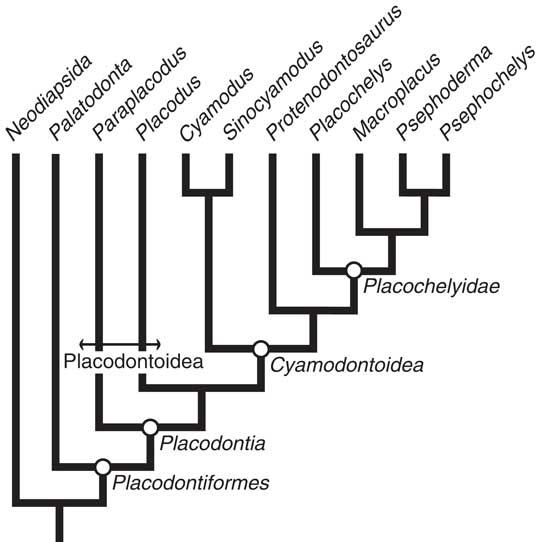
The Placodontiformes (Fig. 2) consist of the placodonts and their sister taxon, Palatodonta bleekeri, which does not have crushing teeth. Instead, the teeth of this species have a more plesiomorphic condition: teeth are tall, narrow, and pointed (Neenan et al. Reference Neenan, Klein and Scheyer2013). This tooth morphology indicates that the earliest common ancestor of Placodontiformes was probably not specialized to eat hard prey but more likely ate soft-bodied prey (Massare Reference Massare1987).
Tree showing the phylogenetic relationships between genera of the Placodontiformes studied in this paper, modified from Neenan et al. (Reference Neenan, Li, Rieppel and Scheyer2015). Major groups labeled at nodes if monophyletic or, in the case of a paraphyletic Placodontoidea, with branches bound by a double-ended arow. It should be noted that other analyses return Paraplacodus and Placodus as sister taxa in a monophyletic Placodontoidea (Neenan et al. Reference Neenan, Li, Rieppel and Scheyer2015).

The Placodontia are typically divided into two groups: the unarmored Placodontoidea and the armored Cyamodontoidea. The placodontoids, generally considered a paraphyletic assemblage though some recent analyses have supported a monophyletic grouping, include two species examined here: Paraplacodus broilii, at the root of the placodont tree, and Placodus gigas, as a sister taxon to the Cyamodontoidea (Fig. 2; Rieppel Reference Rieppel2000; Neenan et al Reference Neenan, Li, Rieppel and Scheyer2015). Both Paraplacodus broilii and Placodus gigas have a set of procumbent premaxillary teeth, thought to be for grasping prey living on top of the substrate, separated by a diastema from the crushing teeth of the maxilla and palatine. While tooth arrangement is similar in these two groups, Paraplacodus broilii has been typically considered to have tooth morphology more reminiscent of the plesiomorphic condition (Mazin Reference Mazin1989; Rieppel Reference Rieppel1995). The battery of crushing teeth in the upper jaw of Paraplacodus broilii consists of seven pairs of maxillary teeth, described as being low or round, each with a persisting central, conical cusp, and four pairs of palatine teeth, described as slightly larger than the maxillary teeth, with an overall low, subspherical profile and similar conical cusps or blunt crowns (Fig. 3A; Mazin Reference Mazin1989; Rieppel Reference Rieppel2000).
Idealized diagrams showing the diversity of placodont tooth arrangement, size and gross morphology. A, Paraplacodus broilii; B, Placodus gigas; C, Cyamodus kuhnschnyderi; D, Sinocyamodus xinpuensis; E, Protenodontosaurus italicus; F, Macroplacus raeticus; G, Psephochelys polyosteoderma; H, Psephoderma alpinum; I, Placochelys placodonta.

Compared with Paraplacodus broilii, Placodus gigas has reduced the number of crushing teeth, most often with four pairs of maxillary teeth and three pairs of palatine teeth, though tooth counts vary slightly by specimen and species (Fig. 3B; Mazin Reference Mazin1989; Rieppel Reference Rieppel1995). Descriptions of the maxillary tooth morphology vary in the literature; some say that they are comparable to the teeth of Paraplacodus broilii, while others describe them as being overall rounded, “subquadrate,” or hemispheric (Owen Reference Owen1858; Mazin Reference Mazin1989; Rieppel Reference Rieppel2000). The gross morphology of the palatine teeth is noticeably different from the maxillary teeth. Palatine teeth are overall larger and are described in the literature as varying in size, with more rostral teeth being smaller and caudal teeth larger. Palatine teeth take on a more triangular or quadrilateral appearance, as compared with the button-like shape of the maxillary teeth, and are described as being flatter overall (Owen Reference Owen1858; Mazin Reference Mazin1989).
Within the armored cyamodontoids there is further subdivision between the Placochelyidae and other cyamodontoids (Fig. 2), which is reflected in changes in dentition. Members of the genus Cyamodus have one to two pairs of reduced blunted premaxillary teeth, two to three pairs of maxillary teeth, and two to three pairs of palatine teeth, though tooth counts vary by species and with ontogeny (Fig. 3C; Mazin Reference Mazin1989). The description of maxillary teeth in Cyamodus spp. ranges from teeth having a full oval crown to being hemispherical (Owen Reference Owen1858; Mazin Reference Mazin1989). Both the maxillary and palatine teeth of Cyamodus vary in size within a tooth row, increasing in size from rostral to caudal, and relative tooth sizes and number can also change with ontogeny (Kuhn-Schnyder Reference Kuhn-Schnyder1959; Mazin Reference Mazin1989). The most rostral palatine teeth resemble the caudal-most maxillary teeth in size and shape, but the caudal-most palatine teeth are much larger and are described as having a full oval, elliptical, or bean shape, with the long axis of the tooth roughly parallel to the line of the maxillary teeth (Owen Reference Owen1858; Mazin Reference Mazin1989; Neenan et al. Reference Neenan, Li, Rieppel, Bernardini, Tuniz, Muscio and Scheyer2014). Sister to the genus Cyamodus is the Chinese species Sinocyamodus xinpuensis. Like the Cyamodus spp., this species has three “small, round” maxillary teeth and two “crushing tooth plates,” the posterior of which is the larger of the two (Fig. 3D; Neenan et al. Reference Neenan, Li, Rieppel and Scheyer2015).
The enigmatic Henodus chelyops has an extremely reduced dentition and an uncertain phylogenetic position among cyamodontoids (e.g., Neenan et al. Reference Neenan, Li, Rieppel and Scheyer2015). There is only one small tooth plate at the caudal extremity of each palatine and dentary and no maxillary teeth, and the premaxilla contains a series of small denticles. It is thought that this taxon was not durophagous, and it has not been included in this study.
Setting them apart from other placodonts, members of the Placochelyidae have lost their premaxillary teeth and instead have an elongated edentulous rostrum. Protenodontosaurus, the sister taxon to the placochelyids (see Figs. 2 and 3E), has a single pair of premaxillary teeth, whereas placochelyids, with the possible exception of an early juvenile specimen attributed to Psephoderma (e.g., Pinna Reference Pinna1979; Nosotti and Pinna Reference Nosotti and Pinna1999) lack premaxillary dentition.
Placochelyids include Placochelys placodonta, Macroplacus raeticus, and Psephoderma alpinum from the western Tethys, and the Chinese species Psephochelys polyosteoderma and Glyphoderma kangi (the latter of which could not be included in this study); all typically have two to three pairs of flat or flattened maxillary teeth that increase in size from rostral to caudal and two pairs of palatine teeth, the rostral of which is similar to the last maxillary tooth. In the literature, the caudal-most palatine tooth is described as being much bigger than the first palatine tooth, elliptical or ovoid, and is often said to be flat (Fig. 3F–I; Mazin Reference Mazin1989). This dental arrangement is sufficiently different from other placodonts that it has led some to hypothesize that placochelyid placodonts were using their rostra to burrow into muddy substrates to find subbenthic invertebrates, likely bivalves, instead of picking epibenthic invertebrates off the substrate (e.g., Mazin and Pinna Reference Mazin and Pinna1993).
While the focus of this work is on changes in form in the palatine and maxillary teeth, it is worth noting that there are similar patterns of change in relative tooth size and shape in the teeth of the dentary as well. In Palatodonta bleekeri, the sister taxon to the placodonts, a single row of peg-like teeth runs the length of the dentary, with overall tooth size increasing caudally. With the jaw closed, this row of dentary teeth would fit between the single row of maxillary teeth laterally and the roughly parallel single row of palatine teeth medially (Neenan et al. Reference Neenan, Klein and Scheyer2013). Within the placodonts, this overall arrangement—dentary teeth positioned midway between the palatine and maxillary tooth rows—hold true. But with the more rounded or flattened placodont tooth morphologies, this tooth arrangement means that placodont dentary teeth occlude with the medial portion of maxillary teeth and the lateral portion of palatine teeth (Owen Reference Owen1858; Mazin and Pinna Reference Mazin and Pinna1993; Rieppel Reference Rieppel1995; Neenan et al. Reference Neenan, Klein and Scheyer2013, Reference Neenan, Li, Rieppel, Bernardini, Tuniz, Muscio and Scheyer2014).
As with the maxillary and palatine teeth of these organisms, there are differences in the number and shape of dentary teeth between groups. In Paraplacodus broilii there are two slender, pointed teeth at the mandibular symphysis followed by a gap, and then seven “subspherical” teeth on each dentary, the posterior of which may have interacted tightly with the associated maxillary and palatine teeth (Rieppel Reference Rieppel2000). There are two procumbent incisors on each Placodus gigas dentary, a diastema, and then three teeth, larger than those of the maxilla, the posterior-most teeth being the largest of the three. In the cyamodontoids there is an even more pronounced difference between the enlarged posterior-most dentary teeth and those anterior, likely due to the constraints of bite mechanics (Rieppel Reference Rieppel2002). Additionally, there are fewer dentary teeth in cyamodontoids, mirroring the patterns seen in the cranial teeth; a small anterior tooth followed by four “crushing” teeth and an enlarged fifth tooth in Sinocyamodus; one enlarged posterior tooth with two smaller anterior teeth in Cyamodus spp. In the placochelyids there are two enlarged teeth on the caudal end of the dentary, the anterior end of which corresponds with the elongated rostrum (Mazin and Pinna Reference Mazin and Pinna1993; Neenan et al. Reference Neenan, Li, Rieppel, Bernardini, Tuniz, Muscio and Scheyer2014; personal observation).
Institutional Abbreviations
BSP, Bayerische Staatssammlung für Paläontologie und Historische Geologie, Munich, Germany; IVPP, Institute of Vertebrate Paleontology and Paleoanthropology, Beijing, People’s Republic of China; MB.R., Museum für Naturkunde, Berlin, Germany; MFSN, Museo Friulano di Storia Naturale, Udine, Italy; MSNM, Museo Civico di Storia Naturale, Milan, Italy; PIMUZ, Paläontologisches Institut und Museum der Universität, Zurich, Switzerland; SMF, Senckenberg Forschungsinstitut und Naturmuseum, Frankfurt am Main; SMNS, Staatliches Museum für Naturkunde, Stuttgart, Germany.
Methods
Given the relatively simple morphology of placodont teeth, occlusal variation can be quantified by fitting a circle or sphere to the occlusal surface and measuring the radius of curvature (RoC; Fig. 4). A large RoC would correspond to a flat or very shallow occlusal surface, with the tooth becoming pointier as the RoC decreases. Positive RoCs correspond to teeth that have an overall convex morphology, and negative RoCs indicate a concavity in the occlusal surface. Only specimens and casts with teeth in situ were used for this study, and we did not take measurements from teeth with obvious damage to the occlusal surface either through wear or taphonomic processes.
Measuring tooth RoC from .stl files (A) using Geomagic. Spheres were fitted to teeth with relatively simple occlusal morphologies by highlighting (B, in gray) the occlusal surface of the tooth delineated by abrupt changes in curvature. This resulted in a single sphere fit to the occlusal surface (D, gray dashed line) and a single RoC measurement. For teeth with more complex morphologies (C), such as the second palatine teeth in the placochelyids, multiple spheres were fitted to the occlusal surface. One sphere, fitted to the concave basin, was defined by placing four offset points (red squares) along the long axis of the tooth (dashed line). A second sphere was defined by placing a staggered set of four points (blue dots) orthogonal to the long axis over the cusp (dotted line). This resulted in a negative “primary RoC” (E1, curve outlined in red squares) and a positive “secondary RoC” (E2, curve outlined in blue circles).

When possible, specimens were scanned with a NextEngine laser scanner (NextEngine, Inc., CA) to collect 3D surface data, with a scan accuracy of 127 μm. While it has been shown that scans at this scale are not accurate enough to measure fine-scale topologies, such as those needed to identify dietary specializations from microwear, this study is concerned with whole-tooth morphology at a scale at which NextEngine scanner data have been shown to be comparable to or more accurate than medical CT scans (Slizewski et al. Reference Slizewski, Friess and Semal2010). RoC was measured using the built-in tool set in Geomagic (3D Systems, Inc., SC) to fit a sphere to the occlusal surface of each tooth, either by highlighting the surface or by placing four points on the surface to be measured. A similar technique has been used to measure the RoC of cusps in hominin teeth by fitting spheres to areas where teeth experimentally came into contact with food items (Berthaume et al. Reference Berthaume, Grosse, Patel, Strait, Wood and Richmond2010). Rather than measuring the curvature at any one particular part of a single tooth, in this study we are concerned with the relative occlusal curvature of different teeth. To that end, when fitting spheres to the highlighted portion of a tooth, we delineated the section of interest by tracing the obvious change in slope that occurs around the perimeter of the tooth. For convex or functionally flat teeth, this was typically where the sides of the tooth appear to fall away in an occlusal view (Fig. 4B). For concave teeth, this was approximately where the curvature of the profile of the concavity changed from negative to positive. We used the four-point sphere-fitting method for teeth with more complex features, typically a cusp projecting into an overall concave surface. For these teeth, we took two measurements: one of the most prevalent occlusal curvature by placing a staggered line of points along the long axis of the tooth, spanning the same portion of the concavity that would be otherwise highlighted; and the second by placing a staggered line of points along an orthogonal axis spanning the convexity of the cusp (Fig. 4C).
In some instances it was not possible to scan specimens, so measurements were taken from 3D surfaces reconstructed using photogrammetry (Falkingham Reference Falkingham2012). Photos were taken with a Kodak Easyshare camera (Z5120), and 3D models were constructed using VisualSFM (Wu et al. Reference Wu, Agarwal, Curless and Seitz2011; Wu Reference Wu2013). Additional specimens were incorporated into the study to expand the geographical range and taxonomic diversity of the study, with measurements taken from computed microtomographic scan reconstructions (Neenan et al. Reference Neenan, Li, Rieppel, Bernardini, Tuniz, Muscio and Scheyer2014).
Results
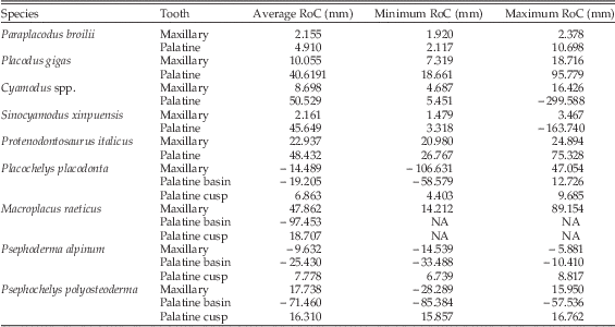
Individual tooth measurements, species names, specimen numbers, and type of data gathered are presented in Supplementary Table 1, and a species-by-species summary of tooth measurements is given in Table 1.
Placodont tooth radius of curvature (RoC) measurements. Measurements reported are the average, maximum, and minimum RoC (mm). In a number of specimens, palatine teeth were overall flatter than maxillary teeth, but the RoC varied between positive and negative. To better represent the overall flatter morphology, the reported values are based on the magnitude of the RoC. Because of this, the average RoC is was calculated using the absolute values for all teeth, and maximum and minimum RoC are similarly based on absolute values. For the placochelyid species, the more complex morphology of the palatine teeth is represented by two RoC measurements: a measurement for the basin that covers most of the occlusal surface and a measurement of the cusp. Measurements for individual teeth/specimens can be found in Supplementary Table 1.

Paraplacodus broilii
The average RoC of Paraplacodus broilii maxillary teeth is 2.16 mm and varies from 1.92 mm to 2.38 mm. Palatine teeth were distinguished from maxillary teeth by the lack of a semiconical cusp and a more rectangular occlusal cross section. The RoC of palatine teeth varies from 2.1 mm to 10.70 mm, but on average is 4.91 mm (Fig. 5A).
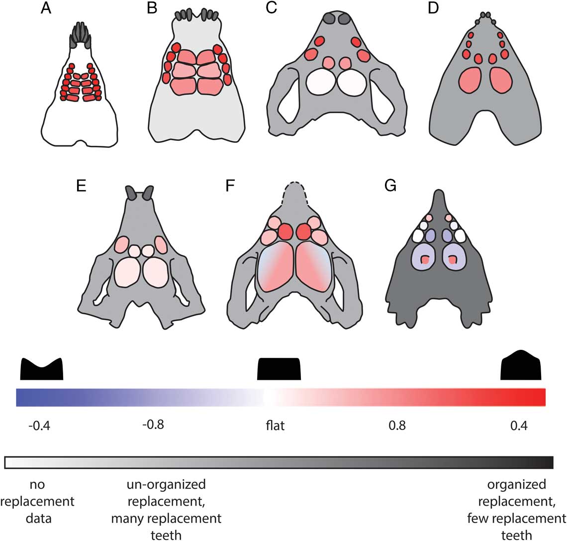
Comparison of placodont tooth RoC between teeth and across taxa. For each tooth, normalized RoCs (normalized by maximum diameter of the tooth to allow for comparison between individual teeth, between taxa, and with canonical models) are averaged between left and right teeth for all teeth at that position in the relevant specimens. Teeth are color-coded by normalized RoC, with reds representing small but positive RoCs; white teeth are approximately flat; and blues represent negative RoCs. Idealized skull outlines are color-coded to demonstrate degree of organization in tooth replacement. Paraplacodus broilii (A) is white to indicate that there is no information for tooth replacement patterns in this taxon. Placodus gigas (B, light gray) has a faster, less organized pattern of tooth replacement; Cyamodus kuhnschnyderi (C, medium gray) and Protenodontosaurus italicus (D, medium gray) have a more organized, slower tooth replacement; and the placochelyids (E, dark gray) have an organized pattern of tooth replacement with very few replacement teeth at any one time (Neenan et al. Reference Neenan, Li, Rieppel, Bernardini, Tuniz, Muscio and Scheyer2014). Cyamodus kuhnschnyderi, shown here as a representative Cyamodus species, has only two maxillary and two palatine teeth, but maxillary and palatine tooth numbers vary between two and three for specimens measured in this study.

Placodus gigas
Placodus gigas teeth, in situ, show a typical pattern of wear, sections of roughened, pitted enamel, noticeably different from the smoother surrounding enamel, where maxillary teeth appear worn on their lingual side and palatine teeth appear worn on the labial side. This may be partially due to the way these teeth come into contact with teeth in the lower jaw or may be a trace of where prey items were processed, serving as an indicator for where RoC may be of functional interest. As with other specimens, we did not measure RoC if teeth were damaged or worn to the point of exposed dentine, but we did include data from partially worn or pitted teeth still affixed in the jaw. We are concerned with the range of functional tooth morphologies, and that includes teeth from the time they erupted to the point when they were lost. On average, maxillary teeth have a larger RoC (10.06 mm) than seen in Paraplacodus broilii maxillary teeth, ranging from 7.32 mm to 18.72 mm. Placodus gigas palatine teeth typically have larger RoCs, on average 40.62 mm, than maxillary teeth, but can range from 18.66 mm to 95.78 mm (Fig. 5B).
Cyamodus spp. and Sinocyamodus xinpuensis
Because they are sister taxa and have similar tooth arrangements and gross morphologies, we have combined the data for all Cyamodus species with data from Sinocyamodus xinpuensis. The maxillary teeth in Cyamodus spp. and S. xinpuensis specimens are relatively small, but have a larger average RoC (14.39 mm) than the maxillary teeth in Placodus gigas. The rostral-most Cyamodus spp. and S. xinpuensis maxillary teeth have smaller RoCs (4.69–8.76 mm) than the caudal-most maxillary teeth (7.86–16.43 mm). The average RoC for the palatine teeth of Cyamodus and S. xinpuensis is 47.40 mm, but there is a noticeable disparity between the caudal palatine teeth and the rostral-most palatine teeth, which often have similar RoCs to the caudal-most maxillary teeth. For the first palatine tooth, RoCs range from as low as 5.45 mm to 30.91 mm. The caudal-most palatine teeth have the largest RoCs, ranging from 30.98 mm to 299.59 mm in magnitude. For most specimens, this means that the caudal-most teeth are shallowly convex; however, in two specimens, Cyamodus sp. (SMNS 91472) and C. rostratus (MB.R.1773), the caudal-most palatine teeth are shallowly concave, with RoCs ranging from −6.53 mm to −299.59 mm (Fig. 5C,D).
Protenodontosaurus italicus and the Placochelyidae
Protenodontosaurus italicus (MFSN 1819 GP) is the sister taxon to the Placochelyidae and has a similarly elongated snout, but with a single pair of premaxillary teeth. For the single pair of maxillary teeth, there is a RoC of 24.89 mm on the left and 22.98 mm on the right. There are two pairs of palatine teeth for P. italicus, and the rostral and caudal teeth differ in size. The rostral-most palatine teeth have a RoC of 33.54 mm on the left and 26.77 mm on the right and are roughly similar in size and shape to the maxillary teeth. The larger, caudal pair of palatine teeth in P. italicus have a RoC of 58.09 on the left and 75.33 on the right (Fig. 5E).
For this study, we measured data from four genera within the Placochelyidae: Placochelys placodonta, Macroplacus raeticus, Psephoderma alpinum, and Psephochelys polyosteoderma, and the teeth of all species followed similar overall patterns, though we have access to only one to two specimens per species (Fig. 5F,G)
The rostral portion of one Placochelys placodonta specimen (MB.R. 1765) was broken off, so we only have data for the last two maxillary teeth. However, we also have data from a cast of an almost compete Placochelys placodonta (MB.R.1767.1) that has three maxillary teeth. In contrast, Psephoderma alpinum (PIMUZ A/III 1491; Neenan and Scheyer Reference Neenan and Scheyer2014) and Psephochelys polyosteoderma (IVPP V 12442) both have only two pairs of maxillary teeth. The first maxillary teeth of Placochelys placodonta have small RoCs, 4.74 mm on the left and 1.15 mm on the right. The first pair of Psephoderma alpinum maxillary teeth are concave and have a RoC of −5.88 mm on the left and −9.38 mm on the right, as opposed to the first pair of Psephochelys polyosteoderma maxillary teeth, which have RoCs of 10.88 mm on the left and 15.95 mm on the right.
Overall, the caudal-most maxillary teeth in all three species have RoCs of larger magnitudes than the teeth that precede them. The concavity/convexity of the last pair of maxillary teeth in Placochelys placodonta varies between the two specimens, though all RoCs are relatively large compared with more anterior maxillary teeth: −98.32 mm and 47.05 mm for the third left maxillary teeth in the two specimens and −106.63 mm and 35.76 mm for the third right maxillary teeth. The last pair of maxillary teeth in Psephoderma alpinum are concave, with RoCs of −14.54 mm for the left tooth and −8.73 mm for the right. Finally, Psephochelys polyosteoderma also has concave causal maxillary teeth, the left has a RoC of −28.29 mm and the right measures −15.83 mm.
In all specimens there are two pairs of palatine teeth, and the rostral palatine teeth are similar to the last pair of maxillary teeth in size and shape, with an average RoC of 20.93 mm, and can be concave, with RoCs varying from −8.45 mm to −24.49 mm, or convex, with RoCs of 12.45 mm to 66.3 mm. The caudal palatine teeth in Placochelys placodonta, Psephoderma alpinum, and Psephochelys polyosteoderma all have a more complex morphology than can be measured with a single RoC (Fig. 4B), though this is more or less obvious in different specimens. These teeth have a shallow, slightly curved furrow or concavity running the length of the long axis of the tooth, and on the lingual–caudal edge of the concavity there is a small cusp projecting from the occlusal surface (Fig. 5G). In Placochelys placodonta, the concavity has a RoC of −39.66 mm, and the cusp has a RoC of 6.86 mm. The cusp/concavity morphology is more obvious in Psephoderma alpinum; the left tooth has a concavity with a RoC of −33.33 mm and a cusp with RoC of 8.82 mm and a concavity of −33.49 on the right with a cusp with a RoC of 6.74 mm. Finally, Psephochelys polyosteoderma cusps are not as pointed as other species, 15.86 mm on the left and 16.76 mm on the right, but are still distinct and are paired with a concavity of −57.54 mm on the left and −85.38 mm on the right.
Macroplacus raeticus has larger teeth than the other placochelyids, most notably the second palatine teeth, and so is discussed separately. The rostral portion of the specimen (BSP 1967 I 324) used in this study is broken and is missing the first of two teeth on the left maxilla. The single rostral maxillary tooth measured from M. raeticus has a RoC of 14.21 mm, with larger RoCs on the two caudal maxillary teeth: 40.22 mm on the left and 89.15 mm for the right. The rostral palatine teeth have RoCs similar to the maxillary teeth, 14.35 mm on the left and 20.49 mm on the right. There is only one caudal palatine tooth present on the M. raeticus specimen that we measured, and instead of a distinct cusp projecting into a concavity, there is a change in RoC from a lingual convexity with a RoC of 18.71 mm to a more concave surface with a RoC of −97.45 mm on the labial half of the occlusal surface (Fig. 5F).
Discussion
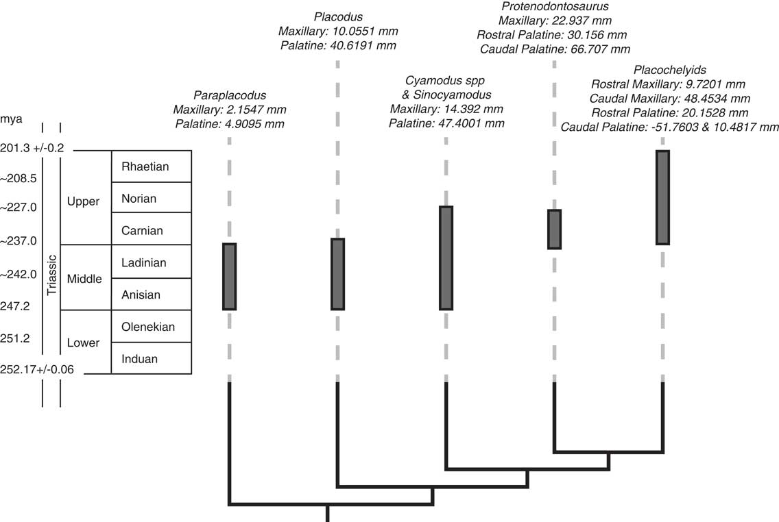
The differences in tooth RoC for nonplacochelyid placodonts measured here overall agree with the qualitative descriptions, where conical or rounded teeth give way to a more flattened shape, as previously described in the literature (Figs. 5 and 6). The maxillary teeth of Paraplacodus broilii have the lowest RoC and are the most similar to the piercing teeth of the sister taxon to the placodonts, Palatodonta bleekeri (Neenan et al. Reference Neenan, Klein and Scheyer2013). The palatine teeth have a larger average RoC than the maxillary teeth, but even these teeth are more pointed than the palatine teeth of other placodonts, though there is some overlap with the RoC of maxillary teeth in other groups. Placodus gigas has maxillary teeth with a larger RoC than the maxillary teeth of Paraplacodus broilii, but still more rounded than the palatine teeth. Based on predictions from previous works, these teeth would have been less likely to break while still effectively crushing prey (Chai et al. Reference Chai, Lee, Kwon, Lucas and Lawn2009; Lee et al. Reference Lee, Constantino, Lucas and Lawn2011; Qasim et al. Reference Qasim, Bush, Hu and Lawn2005; Crofts and Summers Reference Crofts and Summers2014; Crofts Reference Crofts2015). The average RoC of the palatine teeth is much larger than the RoC for the maxillary teeth—approaching a predicted optimal tooth shape for crushing while resisting failure (RoC normalized by tooth width ≥0.8; Supplementary Table 1). In Cyamodus spp., rostral maxillary teeth have smaller RoCs than those that follow, suggesting they may have been less involved with crushing shelled prey. Caudal maxillary teeth, in contrast, have larger RoCs, similar to those measured for the rostral palatine teeth, and were probably moderately involved in breaking prey. The large RoCs of the caudal-most palatine teeth of Cyamodus spp. make these most like the optimal tooth predicted by the physical and finite element analysis models. While most of these teeth are shallowly convex, there were two specimens with concave palatine teeth, but the RoCs were sufficiently large to be functionally flat. Similarly, in Sinocyamodus xinpuensis, the maxillary and rostral palatine teeth have relatively small RoCs and were likely less involved with crushing prey than the caudal palatine teeth, which have RoCs of such large magnitude that they are functionally flat.
Tree showing placodont relationships correlated with occurrence of each taxon and tooth RoC data. Gray bars span time during which taxa occurred, and tree structure is not correlated to time. Dotted lines do not represent ghost lineages but correlate branches of the tree to text. For each genus, average absolute tooth RoC is given for maxillary teeth and palatine teeth, except Sinocyamodus which has been averaged with Cyamodus species, and the placochelyids (Placochelys, Macroplacus, Psephoderma, and Psephochelys) are averaged together. Two measurements are included if there is a difference in tooth RoC between rostral (R) and caudal (C) teeth. In the placochelyids, the rostral and caudal palatine tooth RoCs are reported separately, because the more complex morphology of the caudal palatine tooth requires two RoCs for the tooth concavity (−51.7603 mm) and medial cusp (10.4817 mm).

While placochelyid palatine teeth are often described in the literature as flat, this is not the case. Instead, these teeth have a more complex morphology than seen in other placodonts, with a crescentic furrow running the length of the tooth and a lingual or lingual–caudal cusp, although the relative size and curvature of these features varies between species. This suggests that these organisms may, in fact, have had a different dietary specialization than the other placodonts or have specialized for durophagy in a different way. The shallow concavity may dissipate in-tooth strain and localize any damage that might occur (Qasim et al. Reference Qasim, Bush, Hu and Lawn2005; Crofts Reference Crofts2015), whereas the cusp, which has a relatively small RoC, concentrates stress applied to the prey item (Crofts and Summers Reference Crofts and Summers2014). Alternatively, these teeth may have worked in a way similar to the system that has been proposed for bunodont molars, where stress is applied to food items by a dull cusp that also dissipates in-tooth stress, while three additional cusps, or the concavity in placodonts, serve to hold food items in place (Berthaume et al. Reference Berthaume, Dumont, Godfrey and Grosse2014).
As has been argued above, the ability of teeth to break shells without sustaining damage is a large part of a predator’s success, but to “win” in the predator–prey arms race, predators must also be able to eat their prey. We know from late Olenekian bromolites, inferred to be regurgitates of durophagous predators (possibly sharks, colobodontids, pachypleurosaurs, or placodonts), that these predators were primarily crushing and feeding on bivalves, with some crinoids, gastropods, and even some vertebrates represented as well (Salamon et al. Reference Salamon, Niedźwiedzki, Przemysƚaw, Lach and Surmik2012). Placodus gigas has been found in deposits with a diversity of shelly invertebrate taxa, including moderately mobile/shallowly burrowing bivalves and crinoids, known to be part of Triassic durophagous predator diets, and brachiopods, asteroids, ophiuroids, and gastropods (Assmann Reference Assmann1937; Salamon et al. Reference Salamon, Niedźwiedzki, Przemysƚaw, Lach and Surmik2012). Cyamodus spp. and Placodus gigas can also co-occur in later deposits and are found with some of the same bivalves, ophiuroids, and asteroids as seen in Placodus gigas deposits (Blake and Hagdorn Reference Blake and Hagdorn2003).
However, not all shelled organisms would be potential prey items, as there is a trade-off between energy invested in prey handling and predator success. Mazin and Pinna proposed that only particular areas of the mouth were used to crush prey in Placochelys placodonta, an area overlapping the caudal region of the first pair of palatine teeth and the rostral region of the second palatine teeth, two smaller areas between the palatine teeth and maxillary teeth, and a third small area between the rostral ends of the first palatine teeth (Mazin and Pinna Reference Mazin and Pinna1993). Positioning prey in this way may have resulted in increased handling time and would have led to asymmetric loads applied to the edges of the teeth. Unlike a centrally applied load, which would split the tooth, these off-center loads would be more likely to chip teeth (Chai et al. Reference Chai, Lee and Lawn2011). Off-center loading would especially change the interaction between prey items and concave teeth, with only a portion of the tooth rim transferring force to the prey item. In theory, this should reduce the area of contact and decrease the total applied force, though the RoC of the edge varies around the tooth and between species. In the case of the two Cyamodus spp. with concave caudal palatine teeth, the average RoC of the edges is 16.26 mm, still relatively flat (Supplementary Table 1). In contrast, the edge RoCs in placochelyid species (excluding M. raeticus) tend to be smaller, 5.54 mm, with the labial edges generally having the smallest RoCs and the rostral or caudal RoCs being generally the largest (Supplementary Table 1). This difference in RoC would change the mode of failure for these teeth, or regions of a tooth, with larger RoCs more likely to form small chips at lower loads and smaller RoCs more likely to form large chips or even split (Chai et al. Reference Chai, Lee and Lawn2011; Lawn et al. Reference Lawn, Bush, Barani, Constantino and Wroe2013).
Placodonts may also have been limited in what prey they could have processed. Thicker-shelled or larger animals often present greater resistance to failure than smaller or thinner-shelled alternatives, and predators will be more likely to consume or attempt to consume the easier to break prey or prey that requires less handling time (Kislalioglu and Gibson Reference Kislalioglu and Gibson1976; Boulding Reference Boulding1984). Based on tooth wear facets, the inferred volume between teeth, and morphology of the lever formed by the mandible, it has been predicted that Placochelys placodonta could have eaten prey that was at most 50×25×15 mm (Mazin and Pinna Reference Mazin and Pinna1993). Additionally, given the morphology of a different crushing surface, P. placodonta would have had a minimum prey size of 15×12×10 mm for the small anterior-lateral crushing areas and 35×20×10 mm for the larger medial crushing area involving the caudal-most palatine teeth (Mazin and Pinna Reference Mazin and Pinna1993). If bitten prey were not consumed, due to size refugia or some other defensive trait, and experienced only sublethal damage, they would have been able to repair their shells and pass along whatever trait or traits allowed them to survive the encounter.
While the advances in shell defenses typically associated with the Mesozoic marine revolution (Vermeij Reference Vermeij1977) have traditionally been described as occurring in the Jurassic and accelerating in the Cretaceous, more recent work indicates that these selective pressures may have begun earlier. Toothed hinges and cementing to substrates, for example, appear in the Carnian (Vermeij Reference Vermeij2008), and brachiopod defensive ornamentation also increases throughout the Triassic (Vörös Reference Vörös2010). While environmental factors that may have also driven changes in shelled prey, such as competition for space or environmental stability leading to increased infaunalization in Triassic bivalves (McRoberts Reference McRoberts2001), these changes would have also led to new challenges for predators.
With bivalves increasingly burrowing into the substrate through the Triassic (McRoberts Reference McRoberts2001), later-occurring groups of placodonts may have needed different approaches to obtaining prey. The cyamodontoid lineage may have been capable of creating suction to pull in water, either for feeding or for olfaction (Rieppel Reference Rieppel2002). Within the cyamodontoid group, it is supposed that the placochelyids occupied the same ecological niche as modern rays, using their rostrum and/or buccal suction to root out infaunal bivalves (Rieppel Reference Rieppel2002). Additionally, some placochelyid specimens have been found in deposits that seem to lack bivalves but instead are dominated by epifaunal decapod crustaceans, presenting a possible alternate food source (Stefani et al. Reference Stefani, Arduini, Garassino, Pinna, Teruzzi and Trombetta1992). These potential differences in prey type may account for our observed differences in tooth morphology.
Given the potential differences in diet, one might expect to see variation in bite-force across phylogeny that could also help explain differences in tooth morphology. A durophagous diet requires high bite forces to induce failure in hard-shelled prey items, and in modern taxa we see features like enlarged sites for jaw muscle attachment and modifications to increase mechanical advantage of the jaws in durophagous lizards (Dalrymple Reference Dalrymple1979). Inferred muscle attachments, in both the placodontoids and cyamodontoids, indicate there was a division of labor in jaw closing: the mechanical advantage of the jaw adductors would have been greatest at small gapes, whereas pterygoideus muscles would have provided the most power at wider gape angles. It is likely that these two sets of muscles worked together, depending on the size of the prey item. Additionally the huge superficial pterygoideus may have helped prevent disarticulation of the mandible during strong bites (Rieppel Reference Rieppel2002). In addition to the stabilization provided by the pterygoideus muscles, placochelyids may have been able to use their elongate rostrum to dissipate twisting forces resulting from high bite forces, and even nonplacochelyid cyamodontoids could have also dissipated forces, to a lesser extent, in their short snouts (Mazin and Pinna Reference Mazin and Pinna1993).
Given the ability to generate high bite forces, there should be an increased emphasis on the functional size of teeth in durophagous taxa. Increasing tooth size may serve to prevent tooth failure (Lee et al. Reference Lee, Constantino, Lucas and Lawn2011). With a greater emphasis on durophagy, teeth should increase in size while decreasing in number, and there should be an overall decrease in the replacement rate to maximize the number of functional teeth at any one time (Dalrymple Reference Dalrymple1979). In placodonts, from the placodontoids to the cyamodontoids and placochelyids, we see an overall decrease in tooth number (Mazin Reference Mazin1989) and increasing RoCs that reduce the likelihood of tooth failure. There is also an overall pattern of decreased tooth replacement rates in cyamodontoid lineages compared with placodontoids. Moreover, while all placodont species maintain a functional crushing surface at all times during tooth replacement, the pattern of replacement is more organized in the cyamodontoids compared with the placodontids (Fig. 5; Neenan et al. Reference Neenan, Li, Rieppel, Bernardini, Tuniz, Muscio and Scheyer2014). Replacing worn teeth is important, especially in domed teeth, since increased wear can change the mode of crack propagation through enamel and the load to critical tooth failure (Keown et al. Reference Keown, Bush, Ford, Lee, Constantino and Lawn2012).
While function is not the only factor dictating tooth morphology, the pattern of change across the placodont lineage indicates that the trade-off between breaking prey items while maintaining tooth integrity may influence morphology. In addition to the changes seen in tooth number, replacement, and occlusal morphology, tooth material composition may also change in response to changes in diet, as thicker enamel is better able to resist deformation under high loads than thin, worn enamel (Lucas et al. Reference Lucas, Constantino, Wood and Lawn2008). In this paper we have only addressed changes in teeth of the upper jaw, but these interact with similarly shaped teeth in the lower jaw, the morphology of which will affect how prey items are loaded. This study is a first step in understanding the larger pattern of how tooth morphology changes with the shift to a durophagous diet within a single lineage.
Acknowledgments
Thank you to the curators and research staff at the many museums and collections that were visited in the course of this research; your guidance and assistance were invaluable and your welcomes warm and inviting. Thanks to C. Sidor and G. Wilson for their advice and guidance throughout the process, from looking for collections to drawing conclusions from the data. Finally, we would like to thank our reviewers and S. Marcus for their advice and support. Funding was from the Snyder Award and the University of Washington Department of Biology, the Estes Memorial Grant (Society of Vertebrate Paleontology), and National Science Foundation grant IOS-1256602 to A.P.S., and Swiss National Science Foundation grants P2ZHP3_162102 to J.M.N. and 31003A_149506 to T.M.S.
Supplementary Material
Data available from the Dryad Digital Repository: http://doi:10.5061/dryad.8vs89