It was historically perceived that the genome consists of ‘coding’ regions and other regions that were considered to be ‘junk’ DNA (Ref. Reference Ohno and Smith1). Those regions of the genome thought to have no function are currently perceived as imperative in cellular functioning and control placing microRNAs (miRNAs) at the top of the hierarchy of control over most cellular processes. miRNAs share features in their structure, processing and mode of action, but even though their size is relatively minuscule, the control of a set of a few hundred miRNAs identified to date can extend to almost every intracellular signalling pathway (Ref Reference Lewis2). The sensitive regulation of the expression levels of such small noncoding RNAs is thus of critical consequence for the tight balance of cellular processes, making the study of miRNAs of great value for understanding both physiological and pathological phenomena. Specifically, miRNA-181 is important in cellular development and differentiation, evidenced by its aberrant expression in cancers and autoimmunity (Refs Reference Calin and Croce3, Reference Lashine4), which highlights its responsibility for abnormal cellular functions in such disorders. The miR-181 family has four members (miR-181a, miR-181b, miR-181c and miR-181d) whose seed region is complementary to a few hundred identified mRNAs to date, enabling miR-181 family members to regulate their target genes either by inhibitory binding to their mRNAs or by activating the RNA-induced silencing complex and prompting degradation of the target mRNA (Ref Reference Lim5).
Role of miR-181a in organ development
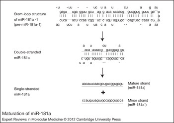
The presence of many matching sites between the mature miR-181a seed region (Figure 1) and the coding sequence for the C2H2 domain of zinc finger (ZNF) genes has facilitated the role of miR-181a in downregulating the expression of ZNFs (Ref. Reference Huang6) (a family of proteins consisting of around 700 members that act as transcription regulators), giving miR-181a access to the regulation of a huge number of cellular pathways, especially developmental ones. For example, Smad-interacting protein 1 (SIP1, also known as ZEB2) is one of the best characterised key players of the ZNF family and is involved in many developmental processes such as neuroectoderm cell fate determination (Ref. Reference Chng7), liver development (Ref. Reference Shaw, Johnson and Kimber8), corticogenesis (Ref. Reference Seuntjens9) and ocular lens development (Ref. Reference Yoshimoto10). More importantly, ZEB2 promotes the process of epithelial mesenchymal transition, which is critical for embryonic development and whose deregulation leads to the pathogenesis of epithelial tumours, by downregulating the cell–cell adhesion molecule E-cadherin and inhibiting the oncoprotein cyclin D (Ref. Reference Peinado, Olmeda and Cano11). In differentiated mouse embryonic stem cells (ESCs), miR-181a was found to be one of the miRNAs that post-transcriptionally downregulate and maintain the low protein expression of silent mating-type information regulation 2 homologue 1 (SIRT1), which regulates processes such as transcription, apoptosis and muscle differentiation by deacetylating key proteins (Ref. Reference Saunders12). Its role in organ development was analysed in a microarray analysis that included different miRNAs, where it showed highest abundance in the brain, followed by the liver and eyes. It also showed high expression levels in the retina during early development (Ref. Reference Ryan, Oliveira-Fernandes and Lavker13). Its specific function in liver development was recently highlighted because of its relative upregulation in embryonic livers and isolated hepatic stem cells compared with adult hepatic cells, and its ability to target different transcriptional regulators of hepatic differentiation along with the Wnt–β-catenin signalling pathway (Ref. Reference Ji14). It has a critical role too in inner-ear development because miR-181a is capable of stimulating proliferation in the basilar papilla, resulting in the production of new hair cells (Ref. Reference Pan15). miR-181a regulates the differentiation of intracardiac skeletal myoblasts (SKMs) by targeting the differentiation repressor homeotic protein A11 (Hox-A11). This fact was used in an attempt to prevent post-SKM-transplantation arrhythmias in rats in an elegant study conducted by Li and colleagues, in which they knocked down miR-181a levels (Ref. Reference Li16). miR-181 along with miR-1 and miR-107 are upregulated on endurance exercise, resulting in the activation of transcription networks that regulate mitochondrial biogenesis, glucose and fatty acid metabolism, and eventually skeletal muscle remodelling (Ref. Reference Safdar17).
Maturation of miR-181a. Development of miR-181a-1 following its transcription, starting with the stem–loop structure (pre-miR) (top), followed by excision of the loop by Dicer resulting in the double-stranded miRNA (miRNA–miRNA* duplex) (middle) and, finally, separation of the double strand into single-stranded mature and minor strands (bottom). Retrieved from the microRNA database (http://www.mirbase.org/).

The function of miR-181a in vascular and lymphatic development has been highlighted recently through its role in targeting PROX1, a homeobox transcription factor that is critical for lymphatic endothelial cell specificity in early embryonic life. Shifting the fate of endothelial cells to a vascular phenotype rather than a lymphatic one is achieved through the upregulation of miR-181a, which leads to PROX1 silencing, an effect that can be reversed on knockdown of miR-181a using its antagomirs (Ref. Reference Kazenwadel, Michael and Harvey18).
Role of miR-181a in haematopoiesis
miR-181a is expressed at the highest levels in the thymus, the primary lymphoid organ and the site of maturation of T lymphocytes (Ref. Reference Chen19). miR-181 is abundantly expressed in the B-lymphoid cells of mouse bone marrow, and its ectopic expression in mouse haematopoietic ESCs increased the fraction of B-lineage cells in adult mice (Ref. Reference Chen19). It is detectable in normal mature human T cells, monocytes and granulocytes, with the lowest concentrations in B cells (Ref. Reference Ramkissoon20). miR-181a is one of the most highly expressed miRNAs in T cells (Ref. Reference Okada, Kohanbash and Lotze21), which could explain its important role in T-cell function and sensitivity, which starts at very early stages of T-cell selection in the thymus (Ref. Reference Ebert22). Its regulation is considered dynamic during thymocyte differentiation, showing the highest expression level in the early stages of T-cell differentiation, especially in both double-positive (DP) and double-negative (DN3) thymocytes (Ref. Reference Neilson23). The ectopic expression of miR-181a in thymic progenitor cells stimulates the development of CD4 and CD8 double-positive cells (Ref. Reference Liu24). On its inhibition by an antagomir, a significant impairment of DP thymocyte sensitivity, and positive and negative selection, is observed, highlighting the role of miR-181a in modulating T-cell sensitivity and selection. This is probably achieved by directly targeting and repressing the expression of BCL2, CD69 and T-cell receptor (TCR) genes, which are key players in the process of positive selection (Ref. Reference Neilson23). miR-181a also modulates TCR-mediated activation, and augmentation of both strength and sensitivity of TCR signalling to strong and weak agonists, and its overexpression can even convert the response of T cells to TCR antagonists into an activating response rather than an inhibitory one (Ref. Reference Li25). This is due to the presence of several putative miR-181a pairing sites in serine phosphatases, protein tyrosine phosphatase nonreceptor type 11 and 22 (PTPN11 and PTPN22) and dual-specificity phosphatases DUSP5 and DUSP6, which act as negative regulators in the TCR signalling pathway (Refs Reference Li25, Reference Dittel26, Reference Stefanova27, Reference Altan-Bonnet and Germain28). In addition, miR-181a controls the clonal deletion of T cells with moderate affinity for MHC class II selecting peptides and ensures that only high-affinity clones undergo maturation (Ref. Reference Ebert22). Interestingly, miR-181a also exerts a role in the determination of myeloid and erythroid progenitor cells to erythroid cell fate (Ref. Reference Choong, Yang and McNiece29).
Role of miR-181a in solid tumours
Several recent studies investigated the expression of miR-181a in tumourigenesis, to discover its possible role in pathogenesis and evaluate its use as a novel biomarker for the diagnosis and prognosis of some cancers. miR-181a levels were investigated in primary squamous lung cell carcinoma (SQCC) and showed significantly downregulated levels (Ref. Reference Gao30). This downregulation of miR-181a was also observed in oral squamous cell carcinoma (OSCC) in a recent study by Shin and coworkers, which also identified the oncogene KRAS as a target of miR-181a, suggesting their combined roles in the oncogenesis of OSCC (Ref. Reference Shin31). This is supported by the fact that abnormal KRAS activity was previously implicated in the pathogenesis of oral tumours (Ref. Reference Caulin32). miR-181a was also found to be downregulated in non-small-cell lung cancer (NSCLC) in a microarray analysis. Such downregulation correlated directly with the poor survival rate of patients, suggesting that miR-181a could be a prognostic biomarker for NSCLC (Ref. Reference Gao33). This is in line with the results of Cheng and colleagues, who found that inhibition of miR-181a in A549 cells leads to increased cell growth (Ref. Reference Cheng34). By contrast, the use of antisense-miRNA-oligonucleotides specific for miR-181a (AMOs-miR-181a) in A549 cells triggered apoptosis and cell cycle arrest at S phase, decreasing the rate of A549 cell growth (Ref. Reference Fei35). Targeting of miR-181a could not modulate the A549 cellular response to cisplatin treatment, although transfection with pre-miR-181a augmented cisplatin-induced cell death by regulating the intrinsic apoptotic pathway, on both the mitochondrial and postmitochondrial levels (Ref. Reference Galluzzi36). miR-181a, together with other members of the miR-181 family, was also found to be downregulated in glioblastoma tumours (Ref. Reference Ciafre37). This was substantiated by the finding that it acts as a tumour suppressor in gliomas, and that its downregulation is responsible for the decreased rate of apoptosis and segregation of glioma invasion (Ref. Reference Shi38). As a more profound explanation for its role in gliomas, evidence from glioblastoma multiforme U87MG cell lines could further explain this effect, where the overexpression of miR-181a targeted and silenced expression of BCL2 and thus sensitised glioblastoma to radiotherapy (Ref. Reference Chen39). What remains unexplained is the level of miR-181a in squamous cell carcinoma of the head and neck (SCCHN); miR-181a is downregulated in human papillomavirus (HPV)-positive SCCHN samples and normal oral keratinocytes, but shows a normal expression profile in HPV-negative cells (Ref. Reference Wald40). By contrast, miR-181a is upregulated in MCF-7 breast cancer cell lines that are either sensitive or resistant to tamoxifen (Ref. Reference Miller41). The role of miR-181a in breast cancer pathogenesis was confirmed recently when it was shown that upregulation of miR-181 family members contributes to the development of cancer stem cell properties, an action that was explained by the negative regulatory effect on its target mRNA ataxia telangiectasia mutated (Ref. Reference Wang42). However, the decreased expression levels of miR-181a among several other miRNAs in breast tumour cell lines could be achieved on oestrogen signalling stimulation, triggered by 17-β-estradiol E(2) (Ref. Reference Maillot43). In hepatocellular carcinoma (HCC) cells, miR-181 family members are also upregulated; here, they target hepatic transcription regulatory factors of differentiation. As much as this profile seems necessary in hepatic stem cells and during embryonic development of the liver, upregulation in adult hepatic cells is abnormal and contributes to the acquisition of cancer stem cell traits and the development of hepatic stem-cell-like HCC (Ref. Reference Ji14).
Role of miR-181a in haematological tumours
Because it is a haematopoiesis-associated miRNA, miR-181a was expected to have a role in the development of haematological neoplastic diseases such as acute myeloid leukaemia (AML), and was shown to exhibit high levels that are correlated with the expression of hox genes involved in AML pathogenesis. Moreover, different morphological subtypes of AML display different miR-181a expression signatures (Ref. Reference Debernardi44). miR-181a expression was examined in myelodysplastic syndrome (MDS) and its progression to AML. Both MDS and AML shared the overexpression of miR-181a in both peripheral blood and bone marrow samples, but AML had significantly higher levels than those observed in MDS, demonstrating the role of miR-181a in AML disease progression (Ref. Reference Pons45), and expression levels also correlated with the survival rate of AML patients (Ref. Reference Wang46). This is, however, contrary to the downregulated expression of miR-181 family members reported in Lyn-mediated imatinib-resistant chronic myelogenous leukaemia (MYL-R) (Ref. Reference Zimmerman47). In line with such upregulation, a recent study correlated miR-181a expression levels and its effect on the histone modifier p300/CBP acetyltransferase (KAT2B, also known as PCAF) expression levels in the pathogenesis of multiple myeloma (MM), confirming the overexpression of miR-181a in most of the MM samples and MM cell lines. miR-181a acts as an oncogene in the case of MM by targeting PCAF. PCAF expression was almost absent in most of the MM cell lines following the increase in miR-181a expression, and therefore downregulated the expression of p53 and decreased the rate of apoptosis (Ref. Reference Pichiorri48). In resistant non-Hodgkin lymphoma, miR-181a was also upregulated and thus contributed to B-cell lymphoma cell resistance to apoptosis by downregulating the proapoptotic protein Bim. Interestingly, follicular dendritic cells are responsible for such upregulation of miR-181a in lymphoma cells by cell–cell contact (Ref. Reference Lwin49). Similarly, samples taken from T-cell canine lymphoma showed the same profile of upregulation of miR-181a (Ref. Reference Mortarino50).
In chronic lymphocytic leukaemia (CLL), miR-181 family members showed decreased expression (Ref. Reference Marton51), which is strongly associated with predictors of disease aggressiveness, such as chromosomal abnormalities including immunoglobulin heavy-chain variable-region (IGHV) unmutated gene, increased expression of zeta-associated protein 70 (ZAP-70) and 17p chromosomal deletion. Additionally, the downregulation of miR-181a specifically in CLL patients is strongly associated with trisomy 12 and is a determinant of disease aggressiveness (Ref. Reference Visone52). These predictors of disease aggressiveness such as the unmutated IGHV gene and overexpression of ZAP-70 are consequences of the increased expression of oncogene T-cell leukaemia/lymphoma 1 (TCL1), later found to be targeted by miR-29 and miR-181 (Ref. Reference Calin, Pekarsky and Croce53). In line with this, the downregulation of miR-181a in CLL samples results in the significant overexpression of pleomorphic adenoma gene 1 (PLAG1) oncoprotein (Ref. Reference Pallasch54). Also, miR-181a is downregulated in response to oxidative stress triggered by 4-hydroxynonenal (HNE) treatment of HL-60 leukaemic cell lines (Ref. Reference Pizzimenti55). Recent studies indicated that miR-181a-transfected K562 leukaemic cells show various alterations in gene expression. Some are involved in tumour suppression, immunity and metabolism, which are consequently upregulated, and others are involved in oncogenesis and cell cycle progression, which are downregulated when compared with levels in untransfected cells (Ref. Reference Zhou56). Controversially, miR-181a upregulation was found to be responsible for the loss of monocytic differentiation and cell cycle progression in HL60 and U937 cell lines by downregulation of CDKN1B (p27/Kip1). These actions can be reversed by treatment with the chemopreventive agent 1,25-dihydroxyvitamin D3 (1,25D), which targets and downregulates miR-181a expression (Ref. Reference Wang57).
Role of miR-181a in autoimmune disorders
The direct effect of miR-181a on a wide range of T cells shows that alteration of miR-181a expression might permit the progress of autoimmune diseases. miR-181a has not yet been widely studied in autoimmune diseases. The rate of apoptosis in autoimmune diseases is expedited rather than attenuated, so it was expected that the miR-181a signature in autoimmune diseases would differ from that in neoplasia, especially in systemic lupus erythematosus (SLE), the autoimmune counterpart of MM. The use of bioinformatics predicted that around 50% of lupus susceptibility genes would be regulated by miR-181, miR-186 and miR-590-3p jointly (Ref. Reference Vinuesa, Rigby and Yu58). In a later microarray analysis, miR-181 was found to be differentially expressed in Epstein–Barr virus immortalised B cells taken from lupus patients (Ref. Reference Te59). miR-181a was analysed quantitatively in SLE peripheral blood mononuclear cells (PBMCs) by qRT-PCR and was shown to be downregulated in paediatric patients, causing upregulation of its target gene PCAF and an increase in the rate of apoptosis (Ref. Reference Lashine4).
Behind the discrepancy
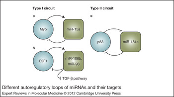
The question yet remains: does miR-181a act as a tumour suppressor or as an oncogene? It is sensible to regard its function in its complete setting, that is the context in which it is acting. However, one interesting possible explanation might lie in its own regulation. The complex and tight manner in which a cell regulates its pathways is further highlighted by regulation of miR-181a by p53 – its own target protein – by means of the p53-responsive element upstream of both of the miR-181a genes (Ref. Reference Boominathan60). Massive parallel sequencing of miRNAs on induction of p53 expression revealed interesting results about the effect of p53 on miR-181a expression, where the sequence corresponding to the complementary arms of its hairpin (miR-181a*) was expressed more abundantly than the mature strand itself, indicating a negative effect of p53 on mature miR-181a levels that needs to be further investigated (Ref. Reference Tarasov61). The existence of autoregulatory loops of miRNAs and their targets has been explored and modelled by Tsang and colleagues, who classified such autoregulatory loops into feedback and feedforward loops, or type I and type II circuits (Ref. Reference Tsang, Zhu and van Oudenaarden62). In a type I circuit, both miRNA and its target are copositively regulated; this type of loop probably functions to provide homeostasis and steady levels of proteins. Alternatively, a type II circuit represents an miRNA with a target that provides a positive-feedback loop (i.e. the miRNA negatively regulates its target, whereas its target directly or indirectly suppresses the transcription of the miRNA), which most likely works for the reinforcement of differentiation commands (Ref. Reference Nuorva63). Although it is presumed that most miRNAs function through each type of circuit depending on the cellular context, according to this classification the above-described relationship between miR-181a and p53 would represent a prototypical type II circuit (Figure 3), which as described by Tsang and coworkers converts a transient signal into a long-lasting cellular response, and thus erroneous switching on this type of circuit results in chronic pathological states, as observed in the diseases discussed above.
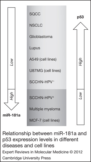
This negative effect can thus be used as a predictor of the levels and mode of action of miR-181a in different diseases, inversely depending on the status of p53 as shown in Figure 2. The neoplastic and autoimmune diseases, SQCC, NSCLC, glioblastoma and lupus, along with their model cell lines such as A549 and U87MG all showed downregulation of miR-181a and also accumulation of p53 protein (Refs Reference Nuorva63, Reference Fontanini64, Reference Gross65, Reference El-Sayed, Farag and Eissa66, Reference Kataoka67, Reference Xu68). More interestingly, SCCHN-HPV+ cell lines differed from their SCCHN-HPV− counterparts by possessing downregulated levels of miR-181a (Ref. Reference Miller41), and this difference is also observable in expression of p53, which was downregulated in HPV+ lines only (Ref. Reference Balz69). This relationship (Figure 3) holds in the other group of diseases possessing upregulated miR-181a levels – MM samples and MCF-7 cell lines – which express low levels of endogenous p53 (Refs Reference Markovic70, Reference Liu71). Additionally, the interesting results of the decrease in miR-181a levels in breast cancer cells on oestrogen stimulation (Ref. Reference Maillot43) can be probably explained by the triggered expression of endogenous p53 as a result of oestrogen signalling (Ref. Reference Bovenkerk, Lanciloti and Chandar72). Nature thus proves its unified stance even on the cellular level: even top regulators are eventually controlled by the base key players, just as in a typical closed ecosystem.
Relationship between miR-181a and p53 expression levels in different diseases and cell lines. miRNA and some of its target mRNA expression levels whether upregulated or downregulated with a consistent inverse association shown in different diseases and model cell lines. Diseases are ordered according to the sequence as mentioned in the text, with their relevant references. SQCC, squamous lung cell carcinoma; NSCLC, non-small-cell lung cancer; SCCHN-HPV−, squamous cell carcinoma of the head and neck (human papilloma-virus-negative); SCCHN-HPV+, squamous cell carcinoma of the head and neck (human papilloma-virus-positive).

Different autoregulatory loops of miRNAs and their targets. Type I and type II circuits. (a) miR-15a inhibits Myb expression, whereas Myb acts as a transcription factor that activates miR-15a expression (Ref. Reference Zhao118). (b) E2F1 expression is inhibited by and at the same time activates the expression of miR-106b and miR-93 (Ref. Reference Petrocca119). (c) p53 has a negative effect on miR-181a expression, which leads to further inhibition of p53.

Therapeutic implications and future directions
To date, the use of miRNAs has been suggested by many studies as biomarkers and as therapeutic targets for different diseases. However, few studies have tackled the design of such therapeutics. The main challenges in the design of therapeutics are (1) efficient delivery to the nucleus, (2) resistance of miRNA to degradation, (3) stability of the miRNA–target complex and (4) tissue specificity of miRNA delivery. Some of the approaches to downregulate the activity of miRNAs include the use of AMOs or antisense oligonucleotides with modified RNA backbones to escape cleavage by nucleases (Ref. Reference Esau73). Locked nucleic acids (LNAs) could also serve as highly promising options for knockdown of miRNAs because of their enhanced resistance to degradation and formation of a stable complex with their targets. Studies using LNAs have already shown long-term effects with low toxicity both in vitro and in vivo (Refs Reference Stenvang74, Reference Naguibneva75, Reference Orom, Kauppinen and Lund76). Cholesterol-conjugated RNA antagomirs were also tested recently and showed promising in vivo effects after intravenous injection into mice. The effects of inhibiting miRNAs using such antagomirs were observed in almost all tissues with the exception of the brain, which might be explained by the specificity of the blood–brain barrier (Refs Reference Esau77, Reference Krutzfeldt78).
Another possible therapy is miRNA sponges, which are inhibitor transcripts that bind the miRNA of interest. Their design includes several tandem binding sites to the miRNA of interest that enable even stronger binding to the miRNA than its targets (Ref. Reference Ebert, Neilson and Sharp79). False-positive miRNAs, also referred to as miRNA masks, are different from other approaches; their use is best suited to inhibit the specific effect of an miRNA on one of its targets rather than the holistic inhibition of the miRNA activity. This is achieved through the specific design of the oligodeoxynucleotides targeting the 3′UTR of the target mRNA and competing with the endogenous miRNA for target binding (Refs Reference Choi, Giraldez and Schier80, Reference Xiao81, Reference Hua82). Ectopic expression of downregulated miRNAs for therapy can be attempted by the use of miRNA mimics. However, the focal challenge in their use is in the development of a suitable delivery system. Viral vectors have been under investigation for a long time. However, one major disadvantage is their pathogenicity. Additionally, in vivo trials by intravenous injection showed limitations of the transfer to specific tissues (Ref. Reference Calin and Croce3). Other or polymer-based nanoparticles, such as 1,2-dioleoyl-sn-glycero-3-phosphatidylcholine, chitosan and ‘reconstituted high-density lipoprotein’ (Refs Reference Landen83, Reference Lu84, Reference Shahzad85). These carriers are better than other viral vectors because of their lack of toxicity and biodegradability. The nanoparticles can also carry ligands on their surfaces that allow tissue (or tumour) specificity and increase miRNA transfer.
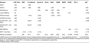
Even though miRNA-based gene therapy seems highly attractive as a mode of therapy because miRNAs are mass regulators of gene expression with a distinguishable deregulated signature in different disorders, an understanding of their networking relationships with target mRNAs, regulating transcription factors and miRNA processors needs further investigation before the design of a safe procedure that redirects the cell towards regularity without causing secondary disorders. Thus, dissection of these tight networks along with linking the expression signature of miR-181a in different diseases to the relevant clinical aspects should be a primary step for designing gene therapy of haematological disorders and solid cancers. Although a sharp correlation can be observed in p53 levels as a determinant to miR-181a levels, such a correlation, however, cannot also be assumed for the relationship of miR-181a with its targets (Table 1). This is nevertheless typical, because of the involvement of a wide range of miRNAs other than miR-181a in the control of such targets. Nonetheless, the control of p53 and its family is not merely limited to miR-181a, but also extends to other miRNAs (Ref. Reference Boominathan60), which makes it sensible to consider the correction of p53 family levels in therapy as a root solution to a multifactorial dilemma that is encountered in such diseases.
Comparison between the relative expression of miR-181a and some of its target mRNAs in different diseases and cell lines

miRNA and some of its target mRNA expression levels in existing literature whether upregulated, shown as ‘ + ’, downregulated, shown as ‘ − ’, or normal levels, shown as ‘N’, represented in different diseases and model cell lines with the relevant references (in parentheses). Methods of validation correspond to a, reporter assay; b, indirect targets through SIP1; c, knockdown experiments; d, overexpression experiments.
*Reporter assay to ZNF domains.
Acknowledgements
We express our gratitude to the referees who painstakingly reviewed this manuscript and whose valuable comments enriched it. We also thank the English Department of the German University in Cairo for help in revising the manuscript.