Introduction
Tonsillectomy and adenoidectomy are two of the most common paediatric surgical procedures performed. As with all surgical interventions, they are associated with some potential complications. However, complications during adenotonsillectomy surgery are rare.Reference Gallagher, Wilcox, McGuire and Derkay1–Reference Glade, Pearson and Zalzal3 In the last few years, different adenoidectomy techniques have been proposed to reduce morbidity and surgical risk, including those involving a microdebrider, bipolar coagulation or coblation.Reference Timms, Ghosh and Roper4–Reference Songu, Altay, Adibelli and Adibelli6 An ideal technique for adenoidectomy would be quick, easy to perform, achieve haemostasis, and minimise post-operative pain and bleeding complications.
Given the large number of adenoidectomies performed, surgeons must pay particular attention to the safety, accuracy and outcomes when choosing from the different surgical techniques. Previous studies have reported coblation tonsillectomy as advantageous over cold dissection, and coblation turbinoplasty has good outcomes in allergic rhinitis.Reference Di Rienzo Businco and Coen Tirelli7, Reference Di Rienzo Businco, Di Rienzo Businco and Lauriello8 Cold curettage potentially minimises morbidities associated with thermal injury. However, haemostasis after dissection is usually obtained either via monopolar or bipolar cautery; these create varying degrees of thermal injury to the surrounding tissue, which in turn may lead to increased post-operative pain. Electrocautery adenotonsillectomy typically affords greater operative speed and less operative blood loss, but may increase the chances of post-operative haemorrhage and pain.
More recently, several additional methods of adenoidectomy have been explored, largely related to advances in surgical instrumentation and equipment. These newer techniques include the use of a laser, harmonic scalpel, microdebrider or coblation wand. Many of these new technologies seek to provide operative speed and haemostasis, without thermal injury to the surrounding tissues imparted by electrocautery. Coblation technology has recently surfaced as a potentially appealing technology for adenotonsillectomy. Operating at much lower surface temperatures (40–70°C) than electrocautery, it enables ablation and tissue dissection, as well as haemostasis.Reference Stoker, Don, Kang, Haupert, Magit and Madgy9–Reference Parsons, Cordes and Comer12
The present study aimed to assess the efficacy and safety of an endoscopic surgical technique using the coblation device compared to cold curettage for adenoidectomy.
Materials and methods
This prospective non-randomised study was carried out for 1 year from June 2014 to June 2015 on 60 patients visiting the ENT department of our institution. The necessary permission and approval of the ethics committee and authority was granted before the study commenced. Informed written consent was obtained from the patients involved in the study according to the protocol approved by the ethics committee of our institution.
The study included all patients aged 5–12 years with a clinical diagnosis of adenoid hyperplasia that was confirmed by a plain X-ray lateral view of the skull. We excluded: patients aged less than 5 years of age or more than 12 years; patients with acute respiratory tract infection; and patients with coagulopathy or any major illness that was a contraindication to surgery.
Data were collected via patient interviews, clinical examination and relevant investigations. A quasi-experimental study design was used, as it was difficult to obtain surgical consent on blinding and randomisation. The patients selected for the study were given the choice of intervention, and segregated into two groups: one group underwent conventional cold curettage adenoidectomy (30 patients), and the other underwent endoscopic assisted coblation adenoidectomy (30 patients). The study does suffer from selection bias because of the lack of randomisation. All patients were operated on by the same surgeon under general anaesthesia, which was administered with the help of an orotracheal tube.
In the conventional cold curettage adenoidectomy technique, the patient was placed in the Rose position. The procedure was performed with a Boyle–Davis mouth gag and St Clair Thomson adenoid curette size 0–5, depending on the nasopharynx volume and area to be reached with the instrument. The adenoid curette with guard was introduced into the nasopharynx until its free edge touched the posterior border of the nasal septum, and was then pressed posteriorly to engage the adenoids. At this point, the head was slightly flexed to avoid injury to the odontoid process. Haemostasis was achieved by compression with a roller gauze pack. Electrocautery was never used in our standard conventional cold curettage adenoidectomy technique, to avoid serious and permanent thermal mucosal injury. Palpation with a finger was used to ensure removal of the adenoid and nasopharyngeal recanalisation.
Endoscopic assisted coblation adenoidectomy was performed using the Coblator II machine and Evac 70 wand system (ArthroCare, Sunnyvale, California, USA), which was malleable to reach the adenoids, designed for the ablation and dissection of soft tissue. The power level was set to 9 for adenoid ablation and to 5 for coagulation, depending on surgeon's preference as judged by the adenoid volume. A nasal endoscope (Karl Storz, Tuttlingen, Germany) was connected to a video camera for direct visualisation of the nasopharynx. A separate suction tube was attached to the coblation wand, and irrigation was provided with normal saline set on the automatic mode. After intubation and throat packing with roller gauze, a Boyle–Davis mouth gag was applied with a properly sized paediatric tongue depressor.
Endoscopic assisted coblation adenoidectomy was performed by putting the patient in a head-up position. The soft palate was retracted by passing a soft rubber catheter via the nasal cavity and tying it across the mouth gag. The nasal cavity was packed, using a solution of 4 per cent lignocaine with adrenaline for decongestion. The adenoid tissue was visualised by passing a 0° nasal endoscope. The scope size (2.7 mm paediatric or 4 mm adult) was determined by the age and the nasal passage of the patient. The oral cavity was kept open using a Boyle–Davis mouth gag. The coblation wand was bent at an angle of 20–30° in such a way that it could be passed under the soft palate, reaching the adenoid tissue.
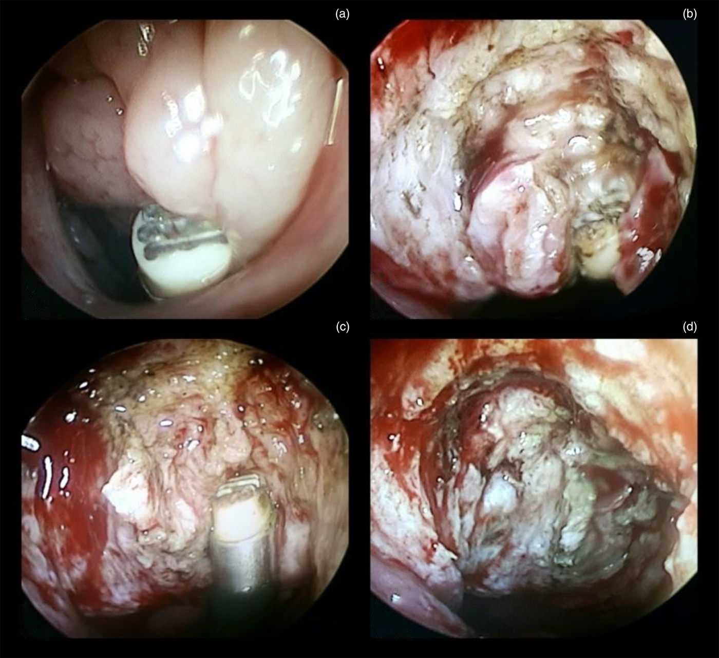
Coblation of the adenoid tissue was performed under visualisation (Figure 1). Adenoid tissue was ablated until the pre-vertebral fascia became visible. Adenoid tissue behind the tubal orifice was also ablated, without damaging the Eustachian tube opening. The absence of any bleeding or mucosal damage was verified. If necessary, bleeding areas were coagulated with the wand by touching the tip directly on the bleeding point for 2–3 seconds.
Coblation adenoidectomy: (a) before the procedure, (b, c) during the procedure, and (d) after the procedure.

Patients of both groups were given a course of antibiotic (amoxicillin plus clavulanate) and analgesic medications, along with saline nose drops, for 5 days post-operatively. Dosage of the analgesic was calculated according to the patient's weight (ibuprofen, 5 mg/kg every 8 hours). Patients were discharged after 24 hours in the event of no complications.
Patients were monitored during and after the procedure to assess: operative time, primary blood loss, post-operative residual tissue and post-operative pain. (1) Specifically, operating time was defined as the time taken for completion of the procedure, from the time the first instrument was introduced in the nasal or oral cavity until haemostasis was achieved after removing all packs. Blood loss was assessed by weighing the roller gauze pieces used to control the bleeding during and after the procedure. The dry weight of the gauze pieces was measured before the start of the surgery and the difference calculated (1 gm = 1 ml of blood loss). In addition, the suction machine fluid collected was subtracted from the total amount of normal saline used for irrigation. The completion of adenoid tissue removal was assessed by nasal endoscopy at the end of the procedure in both groups. Regarding post-operative pain, the Wong–Baker Faces Pain Rating Scale was completed by patients 24 hours after surgery and the pain assessed on a scale of 0–10.
Data were entered into a Microsoft Excel spreadsheet and a master table was created. Further analysis was conducted using IBM SPSS® statistical software, version 20. Relevant graphs and tables were used to present descriptive data. Statistical analysis was performed using the chi-square test and t-test.
Results
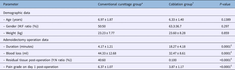
The patients’ ages ranged from 5 to 12 years. The mean age was 6.97 years in the conventional cold curettage adenoidectomy group and 6.33 years in the endoscopic assisted coblation adenoidectomy group. With a standard error of difference of 0.426, the two groups were comparable, as the difference was not statistically significant (p = 0.1389). There were 34 males (56.7 per cent) and 26 females (43.3 per cent), with a male to female ratio of 1:1. The gender difference was not statistically significant and the two groups were comparable. Patients’ mean weight was 23.23 kg (standard deviation (SD) = 7.77) in the conventional cold curettage adenoidectomy group and 23.60 kg (SD = 8.28) in the endoscopic assisted coblation adenoidectomy group. The weight distribution of the two groups was comparable, as the difference was not statistically significant (Table 1).
Summary of results for each intervention group

Data represent means ± standard deviations, unless indicated otherwise. *n = 30; †n = 30. ‡Indicates statistical significance. M = male; F = female; Y = yes; N = no
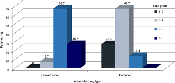
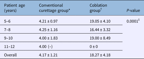
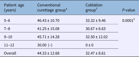
Examination of mean operation duration by age revealed that operative time was statistically significantly longer in the endoscopic assisted coblation adenoidectomy group, across all ages (Table 2). The mean blood loss was 44.33 ml in the conventional cold curettage adenoidectomy group and 32.47 ml in the endoscopic assisted coblation adenoidectomy group (Table 3); this difference was statistically extremely significant (p = 0.0001). The presence of residual tissue was completely absent in the endoscopic assisted coblation adenoidectomy group (p < 0.0001). The mean pain grade was 6.37 (SD = 1.07) in the conventional cold curettage adenoidectomy group and 3.87 (SD = 1.17) in the endoscopic assisted coblation adenoidectomy group (Figure 2). The pain grade was significantly lower in the endoscopic assisted coblation adenoidectomy group.
Pain grades for each adenoidectomy group on day 1 after surgery.

Operation duration for each group according to age

Data represent means ± standard deviations of adenoidectomy duration (in minutes), unless indicated otherwise. *n = 30; †n = 30. ‡Indicates statistical significance
Blood loss for each group according to age

Data represent means ± standard deviations of blood loss (in millilitres), unless indicated otherwise. *n = 30; †n = 30. ‡Indicates statistical significance
Discussion
Adenoidectomy is one of the most frequently performed operations in children. Though not considered a risky surgery, some complications do occur. The most important of these complications is bleeding, with incidence rates ranging from 0.5 to 8 per cent.Reference Tarantino, D'Agostino, Melagrana, Porcu, Stura and Vallarino13 The surgical technique used can have a considerable influence on intra-operative bleeding, post-operative pain and recovery time. Rare complications such as nasopharyngeal stenosis and Eustachian tube stenosis are difficult to treat if they occur.Reference Murray, Fitzpatrick and Guarisco14
Traditionally, adenoidectomy is performed with a curette. The main disadvantage of the conventional cold curettage adenoidectomy method is that it is a relatively blind technique which may lacerate the choanae and torus tubaris, or the nasopharyngeal mucosa. Alternatively, it may leave behind obstructing tissue, particularly at the Eustachian tube orifices, high in the nasopharynx and at intranasal protrusions.Reference Koltai, Kalathia, Stanislaw and Heras15
In an attempt to overcome the shortcomings of the curettage method, various adenoidectomy techniques have been devised. Suction diathermy ablation of the adenoid has been a popular alternative, and is reported to be safe, with minimal blood loss.Reference Owens, Jaramillo and Saunders16, Reference Wong, Moxham and Ludemann17 However, it comes with the risk of cicatrisation and burns to surrounding tissue, as does the use of the carbon dioxide laser, which in addition requires special precautions. Furthermore, suction diathermy ablation may not be able to remove intranasal adenoid tissue.Reference Havas and Lowinger18 Nasopharyngeal stenosis has been reported following adenoidectomy using a potassium titanyl phosphate laser.Reference Giannoni, Sulek, Friedman and Duncan19 Other methods described are radiofrequency adenoidectomyReference Shehata, Ragab, Behiry, Erfan and Gamea20 and use of an electronic molecular resonance tool.Reference Tarantino, D'Agostino, Melagrana, Porcu, Stura and Vallarino13 The microdebrider has been used extensively during endoscopic sinus surgery for tissue removal, with precision, avoiding orbital or intracranial entry.Reference Setliff21 Adenoidectomy using a microdebrider via a transnasal endoscopic approach has been described.Reference Havas and Lowinger18, Reference Yanagisawa and Weaver22 Others have used a mirror for visualisation, in place of an endoscope.Reference Murray, Fitzpatrick and Guarisco14, Reference Stanislaw, Koltai and Feustel23
The use of a rigid endoscope or sinuscope has its advantages. It enables good visualisation, allowing complete removal of adenoid tissue situated even high up in the nasopharynx and intranasally, without damaging surrounding structures. When used transnasally, there is no need to extend the neck, especially in patients with cervical spine instability.Reference Havas and Lowinger18 The camera attachment allows for a better magnified view on the monitor, facilitating recording and trainee teaching. The endoscopic adenoidectomy technique has been described using a rigid telescope for visualisation and forceps for adenoid removal.Reference Cannon, Replogle and Schenk24
Being a minimally invasive, low thermal technology for tissue removal, coblation technology is ideal for ENT procedures such as tonsillectomy, adenoidectomy, hypertrophic nasal turbinates reduction, laryngeal polypectomy, and soft palate and sleep apnoea surgery. While most radiofrequency based surgical products, such as lasers and electrosurgical devices, use imprecise heat-driven processes to remove or cut tissue (with temperatures ranging from 400°C to 600°C), coblation plasma technology creates a controlled, stable plasma field to precisely remove tissue at a low relative temperature (40–70°C), resulting in minimal thermal damage to surrounding soft tissues. Thermal penetration is around 2200 µm with conventional electrosurgical devices, but is less than 1100 µm with coblation devices.Reference Woloszko, Stalder and Brown25, Reference Stalder, Woloszko, Brown and Smith26
Plasma is formed by creating a high density energy field within an electrical conductive fluid, such as saline. A key to sustaining a stable plasma layer is the saline delivery system. Without integrated saline, plasma must be formed using the body's interstitial fluid, which dissipates quickly once energised. Coblation plasma technology uses bipolar energy to help control the amount of energy and heat delivered to the surrounding tissue. By combining bipolar energy with continuous saline delivery, these devices are able to create a stable plasma layer of only 100–200 µm thick around the active electrode. This allows for the precise excision of tissue, while minimising the heat that is transferred to the patient. It also makes sure there is a controlled amount of plasma supplied throughout the entire procedure.
Endoscopic assisted coblation adenoidectomy provides the advantages of both the telescope and the coblator. The Coblator II Surgery System is designed for ablation, resection, and coagulation of soft tissue and haemostasis of blood vessels in ENT surgery. In addition to the wide selection of plasma wands designed for specific ENT surgical applications, the complete system is composed of a controller, a flow control valve unit and cable, a foot control unit, and a hospital-grade power cord. The Evac 70 HP coblation wand is a versatile surgical instrument for the removal of tonsils and adenoids. The triple-wire active electrode configuration removes tissue in both tonsillectomy and adenoidectomy, and the integrated saline and suction ports allow for quick and easy operating theatre setup. The 6 inch (15.24 cm) malleable shaft allows improved access to the choana during adenoidectomy.
The present results demonstrate the ability of endoscopic assisted coblation adenoidectomy to ensure complete and safe removal of adenoid tissue. This is possible because of the small wand tip, which under endoscopic control is able to reach the most cranial part of the adenoid and the adenoid intranasal extension. Another benefit is the ability to use a single instrument to ablate and coagulate tissue. Endoscopic assisted coblation adenoidectomy also allows selective and precise ablation of the adenoid tissue, which is no longer blind but under direct 0° endoscopic vision. Because of the careful preservation of adjacent structures, such as Eustachian tubes, and nasal and pharyngeal mucosa, endoscopic assisted coblation adenoidectomy, as reported by others, is associated with less post-operative neck pain than curette or cautery adenoidectomy.Reference Timms, Ghosh and Roper4
In many countries, it has become common practice to undertake adenoidectomy as a day-care procedure. Despite concerns, a number of reports have demonstrated the safety of day-care adenoidectomy in children.Reference Heppard, Moir, Thomas and Narula27–Reference Siddiqui and Yung29 Treatment for pain following day-care surgery is a great challenge for medical staff. Patients should be pain-free and alert when leaving hospital. However, more than 80 per cent of children need pain medications at home.Reference Kokki and Ahonen30 Kotiniemi et al. showed that behavioural changes were common in children after day-care surgery, and were most common in children aged less than four years. Post-operative pain has proved to be an important factor in their occurrence.Reference Kotiniemi, Rhyanen and Mollanen31 We used ibuprofen in the dosage of 5 mg/kg, taken orally, every 8 hours. The pain grade was significantly lower in the coblation group.
The completeness of adenoid removal was assessed by nasal endoscopy at the end of the procedure in both groups. The difference in incidence of post-operative residual tissue between the two intervention groups was statistically significant. The authors have previously observed that the extent of resection is often incomplete, leading to no improvement in clinical condition or a recurrence.Reference Havas and Lowinger18 The results showed that resection was invariably complete with endoscopic assisted coblation adenoidectomy; in contrast, residual tissue was observed in 40 per cent of the conventional cold curettage adenoidectomy cases. This is comparable to the 39 per cent of cases reported as having residual obstructive adenoids by Havas and Lowinger.Reference Havas and Lowinger18
The main disadvantages of endoscopic assisted coblation adenoidectomy compared with conventional cold curettage adenoidectomy are: the need for a complete paediatric endoscopy setup in the operating theatre, the long preparation time and the decongestion of the nasal cavity with patties. It also involves a specific learning curve in paediatric endoscopy. Another shortcoming is that the coblated tissue is not available for histopathological examination. Furthermore, the equipment cost, with the disposable wand, should be considered. Moreover, we found that our technique requires good training to achieve proficiency.
• Mean operation duration was significantly higher for endoscopic assisted coblation adenoidectomy than conventional curettage adenoidectomy
• Mean blood loss was 44.33 ml for conventional adenoidectomy and 32.47 ml for endoscopic assisted coblation adenoidectomy (p = 0.0001)
• Mean pain grade was 6.37 for conventional adenoidectomy and 3.87 for endoscopic assisted coblation adenoidectomy (p < 0.0001)
• Residual tissue was absent in endoscopic assisted coblation adenoidectomy, whereas 40 per cent of conventional technique cases had adenoid tissue post-surgery
The surgical time was lower in the conventional cold curettage adenoidectomy group; an endoscopic approach to the rhinopharynx also requires nasal decongestion and careful checking for residual tags. However, the additional time aids in the accurate and complete removal of adenoid tissue, compared to ‘en bloc’ curettage. These findings should be considered in the surgical planning; scheduling a series of endoscopic assisted coblation adenoidectomies may save preparation time. Improvement in the learning curve of surgeons and nurses should lead to a reduction in the overall operative time.
Conclusion
Endoscopic assisted coblation adenoidectomy shows significant advantages over conventional adenoidectomy in terms of reduced blood loss, no post-operative residual tissue and a lower pain grade on day 1 post-operation. However, operation duration was significantly higher for endoscopic assisted coblation adenoidectomy. Demographic parameters such as age, gender and weight were comparable in both groups.
Endoscopic assisted coblation adenoidectomy needs to be acknowledged as an alternative to conventional cold curettage adenoidectomy. Its main advantage lies in achieving complete removal of adenoid tissue under vision. However, the need for special equipment and the cost of the procedure have to be considered.
Acknowledgement
I would like to thank the operation theatre staff of the hospital for their co-operation during the study.
Competing interests
None declared