

Nontuberculous mycobacteria (NTM) represent a diverse group of bacterial organisms found in the environment that can cause human disease. Because NTM infections are not reportable, the true burden of illness is unknown; however, recent estimates indicate that it is increasing in the US population.Reference Adjemian, Olivier, Seitz, Holland and Prevots1 Species of NTM have been found in both municipal drinking water distribution systems and the premise plumbing of homes and healthcare facilities and have been associated with true infections or pseudo-outbreaks.Reference Wallace, Brown and Griffith2 NTM have been isolated from waterpipe biofilm of hospital potable water systems, and several reports have described M. fortuitum in ice machines as sources of colonization or infection.Reference Laussucq, Baltch and Smith3–Reference Gebo, Srinivasan, Perl, Ross, Groth and Merz5 The rapid-growing mycobacteria comprising the Mycobacterium fortuitum clade (MFC) include M. boenickei, M. brisbanense, M. cosmeticum, M. conceptionense, M. farcinogenes, M. fortuitum, M. fortuitum subsp. acetamidolyticum, M. houstonense, M. mageritense, M. neworleanse, M. peregrinum, M. porcinum, M. senegalense, and M. septicum.Reference Rocchetti, Silbert, Gostnell, Kubasek, Campos Pignatari and Widen6 Differentiating species within the MFC can be challenging.
Herein, we describe 2 patient cluster investigations linking 3 separate NTM species to water sources at 3 Veterans Affairs medical centers (VAMCs).
Methods
Initial epidemiologic investigation, VAMCs A and B
A total of 4 patients in the medical intensive care unit (ICU) at VAMC B had sputum samples collected for acid-fast bacillus (AFB) culture between February 26, 2015, and March 15, 2015. Isolates were identified by matrix-assisted laser desorption/ionization time-of-flight (MALDI-TOF) mass spectrometry as M. farcinogenes/senegalense, an unusual NTM speciesReference Hamid7 not known to have been isolated previously at this facility. An internal case-control investigation was undertaken by the infection prevention and epidemiology team, revealing no linkages between nebulizer administration or the use of ipratropium bromide/albuterol sulfate.Reference Moritz, Merrick, Bleasdale and Sikka8 No recent construction or engineering problems had been associated with the ICU. All 4 patients had clinical symptoms of pulmonary NTM infection, and 2 had nodular opacities on chest radiograph. However, each had NTM recovered from only 1 sputum sample; thus, current American Thoracic Society (ATS)/Infectious Disease Society of America (IDSA) criteria for pulmonary infection with NTM were not met.Reference Griffith, Aksamit and Brown-Elliott9
Specimens for AFB processing and culturing from VAMC B were sent to a VA regional laboratory (VARL) 91 miles away in an adjacent state. The VARL was also responsible for processing AFB samples from 6 other VAMCs within a tristate region. In November 2015, this VARL notified hospital epidemiologists at VAMC B and VAMC A (located 11 miles away from VAMC B in the same metropolitan area) that cases of both M. farcinogenes/senegalense and M. conceptionense (genome-based taxonomic classification now considers these 2 species to be synonymous)Reference Tortoli, Meehan and Grottola10 had been isolated between April 21, 2015, and October 23, 2015, from respiratory samples of patients housed in the ICUs at both facilities. Thus, the total number of unique patients with either M. farcinogenes/senegalense or M. conceptionenseisolated between February 26, 2015, and October 23, 2015, at either facility was now 14: 6 from VAMC A and 8 from VAMC B. At this point, the Department of Veterans Affairs Public Health Surveillance and Research (PHSR) and Public Health Reference Laboratory (PHRL) were brought in to assist with the investigation.
Expanded case finding and environmental investigation, VAMCs A and B
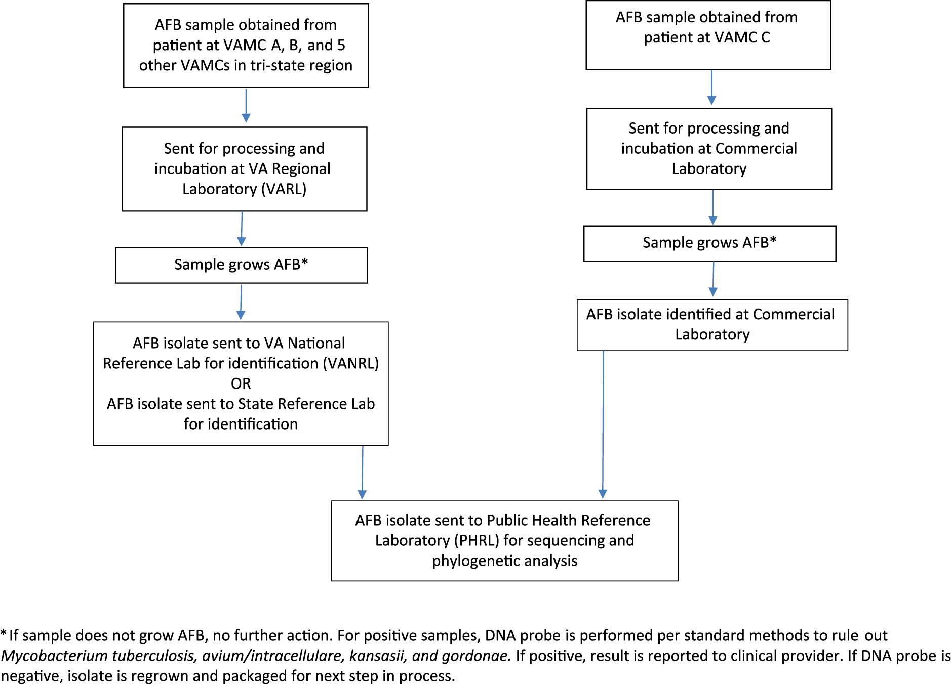
Figure 1 details the processing of patient samples for AFB culture. All samples were sent to the VARL for processing and culture, but not all were sent to the VA National Reference Laboratory (VANRL) for identification; a small number were sent to their state reference laboratory instead. Interestingly, the state utilized their internal protocol for NTM rpoB gene sequencing and identified isolates as M. conceptionense, but the VANRL identified all isolates as M. farcinogenes/senegalense using Bruker MALDI-TOF Biotyper system with Mycobacterium Library.
Acid-fast bacillus processing diagram.

To test a hypothesis that cross contamination of samples occurred at the VARL during processing, we requested that all 14 isolates as well as any clinical MFC isolates from any of the 5 other VAMCs other than A and B processed by and saved at VARL be sent to PHRL for genetic typing (hsp and rpoB gene sequencing, and comparison with reference MFC sequences to determine species and gene relatedness). Isolates were sent to PHRL from the earliest dates that the VARL had isolates stored (ie, 2012), and we monitored for new cases. The total number of MFC clinical isolates tested at PHRL was 58, with collection dates from October 9, 2012, to May 8, 2017: 27 from VAMC A, 21 from VAMC B, and 10 from 2 other VAMCs. To explore the possibility of an environmental source, VAMC engineering staff collected 1-L water samples from ICU sinks and ice machines: 34 from VAMC A in March 2016 and 12 from VAMC B in August 2016 and June 2017.
Epidemiologic investigation, VAMC C
Between March 23, 2018, and June 8, 2018, infection prevention and hospital epidemiology staff at VAMC C identified a cluster of 4 patients with 5 isolates of M. porcinum from clinical samples (1 blood and 4 respiratory). An additional 2 patients had M. porcinum isolated on November 23, 2017, and March 9, 2018, but isolates were not available for typing because they had been sent to a commercial reference laboratory for AFB culture and identification. Of 4 patient samples, 3 were collected on either of 2 acute-care medical units, and 1 was collected at a short-stay nursing home unit. VAMC C infection prevention staff obtained 100-mL water samples from various locations within the potable water system, tracing from the municipal source at which water enters the campus, the water-storage holding tank, to building entries, and sinks and ice machines on inpatient units. Samples were sent to a commercial laboratory certified for environmental testing. In total, 16 water isolates were retrieved from the commercial laboratory where they had been identified and sent along with patient isolates to PHRL for genetic typing.
Microbiologic methods
All clinical MFC isolates involved in the epidemiologic investigation were sent to PHRL from VARL or commercial laboratory on Lowenstein-Jensen slants. Isolates were individually analyzed by hsp and rpoB gene sequencing. DNA was extracted from biomass collected from each slant using Qiagen DNA Blood and Tissue Kits (Qiagen, Chatsworth, CA). From purified DNA, amplicons (720 bp for rpoB, 330 bp for hsp) were generated using Expand High-Fidelity DNA polymerase (Roche Applied Sciences, Indianapolis, IN). The nucleotide sequence of each amplicon was then determined by standard dideoxyterminator sequencing of both the positive and negative DNA strands, and the 2 reads were assembled, edited, and trimmed using Geneious software (Biomatters, San Francisco, CA). To determine species and genetic relatedness between isolates, neighbor-joining phylogenetic trees of PHRL-determined sequences and reference sequences downloaded from GenBank (www.ncbi.nlm.nih.gov/genbank/) were created in Geneious software (Biomatters, Newark, NJ) using Jukes-Cantor distance models and 100 bootstrap replicates. Susceptibility testing was performed on NTM isolates using Sensititre Mycobacterium minimum inhibitory concentration (MIC) plates (Thermo Fisher Scientific, Waltham, MA) according to manufacturer’s instructions.
We qualitatively cultured 1-L water samples from VAMCs A and B at PHRL for NTM using published methods.Reference Covert, Rodgers, Reyes and Stelma11 PHRL received isolates from VAMC C, which had been processed for AFB culture and identification according to standard procedures by a commercial laboratory.12 Recovered NTM isolates were identified using a Bruker Daltonic MALDI Biotyper system (Bruker Scientific, Billerica, MA) 6903, 7331, 7854, with RUO Mycobacterium Library version 5.0, and hsp and rpoB gene sequencing.
Clinical data
We reviewed the electronic health records (EHRs) of patients for demographic data, patient location at time of specimen collection, antimicrobial agents received, predisposing medical conditions, and clinical outcome at 30 days and 1 year after NTM isolation. Classification of pulmonary infection was based on ATS/IDSA criteria: microbiologic results, radiographic findings, and clinical evidence for NTM lung disease in the absence of appropriate exclusion of other diagnoses.Reference Griffith, Aksamit and Brown-Elliott9 The Council for State and Territorial Epidemiologists (CSTE) standardized case definition of nontuberculous extrapulmonary mycobacteria infections was used for nonpulmonary infections.Reference Bancroft, Shih and Cassidy13 We classified infections as healthcare-associated using Centers for Disease Control and Prevention National Healthcare Safety Network (NHSN) Patient Safety Component Manual.14
Results
Initial and expanded epidemiologic investigation, VAMCs A and B
Of 58 clinical MFC isolates tested at PHRL for genetic relatedness, 42 identical M. conceptionense isolates were associated with 41 unique patients whose clinical samples were collected between October 9, 2012, and May 8, 2017, including 27 originally identified as M. farcinogenes/senegalense or M. conceptionense and 14 as M. fortuitum. Of these, 39 were an identical strain (25 from VAMC A, 14 from VAMC B) of M. conceptionense that matched a strain isolated from an ice machine on VAMC A’s medical ICU (Fig. 2, trees 1 and 2). None of the other 45 water samples from either facility contained the endemic strain.
Phylogenetic trees. Trees 1–4: Neighbor-joining phylogenetic trees created from PHRL-determined sequences and reference sequences downloaded from GenBank using Jukes-Cantor distance models and 100 bootstrap replicates. Phylogenetic Tree 1. rpoB phylogenetic tree of Mycobacterium fortuitum clade (MFC) isolates identified from VAMCs A, B, and D. Phylogenetic Tree 2. hsp phylogenetic tree of Mycobacterium fortuitum clade (MFC) isolates identified from VAMCs A, B, and D. Phylogenetic Tree 3. rpoB phylogenetic tree of Mycobacterium fortuitum clade (MFC) isolates identified from VAMC C. Phylogenetic Tree 4. hsp phylogenetic tree of Mycobacterium fortuitum clade (MFC) isolates identified from VAMC C.

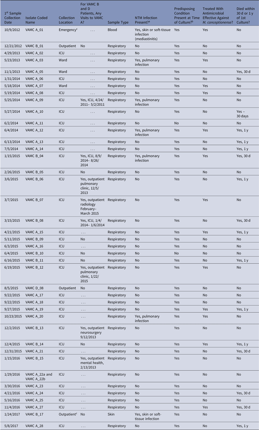
The median age of patients with M. conceptionense was 65 years (range, 50–86); 100% were male; and 34 of 41 (83%) patient samples were collected in the ICU (Table 1). The median number of days in the ICU until AFB culture was 3 (range, 1–24). Only 1 patient was on a ventilator at the time of culture. Of 7 patients with cultures not collected in the ICU, 2 had had a prior ICU stay. Also, 8 VAMC B patients had visits to VAMC A prior to M. conceptionense isolation, 3 of which included an ICU admission (Table 1). Among 41 patient specimen types, 39 (95%) were respiratory, and 5 (12%) M. conceptionense cases met the ATS/IDSA criteria for pulmonary NTM infection. An additional 2 cases had nonpulmonary infection with M. conceptionense: 1 patient with healthcare-associated mediastinitis post-CABG at VAMC A (previously reported)Reference Oda, Ryono and Lucero-Obusan15 and 1 skin or soft–tissue infection in a patient with paraneoplastic Sweet’s syndrome. Both patients were not in the ICU at time of culture but had had prior ICU stays. The remaining 34 of 41 patients (83%) were colonized with M. conceptionense, based on ATS/IDSA criteria. Conditions predisposing for NTM infection were present in 39 of 41 (95%), and 13 of 41 (32%) were administered antimicrobial agents with activity against M. conceptionense, 5 of whom met criteria for NTM infection (4 pulmonary, 1 mediastinitis). Furthermore, 7 patients (17%) died within 30 days of M. conceptionense isolation, and 16 (39%) within 1 year, regardless of treatment status (Table 1). Of the 16 who died within 1 year, 2 met criteria for NTM pulmonary infection.
Characteristics of Veterans With Mycobacterium conceptionense (N = 41)

Note. ICU, intensive care unit; NTM, nontuberculous mycobacteria; VAMC, Veterans’ Affairs medical center.
a Infection categorizations based upon American Thoracic Society/Infectious Disease Society of America official statement criteria for nontuberculous mycobacterial lung disease (Griffith et al,Reference Griffith, Aksamit and Brown-Elliott9 2007), and the Council for State and Territorial Epidemiologists (CSTE) standardized case definition of nontuberculous extrapulmonary mycobacteria infections for nonpulmonary infections (Bancroft et al,Reference Bancroft, Shih and Cassidy13 2018).
b NTM predisposing conditions include: bronchiectasis, pneumoconiosis, cystic fibrosis, COPD, HIV, cancer, tobacco use, genetic factors associated with NTM, steroid therapy, prior TB, esophageal motility disorders, peritoneal dialysis, transplant.
c Patient(s) had a previous ICU stay.
Among these 41 patients, 3 had a strain of M. conceptionense closely related to the VAMC A and B endemic strain (differing by 3 nucleotides, or 0.4%, on the rpoB gene). One isolate (from blood) was collected from a patient readmitted to VAMC A after coronary artery bypass grafting (CABG), another (a skin biopsy sample) was from VAMC B’s dermatology clinic, and the third (a respiratory sample) was from the pulmonary clinic of VAMC D, located 91 miles from VAMCs A and B and serviced by VARL. The latter 2 patients were outpatients at the time of sample collection.
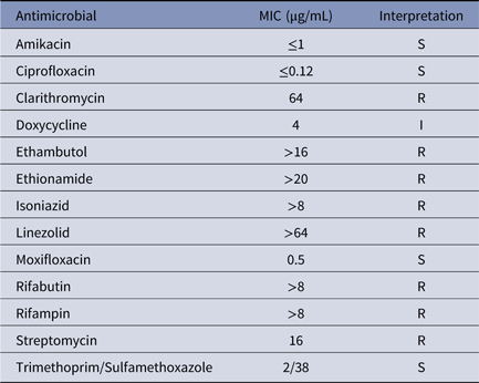
Susceptibility testing performed on 4 random endemic M. conceptionense strains indicated that these isolates were susceptible only to quinolones, amikacin, and trimethoprim/ sulfamethoxazole and were resistant to macrolides, rifabutin, ethambutol, ethionamide, and linezolid according to Clinical and Laboratory Standards Institute (CLSI) guidelines (Table 2).16
Mycobacterium conceptionense Susceptibility Testing

Note. MIC, minimal inhibitory concentration; S, susceptible; I, intermediate resistance; R, resistant (according to Clinical and Laboratory Standards Institute guidelines).
We discovered incidentally that an ice machine from the VAMC B ICU sampled on August 4, 2016, grew M. mucogenicum (Fig. 2, tree 1). Thereafter, we sought any patients with clinical isolates of M. mucogenicum and added them to the total NTM tested at PHRL (2 additional isolates). One M. mucogenicum isolate was identified from a respiratory source of a patient from VAMC B, but we were unable to be type it. Upon EHR review, this M. mucogenicum case met ATS/IDSA criteria for NTM pulmonary infection; however, at the time of culture, this patient was not housed in the VAMC B ICU where the ice machine was located. This patient’s only previous VAMC B ICU stay was 16 months prior. Interestingly, a patient from VAMC A had the identical strain of M. mucogenicum isolated from blood as the VAMC B ice machine strain, despite never having been housed at that facility. This patient was diagnosed as having a healthcare-associated M. mucogenicum central-line–associated bloodstream infection and was treated with azithromycin. Both patients died within 1 year of M. mucogenicum isolation.
Mitigation strategies were employed at affected facilities to prevent further NTM colonization and infection among patients. In VAMC A, the ICU ice machine was removed on November 29, 2016, and was replaced with a new one on December 2, 2016. The VAMC B ICU ice machine was replaced on February 3, 2017 (after most M. conceptionense cases had occurred but prior to any of the M. mucogenicum cases). To our knowledge, bacteriological retentive filters were not placed on the water inlet lines of any of the ice machines. Facilities were required to follow manufacturers’ recommendations for cleaning and disinfection, although documentation was not available. Incidentally, ongoing Legionella control strategies, including hyperchlorination and thermal eradication, were employed by all 3 facilities in compliance with VA policy for control of Legionella in facility water systems.17
Epidemiologic and environmental investigation, VAMC C
In Fig. 2, trees 3 and 4 show results of phylogenetic testing performed on VAMC C patient and environmental NTM isolates. All 5 M. porcinum patient isolates and environmental isolates from 4 patient room sinks in the 2 buildings where patients had been housed were indistinguishable based on sequences of hsp and rpoB genes. Of 4 patients with respiratory M. porcinum, 3 isolates did not meet ATS/IDSA criteria for NTM pulmonary infection and were considered colonized. The fourth had healthcare-associated bloodstream infection, due to a peripherally inserted central catheter, which resolved without antimicrobial treatment once the catheter was removed. Other NTM isolated from potable water samples included M. massiliense, M. chelonae, and M. fortuitum. No patients were found to have M. massiliense; 8 patients had M. fortuitum or M. chelonae isolated from clinical samples (7 respiratory, 1 skin) during the investigation period; however, isolates were not available for typing. The 7 respiratory isolates did not meet ATS/IDSA criteria for infection. The patient with M. fortuitum/chelonae isolated from skin was an outpatient with chronic, draining abdominal infection at the site of insulin injections, thought to be associated with swimming at a non-VA rehabilitation center pool.
Mitigation strategies were performed at VAMC C in part to address concomitant Legionella potable water colonization issues. These included hyperchlorination, replacement of patient shower heads and sink faucets, and replacement of point-of-use mixing valves to ensure maintenance of appropriate water temperature.
Discussion
We retrospectively identified an endemic strain of M. conceptionense isolated from 41 patients over a 5-year period at 2 VAMCs in the same metropolitan area and an additional VAMC located 91 miles away. Most patients were considered colonized; however, 7 (17%) patients were infected with this M. conceptionense strain. All patients who died within 30 days after M. conceptionense was isolated had significant comorbidities, making the role of M. conceptionense in contributing to death unclear. Little has been published on the infectious potential of M. conceptionense, although several case reports have described soft-tissue, pulmonary, and bloodstream infections.Reference Adekambi, Stein, Carvajal, Raoult and Drancourt18–Reference Yaita, Matsunaga and Tashiro25
Despite removal of the VAMC A ICU ice machine that grew M. conceptionense in 2016, an additional case with the identical strain of M. conceptionense occurred on May 8, 2017, in a patient housed in the VAMC A ICU. No definitive M. conceptionense water source for VAMC B was found. However, 8 of 15 VAMC B patients had prior visits to VAMC A; thus, colonization due to direct exposure to M. conceptionense in the VAMC A water supply cannot be ruled out. No reports of this NTM species had been reported by other non-VA hospitals in the local area (Chicago Department of Public Health personal communication). However, methods used to identify NTM isolated in these hospitals are unknown to us. Our hypothesis that VARL had contaminated samples during processing was unlikely, given that 40 cases were from VAMCs A and B and only 1 from VAMC D. M. conceptionense could be more common than previously thought in municipal water supplies. In addition, past clinical identification methods, low environmental NTM sampling frequency and method variability among laboratories may have underestimated its presence.
Potable water in healthcare settings has been implicated as a source for NTM infection or colonization.Reference Laussucq, Baltch and Smith3, Reference Gebo, Srinivasan, Perl, Ross, Groth and Merz5, Reference Fernandez-Rendon, Cerna-Cortes and Ramirez-Medina26–Reference Kline, Cameron and Streifel30M. conceptionense has been reported in suburban potable water sources in Iran but was not found in human clinical isolates.Reference Velayati, Farnia and Mozafari31M. fortuitum and M. porcinum have both been associated with outbreaks and pseudo-outbreaks at medical centers associated with ice machines,Reference Laussucq, Baltch and Smith3, Reference Gebo, Srinivasan, Perl, Ross, Groth and Merz5, Reference Brown-Elliott, Wallace and Tichindelean32 and M. mucogenicum bacteremia has been linked to hospital water supplies.Reference Fernandez-Rendon, Cerna-Cortes and Ramirez-Medina26, Reference Kline, Cameron and Streifel30, Reference Livni, Yaniv and Samra33
Our study has several limitations. Not all environmental and/or water sources at these VAMCs were cultured nor were swabs obtained from sink drains or shower heads, so other hospital water NTM sources could not be definitively ruled out. Environmental samples were collected after many of the patient cases had already occurred. Our investigation was retrospective; thus, we did not have access to pharmaceuticals or other possible common environmental exposures for culture. We did not have access to all NTM isolates ever processed by VARL; therefore, expanded case findings were incomplete. Mitigation strategies were performed by VAMCs in part to control Legionella growth in water systems. Hyperchlorination, commonly used for Legionella control, may do little to control NTM in water supplies as mycobacteria are known to be resistant to chlorine or chloramines.Reference Falkinham34 We did not conduct case-control investigations across affected VAMCs. Because we could not ascertain from EHR documentation any degree of specificity regarding water sources patients may have encountered, specific risk factors associated with infection or colonization were not analyzed in detail. In some cases, only 1 sputum culture was obtained, leading to potentially incorrect classification as colonization versus infection based on ATS/IDSA criteria. Underlying illness and antimicrobial treatment made it impossible to attribute severity of illness to M. conceptionense in many patients. Next-generation sequencing analyses were not performed, which would have provided more granular detail than Sanger gene sequencing to compare species and strain variation.
This investigation highlights the ubiquitous nature of NTM in healthcare facility water systems, and the variation in results obtained in identifying NTM species using different microbiologic methods.Reference Brown-Elliott, Fritsche and Olson35, Reference Busalacchi, Tans-Kersten and Warshauer36 Taxonomic changes to the genus Mycobacterium have been proposed that may make interspecies identification more straightforward.Reference Gupta, Lo and Son37
Early and accurate identification of environmentally associated clusters of NTM supports epidemiologic investigation and potential development of mitigation strategies. However, while epidemiologic linkage of NTM cases to hospital water sources is possible, successful water management programs can reduce, but not eliminate, NTM burden from healthcare system water supplies.Reference Gebo, Srinivasan, Perl, Ross, Groth and Merz5, Reference Kline, Cameron and Streifel30, Reference Williams, Chen and Keane38–Reference Decker and Palmore40
Acknowledgments
We thank the numerous infection prevention, microbiology laboratory, industrial hygiene, and engineering staff who contributed to the investigations at these medical centers. The views and opinions expressed are those of the authors and do not necessarily reflect those of the Department of Veterans Affairs or the US government.
Financial support
This study was funded by intramural funds at the Department of Veterans Affairs.
Conflicts of interest
All authors report no conflicts of interest relevant to this article.