INTRODUCTION
The phylum Apicomplexa includes significant and widespread unicellular pathogens of humans and animals and one of these is the genus Cryptosporidium that is the causative agent of zoonotic disease of the gastrointestinal and respiratory tract, called cryptosporidiosis. In general, the progress of infection in immunocompetent individuals is mild, without fatal consequences, and usually self-curing or self-limiting (Chappell et al. Reference Chappell, Okhuysen, Sterling, Wang, Jakubowski and Dupont1999). The gastric species Cryptosporidium muris parasitizes epithelial cells in the glandular part of the gastric mucosa and was first described in mice by Tyzzer (Reference Tyzzer1907) as a Cryptosporidium species. Uni et al. (Reference Uni, Iseki, Maekawa, Moriya and Takada1987) extended his work and provided new morphometrical data of its developmental stages. Although C. muris is considered to be a predominantly rodent species, occasionally it can be transmitted from its natural host to other animals (Aydin and Ozkul, Reference Aydin and Ozkul1996; Hůrková et al. Reference Hůrková, Hajdúšek and Modrý2003; Pavlásek and Ryan, Reference Pavlásek and Ryan2007; Lupo et al. Reference Lupo, Langer-Curry, Robinson, Okhuysen and Chappell2008) and possibly humans (Katsumata et al. Reference Katsumata, Hosea, Ranuh, Uga, Yanagi and Kohno2000; Guyot et al. Reference Guyot, Follet-Dumoulin, Lelievre, Sarfati, Rabodonirina, Nevez, Cailliez, Camus and Dei-Cas2001; Gatei et al. Reference Gatei, Ashford, Beeching, Kamwati, Greensill and Hart2002; Tiangtip and Jongwutiwes, Reference Tiangtip and Jongwutiwes2002; Palmer et al. Reference Palmer, Xiao, Terashima, Guerra, Gotuzzo, Saldias, Bonilla, Zhou, Lindquist and Upton2003).
The general life cycle, which is well documented for Cryptosporidium parvum parasitizing the intestine (Current and Reese, Reference Current and Reese1986), includes four phases of development: excystation, merogony, gametogony and sporogony. Each phase occurs in a particular chronological period and depends on both the species of Cryptosporidium as well as the host. Diversity in patent and prepatent periods has been reported from various animals (Tarazona et al. Reference Tarazona, Blewett and Carmona1998; Hijjawi et al. Reference Hijjawi, Meloni, Morgan and Thompson2001; Fayer and Santin, Reference Fayer and Santin2009). The infective oocysts are transmitted by the fecal–oral route. The invasive sporozoites, being released from oocysts inside the host, evolve to trophozoites and multiply in the asexual phase involving two types of merogony. Type II merogony produces four merozoites and has been recorded in C. parvum and Cryptosporidium wrairi but not confirmed in C. muris. The sexual part of the life cycle, gametogony, is typified by the occurrence of numerous macrogamonts and microgamonts that produce 16 non-flagellated microgametes. After macrogamont fertilization, the zygote is enveloped by a wall and matures into the oocyst, which sporulates inside the host. A few studies report the existence of two types of oocysts, i.e. thick- and thin-walled (Current and Reese, Reference Current and Reese1986; Uni et al. Reference Uni, Iseki, Maekawa, Moriya and Takada1987).
Due to lack of efficient treatment of cryptosporidiosis, further knowledge on cryptosporidia biology and host–parasite interactions, depending on the host immunological status, is still required. Our aim was to map the complete life cycle of C. muris in two laboratory rodents, BALB/c mice and the southern multimammate rat Mastomys coucha, and to verify the presence of Type II merogony and thin-walled oocysts during the life cycle of C. muris, using a combined microscopic approach, i.e. electron microscopy as well as observations on native and stained preparations using light microscopy (LM), and supplemented with molecular detection. We also attempted to record and re-evaluate the stage-dependent characteristics depicted in the detailed drawings of Tyzzer (Reference Tyzzer1910) and for this reason, we have followed the techniques used in his study. Since the endogenous developmental stages of C. muris attack the stomach tissue, we expected to find some differences in its life cycle when compared with the findings for C. parvum.
MATERIALS AND METHODS
Laboratory animal models
The southern multimammate rat (M. coucha; the Institute of Parasitology Biology Centre, ASCR, v.v.i. in České Budějovice) and BALB/c mice (Anlab s.r.o, Czech Republic) were used for the purposes of this study. Animals were housed in plastic cages (5 animals per cage) with sterile wood-chip bedding and supplied with sterilized food and water ad libitum. The rearing of animals is regulated by Czech legislation (Act No 246/1992 Coll., on protection of animals against cruelty). These documents are consistent with the legislation of the European Commission. All housing, feeding and experimental procedures were conducted under protocols approved by the Institute of Parasitology, Biology Centre of the Academy of Sciences of the Czech Republic and Central Commission for Animal Welfare, Czech Republic (Protocol # 066/2010). The minimum number of animals has been involved to produce statistically reproducible results.
Parasite
Cryptosporidium muris strain TS03 originating from an East African mole rat (Tachyoryctes splendens) previously characterized by Kváč et al. (Reference Kváč, Sak, Květoňová, Ditrich, Hofmannová, Modrý, Vítovec and Xiao2008, Reference Kváč, Kodádková, Sak, Květoňová, Jalovecká, Rost and Salát2011) was used for all experiments in this study. Over 10 years the C. muris TS03 has been passaged through susceptible laboratory hosts, M. coucha and SCID mice, in the Institute of Parasitology Biology Centre, ASCR, v.v.i. in České Budějovice. Fresh rat feces were collected regularly every morning. The presence of oocysts in feces was verified using the staining method according to Miláček and Vítovec (Reference Miláček and Vítovec1985). Oocysts were purified using Sheather's sugar flotation method (Arrowood and Sterling, Reference Arrowood and Sterling1987) and modified caesium chloride gradient centrifugation (Kilani and Sekla, Reference Kilani and Sekla1987). Obtained oocysts of C. muris were either immediately used for experimental inoculations of laboratory animals or mixed with aqueous potassium dichromate (2·5% w/v, final concentration) and stored at 4 °C. Oocysts less than 1 week old were exclusively used for the experiment in this study. Before their use, the residue of aqueous potassium dichromate was removed by centrifugation at 1000 g for 10 min, the supernatant was aspirated, and the pelleted oocysts were resuspended in distilled water. This procedure was repeated three times.
Experimental inoculations and parasitological dissection of laboratory rodents
Rodents, 8–10 weeks old, were inoculated per os using the oesophagus gavage with a dose of 106 viable oocysts of C. muris that had previously been checked for their viability via propidium iodide staining (Sauch et al. Reference Sauch, Flanigan, Galvin, Berman and Jakubowski1991). Inoculations of both rodent models were performed simultaneously. Animals (one per host model), were euthanased by cervical dislocation at 0·5, 1, 6 and 12 h post infection (hpi) and then consecutively every 12 h until 11 dpi, and onwards at 12, 13, 14, 18, 21 and 28 dpi. In M. coucha, additional parasitological examination was performed, of animals kept for 4, 8 and 18 months, and 2 years after inoculation. The negative controls of both models, BALB/c mice and M. coucha, were prepared to evaluate pathological alterations in gastric tissue induced by cryptosporidiosis. A total of 33 individuals of BALB/c mice and 37 individuals of M. coucha were examined. In subsequent repetition of the experiment, performed to detect cryptosporidia during the early phase of parasitization, the infective doses were increased to 108 oocysts of C. muris per animal. Animals were dissected at selected time points after inoculation (a total of 8 individuals of BALB/c mice and 17 individuals of M. coucha).
Excision of the stomach, consisting of both the glandular and non-glandular part was performed. As endogenous developmental stages of C. muris are known to be restricted to the glandular part, this area of the gastric tissue was divided lengthways through the curvatura major and then transversely. The resulting four sections were processed using the various approaches described in the following subchapters. Differences between the course of parasitization in groups of BALB/c mice and M. coucha were tested by the non-parametric Mann–Whitney U test. Testing was performed using the software Statistica® 10.0 (StatSoft CR, Prague, Czech Republic).
Native and stained preparations
As the first step, the blood serum of both BALB/c and M. coucha was obtained from non-infected controls. A total of 10 μL (BALB/c) or 30 μL (M. coucha) of adequate sera were subsequently added to the processed gastric tissue in order to release the C. muris developmental stages. Native preparations, prepared from suspensions obtained from scrapings of the gastric luminal epithelium, were evaluated immediately via LM and checked for the presence of cryptosporidia. Preparations with gastric scrapings were also stained with Lugol's iodine. The protocols according to Giemsa and Wright staining procedures (Tyzzer, Reference Tyzzer1910) were used to visualize the characteristic morphological features of individual C. muris developmental stages. For this purpose, specimens were fixed with a vapour of 2% osmium tetroxide (v/v). Slightly modified staining procedures followed Tyzzer (Reference Tyzzer1910). Preparations were viewed and documented using the light motorized microscope Olympus BX61 equipped with phase contrast optics and Olympus BX51 with Nomarski interference-contrast microscopy (DIC).
Electron microscopy
Samples of gastric tissue were fixed in freshly prepared 3% glutaraldehyde (v/v) in cacodylate buffer and further processed for transmission (transmission electron microscopy (TEM)) and scanning electron microscopy (SEM) as described elsewhere (Valigurová et al. Reference Valigurová, Hofmannová, Koudela and Vávra2007, Reference Valigurová, Jirků, Koudela, Gelnar, Modrý and Šlapeta2008). The grids for TEM were coated with Formvar film at a thickness of 15–25 nm. Ultrathin sections transferred to the Formvar coated copper grids were evaluated using a JEOL 1010 transmission electron microscope. Micrographs from SEM were performed by JEOL JSM-7401F. In the case of negative results obtained during the early phase of infection, samples of gastric tissue already viewed under SEM were broken into smaller pieces to expose the interior of the closed gastric pits, were re-coated with gold and re-evaluated.
Histology
Gastric tissue was fixed in AFA (alcohol – formalin – acetic acid) solution and processed as described previously (Valigurová et al. Reference Valigurová, Jirků, Koudela, Gelnar, Modrý and Šlapeta2008). The blocks in Histoplast II were cut using a Zeiss Hyrax M 300 rotary microtome. The 7 μm thick sections were stained with haematoxylin–eosin (H&E). Preparations were viewed and documented using the light motorized microscope Olympus BX61.
Molecular method; nested PCR
This method was chosen to verify the presence (or absence) of specific DNA of developmental stages of C. muris in the stomachs of both animal models during the early phase of parasitization (up to 3 dpi). In addition, this approach was essential to disprove the localization of C. muris developmental stages in other organs such as the gall bladder, pancreas, oesophagus, spleen, liver, Peyer's patches, duodenum, jejunum, ileum, colon, lung and kidney.
Total DNA was extracted from tissue by bead disruption for 60 s at 5·5 m s−1 using 0·5 mm glass beads in a FastPrep®–24 Instrument (MP Biomedicals, CA, USA), followed by isolation/purification using a commercially available kit according to the manufacturer's instructions (DNeasy Blood and Tissue Kit, Qiagen, Hilden, Germany). Purified DNA was stored at −20 °C prior to being used for PCR. The presence of C. muris was confirmed by sequence analysis of the SSU gene using previously described methods (Jiang et al. Reference Jiang, Alderisio and Xiao2005). The positive (sample containing DNA of C. hominis) and negative control were included in each PCR. Secondary PCR products were detected by agarose gel electrophoresis (2·0%) and visualized by ethidium bromide staining (0·2 μg mL−1).
RESULTS
The effect of parasitization by C. muris on laboratory rodents
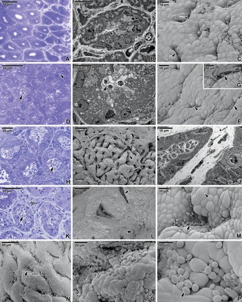
Laboratory rodents exhibited significantly different responses to parasitization and chronology of pathological changes of gastric tissue induced by the parasite, in contrast to the negative control of gastric tissue (Figs 1A–C and 2A, B); nevertheless, the sequence of individual changes during the acute phase of parasitization corresponded in both models. At the beginning of parasitization, the gastric tissue of both host models was irregularly affected by endogenous developmental stages of C. muris in an island-like manner and individual parasitized gastric pits were surrounded by areas of healthy tissue (Figs 1D, F and 2D, F). This specific feature of C. muris hampered the localization of parasites in host stomachs and made the detection of its earliest developmental stages almost impossible in both rodent models. Therefore, a variety of different microscopic approaches along with nested PCR were necessary to confirm the presence or absence of C. muris developmental stages during the early phase of infection.
Pathology of gastric cryptosporidiosis in BALB/c mice. (A) Cross-section of gastric glands; negative control. LM (Histology, H&E). (B) The surface of the gastric epithelium showing empty gastric pits (arrow). Inset shows a detailed view inside an empty gastric pit; negative control. SEM. (C) Cross-section of a gastric gland; negative control. TEM. (D) Longitudinal section of gastric epithelium; gastric pits (arrow) parasitized by C. muris (arrowhead) at 5·5 dpi. LM (Histology, H&E). (E) Cross-section of a gastric gland with C. muris individuals at 3 dpi. TEM. (F) Surface of the gastric epithelium showing gastric pits filled with C. muris (arrowhead) at 7 dpi; an apparently empty gastric pit (arrow). Inset shows a detailed view inside the gastric pit filled by C. muris developmental stages. SEM. (G) Cross-section of enlarged gastric glands after 8·5 dpi (arrow). LM (Histology, H&E). (H) Gastric pits completely filled by C. muris (arrow) at 8·5 dpi; the surface is covered by a mucus layer (asterisk). SEM. (I) Cross-section of a gastric gland completely filled with various developmental stages of C. muris at 8 dpi. TEM. (J) Longitudinal section of a stomach showing parasitized gastric pits (arrow) and glands (arrowhead) at 13 dpi. LM (Histology, H&E). (K) The folds of the enlarged gastric epithelium (arrowhead); C. muris developing at the surface of epithelium (arrows) at 13 dpi. SEM. (L) Hypertrophy and hyperplasia of the gastric tissue; numerous developmental stages of C. muris (arrow) at 13 dpi. SEM.

Pathology of gastric cryptosporidiosis in Mastomys coucha. (A) Cross-section of gastric glands; negative control. LM (Histology, toluidine blue). (B) Cross-section of the gastric gland; negative control. TEM. (C) Surface of the gastric epithelium at 1 dpi; gastric pits (arrowhead). SEM. (D) Cross-section of the parasitized gastric gland (arrow) surrounded by non-parasitized glands (arrowhead) at 5 dpi. LM (Histology, toluidine blue). (E) Cross-section of the gastric gland filled with C. muris (arrow) at 5 dpi. TEM. (F) Surface of the gastric epithelium exhibiting seemingly empty and closed gastric pits (arrowhead) and parasitized gastric pits (arrow) at 5 dpi. SEM. (G) A detailed view inside the gastric pit with stages of C. muris (arrow) at 5·5 dpi. SEM. (H) Cross-section showing an acute phase of parasitization; obvious dilatation of the gastric glands filled with various stages of C. muris (arrow) at 10·5 dpi. LM (Histology, toluidine blue). (I) Surface of the gastric epithelium exhibiting enlarged gastric folds (arrow) covered by mucus (asterisk) at 21 dpi. SEM; (J) Oblique section showing hyperplasia of the gastric gland filled with C. muris at 8 months PI; a massive increase in the volume of the lamina propria (lp). TEM. (K) Cross-section showing hyperplasia of the parasitized gastric glands filled with numerous C. muris individuals (arrow) at 8 months PI. LM (Histology, toluidine blue). (L) Hypertrophy of the gastric glandular part at 8 months PI; enlarged gastric pits (arrowhead), parasite expanding from the pits to the luminal gastric epithelium (arrow). SEM. (M) View inside the dilated gastric pit filled by C. muris (arrow) at 8 months PI; hyperplasia of gastric cells (arrowhead). SEM. (N–P) Heavily modified gastric epithelium covered by numerous C. muris parasites (arrow) during the course of chronic infection after 2 years PI. SEM.

The BALB/c mice showed a significantly shorter prepatent period (P<0·05), in contrast to that of M. coucha, and the first oocysts in their feces were detected at 7·5 dpi with range of 7·5–10 dpi. The patent period in BALB/c mice lasted 10–15 dpi. The first obvious alterations of its gastric surface were noticeable after 3 dpi (Figs 1D–F). Some gastric pits were slightly open and thus enlarged (Fig. 1E). At 8 dpi, the majority of the gastric glandular part was obviously affected (Fig. 1G–I), whereas the non-glandular part exhibited no changes. The most obvious changes were observed at the peak of parasitization around 13 dpi. Pathological modifications of the host tissue induced by the parasite included an intensive epithelial hyperplasia (Fig. 1K) and a mucosal hypertrophy without inflammatory exudates. The gastric surface of the glandular part was markedly deformed due to intense pathological changes, including extension of gastric longitudinal folds, and numerous cryptosporidia were seen to be attached to superficial gastric epithelium outside pits (Fig. 1J–L). The most significant feature that distinguished the progress of parasitization in both host models was the host self-recovery of all BALB/c mice from cryptosporidiosis that occurred after 21 dpi. Similarly, the pathological modifications of gastric tissue in BALB/c mice gradually retreated from 21 dpi onwards. At 28 dpi, the necrotic cells were almost completely replaced by new ones and the gastric tissues appeared to be completely regenerated.
In M. coucha, the prepatent period was significantly longer (P<0·05) and the first oocysts in the feces were detected at 18 dpi. In the early phase of parasitization, the gastric tissue showed no observable changes and no parasites were detected in closed pits evaluated under SEM (Fig. 2C). In the course of parasitization, individual gastric pits were progressively invaded by parasites and thus became slightly dilated at 5 dpi (Fig. 2D–G). Subsequently, the dilatation of gastric glands and pits became increasingly noticeable. At the peak of parasitization at 10–14 dpi, almost all pits and glands were filled by C. muris (Fig. 2H). The further progress of parasitization caused a distinctive diffuse mucosal hypertrophy characterized by the formation of enlarged or giant gastric folds (Fig. 2I, L and N), without inflammatory exudates, and subsequently an intensive epithelial hyperplasia occurred (Fig. 2M). The massive increase in the volume of the lamina propria, located beneath the epithelium, caused an enlarged distance between individual affected gastric glands (Fig. 2J–K) and longitudinal folds were twisted and deformed. When the infection entered a chronic phase, all the above-described pathological alterations of parasitized gastric tissue became much more obvious (Fig. 2N–P) and the feces of M. coucha remained positive on the presence of C. muris oocysts.
The life cycle of C. muris
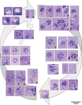
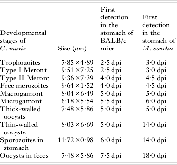
The life cycle of C. muris was studied and documented in detail at set time intervals after inoculation of both host models; BALB/c mice and M. coucha. Oocysts released active sporozoites (Figs 3A and 7O, P), which rapidly penetrated deep into the bottom of the pits of the gastric glands to avoid the adverse conditions in the host stomach. The first documented attached developmental stages of C. muris in both hosts (for chronology and dimensions see Table 1) were oval to ovoid young trophozoites observed at 2·5 dpi in BALB/c mice and at 3 dpi in M. coucha (Figs 3B and 4A–F). In preparations stained with Giemsa and Wright, the apical part of the attachment site of the parasitophorous sac (PS) enveloping the trophozoite stained intensively pink, thereby confirming its host cell origin, and the cytoplasmic region packed with lamellae of the feeder organelle appeared transparent (Fig. 3B–D). The alteration of the gastric microvillous surface and the gradual formation of a PS from the host cell plasma membrane were observed (Fig. 4A–C). The formation of lamellae of the feeder organelle from the membrane of an anterior vacuole was observed exclusively in the earliest trophozoites (Fig. 4B and C). The apical part of the mature trophozoites enveloped by the PS, which were mechanically detached from the host tissue, exhibited strong adhesive properties, causing the clustering of trophozoites and subsequent formation of rosettes (Fig. 3C). A slight elongation of host cell microvilli surrounding some of the attached parasites was observed. Further observations on early development of C. muris were described in detail previously (Valigurová et al. Reference Valigurová, Hofmannová, Koudela and Vávra2007, Reference Valigurová, Jirků, Koudela, Gelnar, Modrý and Šlapeta2008) and hence not reported again.
The life cycle of Cryptosporidium muris as observed in smear preparations stained with Wright. (A) Free, infective sporozoites. (B–D) Trophozoite stage; gradually maturing trophozoites (B), clustering of trophozoites (C), trophozoite attached to a host cell (D). (E–G) Type I merogony; clusters of chromatin in the cytoplasm at the periphery of parasite (E), budding of merozoites from the meront cytoplasm (F, G). (H–I) Free, motile Type I merozoites. (J–K) Type II merogony; first mitotic division (J), four daughter nuclei formed following the second mitotic division (K). (L) Type II merozoites inside the parasitophorous sac. (M–N) Macrogamont stage; maturing macrogamonts (M) transforming into mature ones (N) typified by an increased number of amylopectin granules, giving their cytoplasm (purple) a foam-like appearance; (O) Mature macrogamont and microgamont exhibiting multiple divisions of nuclei. (P) Microgamont stage; small compact nuclei localized at the cytoplasm periphery or at the distal end of the microgamont. (Q–R) Thick-walled oocysts; zygote (left) transforming into an oocyst (right) still enveloped by the parasitophorous sac (Q), more advanced stages of oocyst sporulation following release from the sac (R). (S–T) Thin-walled oocysts enveloped by parasitophorous sacs, one of them releasing four sporozoites (T).

Trophozoite stage. (A) Longitudinal section of an attached zoite gradually becoming enveloped by the parasitophorous sac (ps); endoplasmic reticulum (er), microvilli (mv), parasite nucleus (n). TEM. (B) Section of an early trophozoite enveloped by the parasitophorous sac (ps); anterior vacuole (av), endoplasmic reticulum (er), microvilli (mv), parasite nucleus (n). TEM. (C) Detailed view of an anterior part of a young trophozoite. Note the folded membrane of the anterior vacuole and its gradual transformation into the lamellae of the feeder organelle (arrows); parasitophorous sac (ps). TEM. (D) Young trophozoite. LM (native preparation). (E) Trophozoite enveloped by the parasitophorous sac. LM (native preparation); (F) View of a maturing trophozoite completely enveloped by the parasitophorous sac; elongated host cell microvilli (mv). SEM.

The occurrence of specific developmental stages of C. muris

dpi = days post-infection; hpi = hours post-infection.
The progress of the asexual phase into Type I merogony was characterized by clusters of chromatin localized in the peripheral cytoplasm of the parasite (Fig. 3E). Daughter nuclei formed inside a Type I meront undergoing a nuclear division (Fig. 5B–D). The asynchrony of nuclear division often resulted in an odd number of chromatin clusters. By the process of multiple budding from the residuum still attached to the host cell (ectomerogony), motile and invasive merozoites differentiated (Figs 3F–G and 5A, E–O). Almost the entire cytoplasm of the mother meront appeared to be used up for merozoite formation. Type I merogony usually resulted in the formation of 6–8 merozoites, most likely depending on the number of nuclear divisions (Figs 3H, I and 5K, L). Occasionally, five merozoites still attached to the residual body were observed (Fig. 5O). Type I merozoites (9·64×1·52 μm), which were released from the PS, either recycled to form another generation of Type I meronts, or transformed into the Type II meront stage and produced four merozoites. Observation on Type II merogony is well documented by scanning electron micrographs showing the ruptured PS releasing four merozoites and further confirmed by native/stained smear preparations (Figs 3J–L and 5P–S). The Type I meronts (9·51×7·25 μm) were observed in both hosts at the same time as the trophozoites; however, Type II meronts (9·36×7·39 μm) were first observed at 4 dpi in BALB/c mice and at 4·5 dpi in M. coucha.
Merogony. (A) Meront with six merozoites. LM (native preparation). (B–D) Asynchronous mitotic division of nuclei in a meront; cytoplasm with abundant endoplasmic reticulum (er), filamentous cytoplasm (asterisk), lamellae of feeder organelle (fo), nucleoli of future merozoites (n), parasitophorous sac (ps), pellicle of meront (arrow). TEM. Fig. D shows a meront with five daughter nuclei in cross section. TEM. (E) Meront with prominent endoplasmic reticulum (er), feeder organelle (fo) and lipid inclusion (asterisk). Note the formation of merozoites from the cytoplasm of the mother meront (arrowhead) still enveloped by an intact parasitophorous sac (ps). TEM. (F) Merozoite still attached to residual body with lipid inclusion (arrow); released merozoite (arrowhead). LM (native preparation). (G) Type I meront with budding merozoites (m). SEM. (H) Ruptured parasitophorous sac (ps) revealing more advanced stages of budding merozoites (m). SEM. (I) Daughter merozoites budding from a Type I meront still enveloped by a parasitophorous sac (ps); apical ends of merozoites (arrows), endoplasmic reticulum (er), nuclei (n). TEM. (J) Detailed view of budding merozoite from Fig. 5I; residual body of meront, nucleus (n), rhoptries (arrows). TEM. (K) Two parasitophorous sacs filled with Type I merozoites (arrow) inside and one free merozoite. LM (native preparation). (L) Merozoites being released from the parasitophorous sac. LM (native preparation). (M–N) Formation of merozoites (m) from the cytoplasm of a meront; ducts of merozoite rhoptries (arrows), large residual body (rb) with endoplasmic reticulum (er) and lipid inclusion (asterisk), parasitophorous sac (ps). TEM. (O) Odd numbers (5) of mature Type I merozoites attached to the residual body. SEM. (P–Q) Type II merogony producing four shorter merozoites. LM (native preparation). (R) Cross-section of a Type II meront; four merozoites with nucleus (n) and rhoptries (arrows). TEM. (S) Ruptured parasitophorous sac (ps) releasing four Type II merozoites (m). SEM. (T) Mature merozoites in a parasitophorous sac in longitudinal section; dense granules (×), micronemes (arrowhead), rhoptries (arrows).

Merozoites exhibited a typical subcellular organization (Fig. 5M and T), including presence of subpellicular microtubules, however no mitochondria were observed. In both host models, the Type I merozoites were usually present simultaneously with Type II merozoites, the latter being slightly shorter and broader. Merozoites seemed to possess several rhoptries (Fig. 5J and T).
Released merozoites (it is generally assumed that Type II merozoites) initiated sexual multiplication, called gametogony, via differentiating into either female (Fig. 3M–O) or male microgamonts (Fig. 3O and P). Macrogamonts (8·04×6·49 μm) were detected in both hosts at 5 dpi and microgamonts (6·18×5·54 μm) were successfully documented slightly later, i.e. at 5·5 dpi in BALB/c mice and at 6 dpi in M. coucha. The mature macrogamonts were easily recognized, as their cytoplasm was packed with numerous oval-shaped amylopectin granules, the quantity of which was directly proportional to the maturation stage (Figs 3M–O and 6A–F, I). Their cytoplasm possessed typical wall-forming bodies localized just beneath the pellicle (Fig. 6D, E).
Sexual multiplication. (A–B) Macrogamonts packed with numerous amylopectin granules giving them a foam-like appearance, feeder organelle (white arrowhead). LM (native preparation). (C) Maturing macrogamont with a few amylopectin granules (ag), prominent endoplasmic reticulum (er) situated in the centre, feeder organelle (fo), filamentous projection (asterisk), lipid inclusion (x), nucleus (n), pellicle of macrogamont (arrowhead). TEM. (D) Macrogamont with wall-forming bodies (arrow) localized just beneath its pellicle (arrowhead); the lamellae of the feeder organelle (fo), cytoplasm with prominent amylopectin granules (ag), endoplasmic reticulum (er), parasitophorous sac (ps). TEM. (E) Macrogamont with numerous wall-forming bodies (arrow), feeder organelle (white arrowhead). LM (native preparation). (F) Numerous macrogamonts at different stages of development. LM (native preparation). (G,H) Microgamonts with budding microgametes localized at their distal ends or periphery (arrowhead), feeder organelle (white arrowhead). LM (native preparation). (I) Various stages of mature macrogamonts (ma) and one microgamont (mi) with microgametes (arrowhead); host cell (hc). TEM. (J) Microgamont showing an asynchronous budding (arrows) of microgametes (mi); feeder organelle (fo), parasitophorous sac (ps), residual body (rb).TEM. (K) Microgamont exhibiting a formation of microgametes (mi) inside a parasitophorous sac (ps); dense band (arrowhead), feeder organelle (fo), residual body (rb). TEM. (L) Two microgamonts (mi) at different developmental stages stained with Lugol's iodine; free bullet-shaped microgametes (right). LM. (M) Higher magnification of three microgametes in longitudinal section. Note the apical cap (asterisks) and the elongated nucleus (n) surrounded by microtubules (arrow). TEM. (N) Higher magnification of a microgamete in cross-section; microtubules (arrow) surrounding the nucleus (n). TEM. (O) Microgamete in tangential section; apical cap (asterisk) showing the invagination of the inner single membrane (arrowhead) attached to an anterior conical structure; microtubules (arrow), nucleus (n). TEM. (P) Further microgamete in longitudinal section; concentric lamellae located posteriorly to the apical cap (white arrow), microtubules (arrows), microtubule forming a loop (arrowhead) around posterior end of nucleus (n). TEM. (Q) The microgamete (arrowhead) penetrating the parasitophorous sac most likely enveloping a macrogamont (ma). SEM. (R) Free microgametes (mi) close to the macrogamont (ma), apical caps of microgametes (asterisks). TEM.

Non-mature microgamonts in stained smears resembled the meront stage but contained smaller and more compact nuclei (Fig. 3O). The nucleus of the microgamonts divided four times and gave rise to the 16 clusters of chromatin usually localized at the distal end or periphery of microgamonts (Fig. 3P). After nuclear division, daughter nuclei migrated out of the microgamont cytoplasm into the space of the PS and subsequently developing microgametes budding from the surface of the microgamont were observed (Figs 3P and 6G–K). The beginning of microgamete budding was accompanied by an obvious evagination of the microgamont pellicle (Fig. 6J and K). The formation of microgametes appeared to be asynchronous (Fig. 6J and K) and resulted in the formation of up to 16 microgametes. After separation of the mature microgametes, a large residual body remained attached to the host cell via feeder organelles (Fig. 6K). The mature non-flagellated, bullet-shaped microgametes exhibited a slightly flattened, expanded anterior region and possessed an elongated, condensed nucleus (Fig. 6L–P). Microgametes were covered by a relatively thick plasma membrane and an additional single membrane was observed beneath this in the anterior half of the body. This single membrane formed an invagination beneath the apical cap and appeared to join the anterior conical structure (Fig. 6O). Originating at the anterior conical structure localized just beneath the apical cap, numerous (at least 10) parallel-arranged microtubules extended posteriorly surrounding the nucleus (Fig. 6M–P). The microtubule forming a loop was observed (Fig. 6P) behind the posterior end of nucleus and it might play a role in pushing out the nucleus from the microgamete towards the macrogamont nuclear envelope during the fertilization process.
After macrogamonts became fertilized by microgametes (Fig. 6Q–R) their wall appeared thicker. Through a transient stage of the diploid zygote (Figs 3Q and 7A, C), they rapidly developed into oocysts (Figs 3R and 7A–M). The oocyst stage occurred within the same time period as macrogamonts, i.e. after 5 dpi in both hosts. In the course of sporulation, oocysts formed four sporozoites that were coiled around a large residual body (Fig. 7F–I). Based on TEM observations, oocysts appeared to sporulate in situ whilst being enveloped by the intact PS, as they exhibited already well-differentiated sporozoites (Fig. 7F, H and I). Both types of oocysts were observed: the typical thick-walled ones and a second type resembling the so-called ‘thin-walled’ oocysts (Figs 3R–T, 7H and I). Nevertheless, variability in oocyst wall thickness was very high. The suture present on the surface of a well-developed oocyst marks the place where sporozoites were released (Fig. 7D, J–L and N). As the ‘thin-walled’ oocysts found in BALB/c mice at 5 dpi appeared to open inside the host stomach, they are presumed to be responsible for the presence of free sporozoites (11·72×0·98 μm) observed in scrapings of gastric mucosa at 6 dpi (Fig. 7P) onwards and they appeared to result in autoinfection (Fig. 7O). In M. coucha, no ‘thin-walled’ oocysts were detected in the initial phase of parasitization, but they were observed from 14 dpi onwards, and the free sporozoites occurred simultaneously with them. In BALB/c mice, thick-walled oocysts (7·48×5·86 μm) that were shed into the environment in host feces were detected at 7·5 dpi, whereas this did not occur in feces of M. coucha until 18 dpi.
Oocyst sporulation and excystation. (A) Various developmental stages from zygotes to oocysts. LM (native preparation). (B) Non-sporulated oocysts possessing a large residual body (arrow). LM (native preparation). (C) Zygote with numerous amylopectin granules (ag) after fertilization; developing oocyst wall (arrow), parasitophorous sac (ps). TEM. (D) Mature oocyst inside parasitophorous sac. SEM. (E) Oocyst in retreating parasitophorous sac (ps). SEM. (F) Longitudinal section of an oocyst in parasitophorous sac (ps); feeder organelle (fo), large residual body (rb), formed sporozoites (s), oocyst wall (arrow). TEM. (G) Excystation of sporozoites from the oocyst. LM (native preparation). (H) Thin-walled oocyst with sporozoites (s) inside a complete parasitophorous sac (ps); residual body (asterisk) with amylopectin granules (ag), wall of oocyst (arrowhead). TEM. (I) Thick-walled oocyst inside an intact parasitophorous sac (ps), amylopectin granules (ag), sporozoites (s). TEM. (J) Oocyst equipped with a typical suture (arrow). SEM. (K) The excystation of sporozoites from the oocyst. LM (native preparation). (L) Empty oocyst and free sporozoites. LM (native preparation). (M) Oocyst with an even surface considered to be thin-walled. SEM. (N) The ruptured empty oocyst. SEM. (O) Free sporozoite invading the gastric epithelium. SEM. (P) Invading sporozoite. LM (native preparation).

To detect early infection by C. muris, the experiment was repeated using a higher infective dose of 108 oocysts per host. Subsequently, additional microscopic and molecular analyses were performed. Based on PCR results, C. muris DNA was detected in the stomach of BALB/c mice at 1, 6, 12, 36, 48 and 72 hpi. Using the combined approach of SEM and PCR, other organs from BALB/c mice including pancreas, oesophagus, spleen, liver, Payer's patches, kidneys, lungs, duodenum, jejunum, ileum, gall bladder and feces were examined for the presence of C. muris developmental stages and specific Cryptosporidium DNA but none was found. The parasitization of the stomach in M. coucha was confirmed at 6, 48 and 72 hpi. The same organs as in BALB/c mice (except for the gall bladder) were analysed and showed positive results in the duodenum at 48 hpi and in the jejunum at 72 hpi. Using molecular tools, C. muris developmental stages were also found in the feces of M. coucha at 36 and 72 hpi.
DISCUSSION
Understanding the life cycle of parasitic organisms is fundamental to explain their pathogenicity. Parasitic strategies, such as the ability to enter into the host and avoid the host defence mechanisms, must be closely analysed. The essential factors influencing infectivity are morphological structure and the physiological processes occurring inside the parasite and host. The sensitivity of hosts to C. muris depends on its specific isolate or strain (Xiao et al. Reference Xiao, Escalante, Yang, Sulaiman, Escalante, Montali, Fayer and Lal1999; Kváč et al. Reference Kváč, Sak, Květoňová, Ditrich, Hofmannová, Modrý, Vítovec and Xiao2008; Kodádková et al. Reference Kodádková, Kváč, Ditrich, Sak and Xiao2010). Our observations on the life cycle of C. muris (strain TS03) in different rodent models showed a dissimilar chronology with respect to the occurrence of individual developmental stages. As the physiology and immune response to parasitization of both host models varies in many aspects, the host anatomy and tissue morphology together with enzymatic secretion appear to be important in the understanding and evaluation of differences in parasitization of both hosts with C. muris.
Localization
The gastric pathogen C. muris clearly prefers the acid environment of the stomach and the common use of hydrochloric acid to accelerate the in vitro excystation of its oocysts also serves as proof of this. The pH values in the gastrointestinal tract of a rat or mouse are about 5·5–6 in the mouth and intestinal parts, whereas those in the stomach rapidly decrease to 2–4 (Ward and Coates, Reference Ward and Coates1987; McConnell et al. Reference McConnell, Basit and Murdan2008). The stomach is comprised of a non-glandular and a glandular part. As the mucosa of the non-glandular stomach is lined by keratinized, stratified squamous epithelium without secretory activity, its surface is unsuitable for parasite attachment and development. In contrast, the glandular part forms gastric pits lined by a simple columnar epithelium. Simple tubular gastric glands, found on the bottom of the gastric pit, are dedicated to the production of acids to maintain the low pH level of the stomach juice and proteases, as well as mucous substances. The wall of the glandular part of the stomach is thicker and creates longitudinal folds (Ward and Coates, Reference Ward and Coates1987; Ghoshal and Bal, Reference Ghoshal and Bal1989). Considering all these morphological features, despite its extremely acid environment providing less hospitable conditions for parasites than the intestine, the gastric glandular part appears to be more suitable for Cryptosporidium development when compared with the non-glandular part. Hence, the question arises how the invasive stages of these minute parasites avoid the effects of gastric juices containing strong acids. Matsubayashi et al. (Reference Matsubayashi, Ando, Kimata, Takase, Nakagawa, Furuya, Tani and Sasai2011) showed that the viability of sporozoites from oocysts excysted in medium at pH 7·0 is approximately 90% in the gastric species, Cryptosporidium andersoni, in contrast to 56% viable sporozoites for the intestinal species C. parvum. At a lower pH, however, the viability of gastric sporozoites gradually declined to 61% after 3 h incubation at pH 2. Consequently, in comparison with intestinal cryptosporidia, the better adaptation of gastric species to an acidic environment enables them to survive for longer in the stomach (Matsubayashi et al. Reference Matsubayashi, Ando, Kimata, Nakagawa, Furuya, Tani and Sasai2010).
In general, cryptosporidia have evolved efficient strategies to avoid the potential harmful effects of the gastrointestinal tract, e.g. direct exposure to digestive enzymes, possible mechanical damage or their peeling off from the host tissue. Probably one of the most important of these strategies is enveloping by the PS of host cell origin (Valigurová et al. Reference Valigurová, Hofmannová, Koudela and Vávra2007, Reference Valigurová, Jirků, Koudela, Gelnar, Modrý and Šlapeta2008). Another is invading a proper shelter (e.g. gland or pits) and becoming covered by a mucus layer, especially in gastric species. To prevent too-rapid erosion of the gastric mucosa due to a hostile stomach environment, the surface cells and cells of the gland necks produce mucous secretions that form a relatively thick protective layer, which often hampers the ability to view inside the gastric gland using SEM. Even very careful washing of gastric tissue is often not sufficient to clear the entire surface from mucus, which forms a compact and non-transparent layer after chemical fixation. As even this protection is not perfect, the shedding of dead cells from the gastric pits into the lumen is often observed and might also be one of the reasons for the occasional detachment of PS filled with cryptosporidia and their subsequent presence in the host intestine or feces.
Another curiosity is the progress and mode of parasitization by C. muris as well as its dispersal throughout the gastric tissue in an irregular, island-like pattern. Obviously, invasive stages preferred regions that were already occupied by C. muris. This evidently non-competitive behaviour could be understood as exploiting the colonization of epithelial tissue (cell) already modified by the parasite. Stained smear preparations, showing parasites oriented by their apical ends to each other, confirmed this speculation.
Pathological changes
Both host models displayed the same pathological manifestations, but significantly differed in the chronology of parasitization. Taylor et al. (Reference Taylor, Marshall, Green and Catchpole1999) observed the correlation between increased infective dose and subsequent shortening of the prepatent period of mice as well as the increased shedding of oocysts. In this study, increased infective doses used for inoculation did not influence the success of parasite detection in the host stomach in the early course of parasitization, even if all mentioned detection methods including molecular tools were used.
The pathological alterations induced by the multiplication of cryptosporidia in BALB/c mice, were compared with other studies (Ozkul and Aydin, Reference Ozkul and Aydin1994; Aydin and Ozkul, Reference Aydin and Ozkul1996; Taylor et al. Reference Taylor, Marshall, Green and Catchpole1999; Kváč et al. Reference Kváč, Sak, Květoňová, Ditrich, Hofmannová, Modrý, Vítovec and Xiao2008). Our observations correspond with previous reports on the self-curing process in BALB/c mice infected with different strains of C. muris linked with migration and proliferation of T-cells in the site of infection (Jalovecká et al. Reference Jalovecká, Sak, Kváč, Květoňová, Kučerová and Salát2010; Kváč et al. Reference Kváč, Kodádková, Sak, Květoňová, Jalovecká, Rost and Salát2011).
In M. coucha, the immune system obviously failed and modifications of gastric tissue persisted during the entire chronic phase of infection. The parasite continued to expand from affected gastric pits to the surrounding healthy gastric epithelium and these observations are in concordance with previous study (Valigurová et al. Reference Valigurová, Jirků, Koudela, Gelnar, Modrý and Šlapeta2008). Kváč et al. (Reference Kváč, Sak, Květoňová, Ditrich, Hofmannová, Modrý, Vítovec and Xiao2008) and Kváč and Vítovec (Reference Kváč and Vítovec2003) observed the dispersal of parasitization in the abomasum of domestic ruminants from the region of the curvature major. Pathological alterations in this study concur with their histological observations. In contrast to Taylor et al. (Reference Taylor, Marshall, Green and Catchpole1999), the infiltration of the lamina propria was not observed and these findings correspond with the study of Kváč et al. (Reference Kváč, Sak, Květoňová, Ditrich, Hofmannová, Modrý, Vítovec and Xiao2008).
Life cycle
In general, excystation is a very rapid process (Lumb et al. Reference Lumb, Smith, O'Donoghue and Lanser1988). As it was almost impossible to record invading stages and the first observed stages were usually trophozoites, more sensitive molecular tools were used to detect C. muris DNA. Considering the irregular manner of parasitization, negative results of PCR for the presence of C. muris in stomachs of M. coucha could be the consequence of tissue excision from the non-parasitized part. Although molecular methods specifically proved the presence of C. muris DNA in the feces or stomach, they did not provide information concerning the presence of its concrete developmental stages, i.e. residual oocysts passing through the body vs real infection. For example, after unsuccessful attachment of zoites to the epithelium or due to the mechanical detachment of the PS in the area of the dense band reported previously (Valigurová et al. Reference Valigurová, Jirků, Koudela, Gelnar, Modrý and Šlapeta2008), various stages apart from oocysts might be present in host feces. This could also explain positive testing of M. coucha duodenum and jejunum. Miller and Schaefer (Reference Miller and Schaefer2007) claim that intact or excysted oocysts in the first hours following inoculation were neither detected in the host (stomach, caecum and colon) nor in its feces. They suggested that oocysts were destroyed during normal transit through the gut to the feces. Considering the persistence of sporulating oocysts released from the stomach and passing through the intestine to the feces, it is questionable whether well-developed and resistant oocysts used for inoculation could be destroyed by digestion if not excysted. Acknowledging such a possibility, sporulating oocysts leaving the host should also be destroyed.
Several studies highlight extraordinary epicellular development of cryptosporidia and their phylogenetic affinity with gregarines (e.g. Barta and Thompson, Reference Barta and Thompson2006; Valigurová et al. Reference Valigurová, Hofmannová, Koudela and Vávra2007; Valigurová et al. Reference Valigurová, Jirků, Koudela, Gelnar, Modrý and Šlapeta2008). Despite few studies reporting a complete development of cryptosporidia in cell-free cultures (Hijjawi et al. Reference Hijjawi, Meloni, Morgan and Thompson2001, Reference Hijjawi, Estcourt, Yang, Monis and Ryan2010; Borowski et al. Reference Borowski, Thompson, Armstrong and Clode2010), we did not noticed any extracellular stages resembling those of gregarines and developing without host cells. This study did not focus on the formation of the attachment site and the epicellular localization of C. muris as newly obtained data confirmed our previous published findings (Valigurová et al. Reference Valigurová, Hofmannová, Koudela and Vávra2007, Reference Valigurová, Jirků, Koudela, Gelnar, Modrý and Šlapeta2008). Because of the unique niche of C. muris developing within the PS, this parasite requires a more sophisticated defence strategy against the unfriendly host environment when compared with typical intracellular coccidians. The PS is a product of the host cell extensions enveloped by the plasma membrane, modified by products of the parasite (Robert et al. Reference Robert, Antoine, Dreze, Coppe and Collard1994; Bonnin et al. Reference Bonnin, Gut, Dubremetz, Nelson and Camerlynck1995; McDonald et al. Reference McDonald, McCrossan and Petry1995; O'Hara et al. Reference O'Hara, Yu and Lin2004; Valigurová et al. Reference Valigurová, Hofmannová, Koudela and Vávra2007, Reference Valigurová, Jirků, Koudela, Gelnar, Modrý and Šlapeta2008) and thus perfectly masks the parasite from host immune mechanisms (McDonald et al. Reference McDonald, Deer, Uni, Iseki and Bancroft1992, Reference McDonald, Robinson, Kelly and Bancroft1996). Although we noticed the formation of several layers of PS in early stage of trophozoites, we did not observe the pre-parasitophorous vacuole described by Huang et al. (Reference Huang, Chen and LaRusso2004).
Generally, the occurrence of two generations of merogony is described for cryptosporidia (Vetterling et al. Reference Vetterling, Takeuchi and Madden1971; Current and Reese, Reference Current and Reese1986) and even merogony including three generations has been observed for C. baileyi (Current et al. Reference Current, Upton and Haynes1986). Only one published report on Type II merogony in C. muris exists to date (Aydin, Reference Aydin1999), but because this is exclusively based on TEM, it cannot be considered reliable. This observation might be influenced by the plane of sectioning and only serial sectioning or a complete view of the entire parasite (as obtained by LM or SEM) would avoid any possible misinterpretations. This study, however, succeeds to reliably document the presence of both the Type I and Type II merogony. Considering three cycles of mitotic divisions in Type I meronts, it might be possible that Type II merogony comprises only two generations of nuclei, resulting in the formation of four merozoites. In addition, an anomaly in the final number of Type I merozoites often occurred. In accordance with a previous TEM study on C. muris (Uni et al. Reference Uni, Iseki, Maekawa, Moriya and Takada1987), we observed subpellicular microtubules in merozoites. The failure of studies on C. parvum (Current and Reese, Reference Current and Reese1986) and C. wrairi (Veterling et al. Reference Vetterling, Takeuchi and Madden1971) to document subpellicular microtubules, might be due to the known instability of these structures and to their frequent damage due to improper fixation. In addition, as the basic mechanism of apicomplexan motility is expected to be based on the orientation of the actomyosin motor by subpellicular microtubules (Dubremetz et al. Reference Dubremetz, Garcia-Reguet, Conseil and Fourmaux1998), it is not likely that motile merozoites lack these structures.
Concerning the sexual phase of the life cycle, the most easily recognized stages were macrogamonts. Microgamonts are often found in close proximity to macrogamonts, probably to facilitate macrogamont fertilization, considering that microgametes are non-flagellated. The ratio of their occurrence in comparison to that of macrogamonts, however, was much lower. This might be the consequence of a large number of microgametes (up to 16) produced by a single microgamont, that are sufficient to fertilize the surrounding macrogamonts. The presence of minute microgamonts in low numbers might be the reason for our failure to detect them in the early phase of parasitization (they were detected later than oocysts). Fertilization as described previously (Current and Reese, Reference Current and Reese1986; Aydin and Ozkul, Reference Aydin and Ozkul1996), was not observed using TEM, but we observed migrating microgametes towards the macrogamont surface and found similar coupled individuals in smears stained with Lugol's iodine as well as in SEM preparations.
Endogenous stages of thick-walled oocysts were detected in the stomachs of both hosts. In the same time period, oocysts enveloped by an obviously thinner wall were noticed in BALB/c mice, whereas similar ‘thin-walled’ oocysts in M. coucha were absent. We speculate that sporulated oocysts might also have been present during earlier phases of infection in M. coucha, but stomach dimensions and anatomical features probably did not allow their detection. Since the first microscopic detection of sporulated oocysts of C. muris in host feces was possible only after several hours or even days, the destiny of oocysts found inside the host stomach and identified as thick-walled oocysts remains enigmatic. In the course of early parasitization, we repeatedly observed structures resembling empty oocysts in mucosal scrapings or fecal smears, and often, the wall of these structures appeared to be very thin. We consider them to be oocysts, but since the identification of developmental stages based exclusively on native (unstained) preparations is problematic and usually not reliable, we cannot conclude this with absolute certainty. Current and Reese (Reference Current and Reese1986) claimed to show endogenous thick-walled oocysts releasing sporozoites (Fig. 23 in their study), but this stage more resembles an earlier stage of merogony than an oocyst. Numerous released sporozoites found in mucosal scrapings 6 dpi in BALB/c mice and 14 dpi in M. coucha support the existence of oocysts that release sporozoites in an early phase of infection. Considering the variability of wall thickness in mature oocysts observed in ultrathin sections during this study, several conflicts arose. Firstly, it appears almost impossible to strictly identify oocysts as thick- or thin-walled, even via electron microscopy. Furthermore, the size of the oocyst wall in cryptosporidia is not uniform and varies significantly during oocyst maturation. The study that reported the existence of so-called ‘thin-walled’ oocysts (Current and Reese, Reference Current and Reese1986; Aydin, Reference Aydin1999), described them as structures comprising a single unit membrane, in contrast to that of thick-walled oocysts that contain both the inner and outer membrane. Despite the high-quality micrographs showing the wall in maturing oocysts in detail, the magnification in micrographs showing sporulated oocysts in the mentioned study is not sufficient to evaluate the true number of oocysts in membranes. Moreover, we often observed detached PS containing oocysts in various stages of sporulation and the outer layer reported to be the outer membrane of the oocyst wall in Fig. 45 in Current and Reese (Reference Current and Reese1986) actually more resembles the PS shown in micrographs showing attached stages. Whether the variability in the thickness of the oocyst wall found in the stomach is the consequence of oocyst structural disorder, their malfunction or simply misinterpretation of observations caused by the oblique plane of ultrathin sectioning, remains open. The ‘thin-walled’ oocysts are believed to be responsible for autoinfection. Nevertheless, oocysts release only four sporozoites equipped with a single rhoptry and thus having only a single attempt for successful attachment to the suitable host cell (Tetley et al. Reference Tetley, Brown, McDonald and Coombs1998; Petry and Harris, Reference Petry and Harris1999; Blackman and Bannister, Reference Blackman and Bannister2001; Petry, Reference Petry2004; O'Hara et al. Reference O'Hara, Huang, Chen, Nelson and LaRusso2005). In contrast, merogony produces six to eight merozoites, which seem to possess several rhoptries and numerous micronemes (Figs 5J and 5T in this study), providing them more chances for successful attachment (Current and Bick, Reference Current and Bick1989; Tetley et al. Reference Tetley, Brown, McDonald and Coombs1998; Jirků et al. Reference Jirků, Valigurová, Koudela, Křížek, Modrý and Šlapeta2008). Since the cyclic multiplication of parasites during Type I merogony (asexual division resulting in identical daughter cells) is undoubtedly more than sufficient for the infestation of the host tissue by C. muris and the zygote is the only heterozygous diploid stage in the life cycle of cryptosporidia (McLauchlin and Nichols, Reference McLauchlin, Nichols, McLauchlin and Nichols2002), the multiplication via autoinfective oocysts (sexual stage) appears to be of benefit to increase the genetic variability of the parasite and thereby its fitness and probably infectivity.
CONCLUSIONS
A combined approach used in this study was crucial to achieve reliable and complex data on the life cycle of C. muris in two host models. The most problematic issue proved to be the detection of parasite developmental stages in gastric pits during the early phase of parasitization. Light micrographs helped to record the presence of individual developmental stages of C. muris in host stomachs and to visualize their typical morphological features in staining preparations, and these are in accordance with Tyzzer's detailed depictions shown in his work (Tyzzer, Reference Tyzzer1910). The Wright staining procedure has been evaluated to be the most suitable method, since it allows a reliable and relatively rapid identification of all developmental stages found in scrapings from host stomach.
ACKNOWLEDGEMENTS
The authors are greatly indebted to Ing. Lenka Hlásková and RNDr. Dana Květoňová from the Laboratory of Veterinary and Medical Protistology, and to the staff of the Laboratory of Electron Microscopy (both groups from the Institute of Parasitology, Academy of Sciences of the Czech Republic, in České Budějovice), for their valuable advice and help. Many thanks to Monika Nechvílová from the Department of Pathological Morphology and Parasitology, University of Veterinary and Pharmaceutical Sciences, for her help with laboratory animals.
FINANCIAL SUPPORT
Financial support was provided by a postdoctoral grant GPP506/10/P372 from the Czech Science Foundation (Investigator: A.V.). J.M., J.I. and B.S. were partially supported by the project GAP505/11/1163 from the Czech Science Foundation and M.K. by a grant from the Ministry of Education, Youth and Sports of the Czech Republic (LH11061). The authors acknowledge support from the Department of Botany and Zoology, Faculty of Science, Masaryk University, in the preparation of this manuscript.