Introduction
Micas are widely distributed phyllosilicate minerals commonly found in rocks and soils. Individual crystals can reach sizes of tens of centimeters across with a platy structure. In mine settings, micas are predominantly found as a silt-sized waste material. With the general chemical formula KAl2(AlSi3)O10(OH)2, mica is classified as a 2:1 clay mineral. The crystal structure of mica is a stack of unit layers of an Al-O-Al octahedral (O) sheet sandwiched between two Si-O-Al tetrahedral (T) sheets. Replacement of Si(IV) by Al(III) in the tetrahedral sheet, and Fe(III) or Mg(II) and Ca(II) for Al(III) in the octahedral sheet leads to a permanent net negative charge on the basal surfaces. This charge deficit is balanced by interlayer K+ which becomes adsorbed between the T-O-T layers, fitting closely into the hexagonal spaces of the Si-O-Al layer. The interlayer cations such as K+ strengthen the bonding between basal planes of T-O-T layers, which are normally held by attractive Van der Waals forces, through the attractive electrostatic interactions (Nosrati et al., Reference Nosrati, Addai-Mensah and Skinner2009).
The commonly occurring natural micas are phlogopite, biotite, and muscovite. These are abundant, cost effective, and ecofriendly, making them effective adsorbents for the removal of potentially toxic metals and metalloids. Among the natural micas, muscovite (KAl2(AlSi3O10)(OH)2) has a dioctahedral structure with potassium as the interlayer cation and studies have shown that it is difficult to dissolve and exchange the potassium in the mica interlayers with other cations (Scott and Smith, Reference Scott and Smith1966). Such strong interlayer cation retention in turn leads to small cation exchange capacities and weak sorption abilities to potentially toxic metals and metalloids (Wu et al., Reference Wu, Fan, Xu, Niu and Lu2007). Unlike muscovite, biotite (K(Mg,Fe)3AlSi3O10(OH)2) has a trioctahedral structure, rich in magnesium and iron, and exhibits a large cation exchange capacity. According to Farquhar et al. (Reference Farquhar, Vaughan, Hughes, Charnock and England1997) and Chakraborty et al. (Reference Chakraborty, Wolthers, Chatterjee and Charlet2007), biotite is able to provide more reactive sites than muscovite for the sorption of Pb(II), Cd(II), Cu(II), As(III), and As(V). Studies also showed that sorption in micaceous minerals occurs mainly through cation exchange and selective binding at frayed edge and interlayer sites, with the process influenced by mineral type, degree of weathering, competing cations, solution chemistry, and structural factors, leading to both reversible and strong fixation of ions (Kitayama et al., Reference Kitayama, Yanai and Nakao2020; Wick et al., Reference Wick, Baeyens, Fernandes, Göttlicher, Fischer, Pfenninger, Plötze and Voegelin2020; Dubus et al., Reference Dubus, Leonhardt and Latrille2023). However, it has also been reported that the sorption capacity of micas could be enhanced by surface functionalization, thermal or chemical activation, microbial interactions, and modification of structural charge or interlayer composition (Pazos et al., Reference Pazos, Castro, Cota, Osuna, Pavón and Alba2017; Hussein and Vegi, Reference Hussein and Vegi2020; Luo et al., Reference Luo, Geng, Wang, Ding, Qiang, Liang, Li, Zhang and Fan2021; Tian et al., Reference Tian, Cao and Li2022; March et al., Reference March, Hand, Morrissey and Kelsey2025).
India is rich in mica deposits; after extraction, ~75% are discarded as waste during the cleaning and processing stages and currently have no practical use. The handling of waste mica has garnered interest recently due to the large amount produced. As well as mining of mica, quarrying of coarse materials such as granite (Hojamberdiev et al., Reference Hojamberdiev, Eminov and Xu2011), phyllosilicates such as china clay (Zografou et al., Reference Zografou, Heath and Walker2014), and valuable materials such as copper, uranium, gold, and platinum (Nosrati et al., Reference Nosrati, Addai-Mensah and Skinner2009; Basak, Reference Basak2019) are the important sources of waste mica. During the dressing of raw mica, ~75% of the material is discarded as waste close to the mica mines, which consist primarily of biotite (black mica) and muscovite (white mica) (Villaros and Pichavant, Reference Villaros and Pichavant2019). These wastes are generally homogeneous, fine, and loose, with little moisture-holding capacity and small bulk density; they lack nutrients and can also contaminate the environment during monsoons and strong winds (Kumar and Maiti, Reference Kumar and Maiti2015; Monnier et al., Reference Monnier, Salvi, Melleton, Lach, Pochon, Bailly, Béziat and De Parseval2022). Currently, the re-use of waste mica has been limited, primarily in applications such as flux for ceramic glazes and as a mild abrasive (Hojamberdiev et al., Reference Hojamberdiev, Eminov and Xu2011; Zografou et al., Reference Zografou, Heath and Walker2014; Basak, Reference Basak2019). Because of the large amount involved, waste mica is currently used for landscaping purposes and as backfilling material in construction (Dudeney et al., Reference Dudeney, Chan, Bouzalakos and Huisman2013; Karlsson et al., Reference Karlsson, Kauppila, Lehtonen, Wolkersdorfer, Sartz, Sillanpää and Häkkinen2017). It is important to investigate how this material can be modified to enhance its properties for environmental remediation.
The modification of the mineral structure and surface properties as a result of artificial weathering has been studied extensively through various chemical methods. Clay mineral modification with mineral or organic acids is one of the most common and widely used chemical methods to enhance specific surface area, acidic sites, and functional groups, and to produce materials with desirable properties (Boudriche et al., Reference Boudriche, Calvet, Hamdi and Balard2011). Acid-modified biotite is more effective than raw biotite at intercalating molecules due to the increased surface area, expanded interlayer spacing, and enhanced adsorption sites (Yu et al., Reference Yu, Kou, Xing and Sun2020; Kwon et al., Reference Kwon, Kim and Roh2021; Huang et al., Reference Huang, Xia, Liang, Xie, Kong, Fu, Dou, Yang, Suo, Zhang and Meng2022). The two most frequently utilized acids in acid activation are hydrochloric acid and sulfuric acid, as they exhibit strong affinities for the process parameters and have superior modification effects on adsorption capacity, specific surface area, and porosity (Siddiqui, Reference Siddiqui1968; Jozefaciuk and Bowanko, Reference Jozefaciuk and Bowanko2002; Tiwari et al., Reference Tiwari, Choi and Lee2014; Komadel, Reference Komadel2016; España et al., Reference España, Sarkar, Biswas, Rusmin and Naidu2019). Leaching cations such as Al(III), Fe(II), or Mg(II) from the octahedral sheet by the acid would dissolve the crystalline clay structure partially during activation, and exchange the acid protons for the interlayer exchangeable cations, resulting in a modified clay mineral with high porosity, larger specific surface area, and greater surface acidity than the original mineral (Komadel and Madejová, Reference Komadel, Madejová, Bergaya and Lagaly2013; Krupskaya et al., Reference Karlsson, Kauppila, Lehtonen, Wolkersdorfer, Sartz, Sillanpää and Häkkinen2017; Al-Essa, Reference Al-Essa2018). Acid-activated clay minerals find applications in environmental control as they are applied in the removal of heavy metals, dyes, and phenolic compounds from wastewater that could otherwise contaminate the environment (Srinivasan, Reference Srinivasan2011; Al-Essa, Reference Al-Essa2018; España et al., Reference España, Sarkar, Biswas, Rusmin and Naidu2019). Bhattacharyya et al. (Reference Bhattacharyya and Gupta2011) investigated the performance of kaolinite, montmorillonite and their acid-activated equivalent clays for the removal of copper ions from aqueous solutions by adsorption-mediated surface immobilization. Kwon et al. (Reference Kwon, Kim and Roh2021) reported that the acid activation of biotite improved significantly its ability to adsorb cesium ions by way of creation of additional exchange sites and increase in surface area. Even though the acid activation of micaceous minerals has been studied, there exist serious gaps on the specific types of mica wastes derived from mining operations and the acid modification effects on the structural and surface characteristics of this waste mica. The specific aim of the study was to determine the effect of acid and varying process conditions on the physicochemical and structural transformations of the waste mica minerals.
Materials and methods
Materials
The waste mica from mica-mining sites located in Andhra Pradesh, India, was purchased from a commercial supplier. The material obtained was in the form of flakes (1–2 mm), and represented a composite of waste generated from different mica mines. Prior to acid activation, it was crushed gently and ground to a particle size that passed through a 0.2 mm sieve. The waste mica was dried at 80°C, ground to <55.1 nm, i.e. nanoform, in a high-energy ball mill (SPEX Sample Prep 8000-series Mixer/Mills) (top-down approach) at 1425 rpm (230 V) for 10 min, and stored for further use. Analytical-grade (purity >98%) sulfuric acid (H2SO4) supplied by Merck (Germany) was used for acid activation. Deionized water was used for all dilution and washing procedures throughout the experiments to avoid interference from dissolved ions.
Preparation of modified mica
Acid activation of waste mica involved mixing 10 g of the nanoform mica with 100 mL of H2SO4 of various concentrations (1 M, 2 M, and 3 M) under continuous shaking for 24, 48, and 72 h at room temperature. The acid-activated mica samples were centrifuged at 3500 rpm (1437×g) for 3 min, followed by multiple washes with deionized water until no sulfate
 $ \left({\mathrm{SO}}_4^{2-}\right) $
was detectable. The final products were dried at 80°C, and named 1M 24H, 1M 48H, 1M 72H, 2M 24H, 2M 48H, 2M 72H, 3M 24H, 3M 48H, and 3M 72H, respectively.
Characterization of acid-activated mica
The nanoform and acid-activated waste mica samples were characterized using various analytical techniques detailed in the subsequent sections.
Chemical composition and elemental formula
Elemental analysis of the samples was carried out using a SPECTRO XEPOS X-ray fluorescence (XRF) spectrometer (X-ray tube with a thick binary Pd/Co alloy anode and air-cooling operated at 50 kV and 50 W). The samples were prepared using the fundamental parameters (FP) oxides cups method (Jenkins, Reference Jenkins1999), in which finely ground powders were placed in prolene thin-film sample cups. The final oxide concentrations were obtained after correction for impurity values, ensuring accurate representation of the sample composition. The chemical formulae of the samples were calculated from the chemical compositions following the method proposed by Ross and Hendricks (Reference Ross and Hendricks1945), with modifications as described by Datta (Reference Datta2022).
Specific surface area analysis
An Autosorb Brunauer-Emmett-Teller (BET) apparatus from Quantachrome USA was used to assess the specific surface area at a liquid nitrogen temperature of –196°C. The analytical process used the static volumetric approach and was automated. The samples were subjected to an outgassing pre-treatment for 3 h at 250°C before each measurement.
X-ray diffraction
The X-ray diffraction (XRD) patterns of waste mica and acid-modified waste mica samples were obtained using an X-ray diffractometer (Rigaku Miniflex 6G) with CuKα radiation (λ = 0.154 nm) and operating at 40 kV and 15 mA. The XRD patterns were recorded in the range 3–60°2θ with a scanning speed of 2° min–1 and a step size of 0.01°. Waste mica samples were saturated with: (1) Ca and solvated with ethylene glycol (EG); (2) K at 25°C and heated to 110°C, 300°C, and 550°C. Furthermore, the waste mica and acid-modified waste mica samples were subjected to randomly oriented powder XRD. Molecular modeling was done using VESTA (Visualization of Electronic and Structural Analysis) software, a 3D visualization system for crystallographic studies, structural models, and electronic state computations (Momma and Izumi, Reference Momma and Izumi2011).
Fourier-transform infrared spectroscopy
Attenuated total reflectance (ATR) with a diamond crystal accessory was used for Fourier-transform infrared spectroscopy (FTIR) spectroscopy characterization (Spectrum Two model; Perkin Elmer Inc., USA). The measurement of spectra was over the range 4000 to 450 cm−1 with 16 scans at a resolution of 4 cm−1 at room temperature. For FTIR–ATR analysis, the powdered samples were used directly without preparation.
Scanning electron microscopy
The morphological features of the waste mica and acid-modified mica were captured using scanning electron microscopy (SEM) (JSM-6390LV; JEOL, Tokyo, Japan). A small portion of each sample was smeared over a multi-sided adhesive carbon tape fixed onto specimen stubs and coated with gold using a JFC-1600 ion-sputtering device, which ensured rapid and uniform coating for surface visualization. Observations by SEM were made at an accelerating voltage of 20 kV, at various magnifications.
Zeta potential
The zeta potential of the waste mica and acid-activated waste mica were determined using a nano particle analyzer (HORIBA SZ-100) using a DPSS LASER 532 nm. The samples were dispersed in deionized water to form a stable suspension and sonicated for 15 min before analysis.
Thermogravimetric analysis/differential scanning calorimetry
Thermogravimetric analyses were carried out by heating a known weight of the powdered sample in a silica crucible at a heating rate of 4°C min−1 in a N2 atmosphere from 0 to 1000°C (SHIMADZU DTG-60/60H).
Results and discussion
Elemental composition
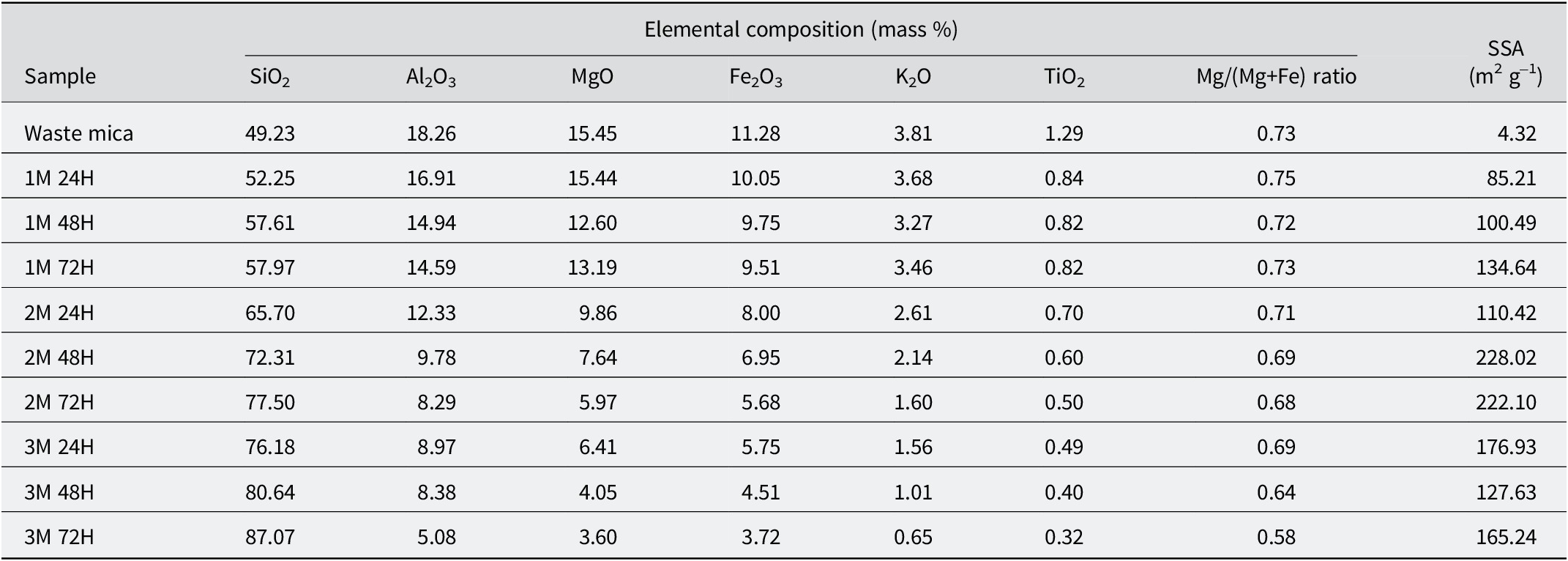
The XRF analysis (Table 1) was used to calculate the structural formulae (Table 2) of the samples and revealed that waste mica contained 49.23% SiO2, 18.26% Al2O3, 15.45% MgO, 11.28% Fe2O3, 3.81% K2O, and 1.29% TiO2. In the waste mica, the Mg/(Mg+Fe) ratio of 0.73 (Mg = 24.31 g mol−1, Fe = 55.85 g mol−1) indicated that biotite, rather than muscovite, was the dominant mica type in the samples, based on the classification of Rieder et al. (Reference Rieder, Cavazzini, D’yakonov, Frank-Kamenetskii, Gottardi, Guggenheim, Koval, Muller, Neiva, Radoslovich, Robert, Sassi, Takeda, Weiss and Wones1998). Following the acid treatment of waste mica, the Mg/(Mg+Fe) ratio decreased to 0.58 for the greatest acid concentration and longest treatment. This variation results from acid leaching of octahedral cations with the Mg removed more rapidly than Fe (Corma et al., Reference Corma, Mifsud and Sanz1987). Being relatively resistant to acid treatments, the silica content increased from 49.23% in the waste mica to 87.07% after treatment with 3 M sulfuric acid for 72 h, indicating that acid modification produces Si-rich structure, consistent with earlier studies (Myriam et al., Reference Myriam, Suarez and Martin-Pozas1998; Zhang et al., Reference Zhang, Wang, Chen and Wang2010).
Elemental composition and SSA of waste mica before and after acid treatments

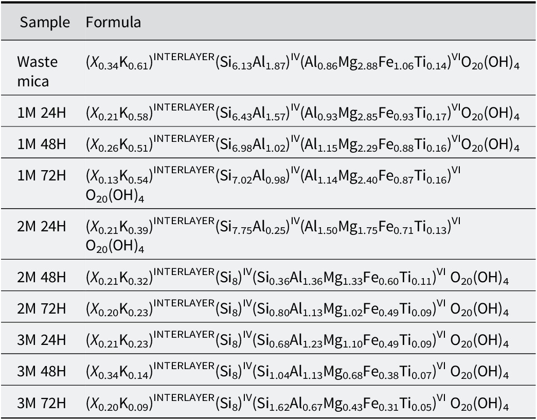
Variations in the elemental formula of waste mica after acid treatments for various time periods

The acid treatment of waste mica using varying molarities (1 M, 2 M, and 3 M) and durations (24, 48, and 72 h), was found to induce significant structural and compositional changes within the trioctahedral crystal structure (Table 2). These changes were attributed to the breaking of bonds, recrystallization, and structural rearrangement, as evidenced by the systematic trends in the interlayer, tetrahedral, and octahedral regions (Lapides, Reference Lapides1994; Bickmore et al., Reference Bickmore, Bosbach, Hochella, Charlet and Rufe2001). The amount of K+ in waste mica, K0.61, was reduced to K0.09 after 3 M treatment for 72 h. The leaching of K+ with increasing acid strength and duration weakens the electrostatic forces that bind the negatively charged tetrahedral sheets. In turn, this leaching process would facilitate layer delamination or exfoliation and tetrahedral reorganization, and thereby increase the surface area and reactivity of the material significantly (Komadel and Madejová, Reference Komadel and Madejová2006). In the tetrahedral sheets, de-alumination occurred, breaking weaker Al–O bonds and shifting the tetrahedral composition from Si(6.13)Al(1.87) to Si(8)). The resultant silica-rich framework was dominated by stronger Si–O bonds, which would be expected to increase the chemical and thermal stability and reduce the ion-exchange capacity by depleting reactive Al sites (Rodriguez et al., Reference Rodriguez, González and Munoz1995a; Komadel et al., Reference Komadel, Madejová, Janek, Gates, Kirkpatrick and Stucki1996; Pentrák et al., Reference Pentrák, Madejová and Komadel2010). The amounts of Mg, Fe, and Ti in the octahedral sheets decreased from Mg(2.88)Fe(1.06)Ti(0.14) in untreated mica to Mg(0.43)Fe(0.31)Ti0.05) after 3 M treatment for 72 h. Extensive cation leaching from the octahedral sheets of the waste mica during acid activation indicated disruption of metal–oxygen bonds, resulting in partial collapse of the octahedral sheet. This structural destabilization generates defects and enhances the porosity of the modified mica (Komadel and Madejová, Reference Komadel, Madejová, Bergaya and Lagaly2013; Krupskaya et al., Reference Krupskaya, Zakusin, Tyupina, Dorzhieva, Zhukhlistov, Belousov and Timofeeva2017; Al-Essa, Reference Al-Essa2018). The results showed that acid treatment promoted recrystallization, leading to a stable new framework dominated by Si–O bonds. In contrast, regions subjected to extensive cation leaching underwent partial amorphization, thereby enhancing surface reactivity in modified mica (Fordham, Reference Fordham1990; Jozefaciuk and Bowanko, Reference Jozefaciuk and Bowanko2002). The removal of Al and interlayer cations further generated acidic sites through unsaturated oxygen bonds or protonation of hydroxyl groups, that would enhance the acid catalytic activity of the material (Boudriche et al., Reference Boudriche, Calvet, Hamdi and Balard2011). Collectively, these modifications produced a porous, reactive material with greater surface area, acidity, and exfoliation potential (Zhuravlev, Reference Zhuravlev2000).
Specific surface area
BET analysis (Table 1) showed that the surface area increased from 4.32 m2 g−1 in the waste mica to 85.21 m2 g−1 after treatment with 1 M sulfuric acid for 24 h. The maximum specific surface area (SSA) value was obtained for the waste mica activated with 2 M sulfuric acid for 48 h (228.02 m2 g−1); the next highest value was for waste mica activated with 2 M sulfuric acid for 72 h (222.10 m2 g−1). Even though 3 M sulfuric acid increased the SSA to 176.93, 127.63, and 165.24 m2 g−1 for 24, 48, and 72 h, respectively, the SSA values were less than those obtained using 2 M acid. Treating the waste mica with 2 M sulfuric acid for a longer duration (48 h or 72 h) would have led to an etching or leaching of components from the mica structure, and thereby enhanced the SSA in the modified mica.
Studies have shown that silicates such as smectites (Komadel et al., Reference Komadel, Schmidt, Madejová and C̆íc̆el1990; Tkáč et al., Reference Tkáč, Komadel and Muller1994; Rodriguez et al., Reference Rodriguez, González and Munoz1995a; Komadel et al., Reference Komadel, Madejová, Janek, Gates, Kirkpatrick and Stucki1996; He et al., Reference He, Guo, Xie, Lin and Li2002), palygorskite (Barrios et al., Reference Barrios, González, Rodríguez and Pozas1995), sepiolite (Rodriguez et al., Reference Rodriguez, Gonzalez and Muñoz1995b), and mixed illites (Pentrák et al., Reference Pentrák, Madejová and Komadel2010), typically produce protonated, porous, amorphous, and hydrated silica with a cross-linked three-dimensional structure (Komadel, Reference Komadel1999). In biotite, the acidic solution swells the step edges leading to progressive peeling of the layers. This also initiates a sequence of structural and chemical transformations, including layer hydration and preferential leaching of cations such as Al, Mg, K, and Fe from the biotite layers. The combined effects ultimately led to the formation of a highly condensed Si-rich phase (Turpault and Trotignon, Reference Turpault and Trotignon1994) with potentially greater surface area and adsorption sites than the original waste mica. The enhancements in SSA observed could also be ascribed to the removal of impurities or layers of the mica, thereby providing additional accessible surface area for adsorption (Jozefaciuk and Bowanko, Reference Jozefaciuk and Bowanko2002). Acid treatment replaces interlayer cations (K+, Na+, and Ca2+) with H+ and removes Mg(II), Fe(III), and Al(III) from tetrahedral and octahedral sheets (Komadel et al., Reference Komadel, Janek, Madejová, Weekes and Breen1997). However, at greater acid concentrations (>2 M sulfuric acid), the silica released from the initial breakdown of the tetrahedral sheet undergoes polymerization (Pesquera et al., Reference Pesquera, González, Benito, Blanco, Mendioroz and Pajares1992). The resulting polymerized silica is deposited on the remaining silicate fractions, shielding them from further degradation, and thereby reducing the surface area. The BET results indicated clearly that the acid activation, up to a specific concentration and duration, enhanced the surface area of waste mica significantly.
XRD analysis
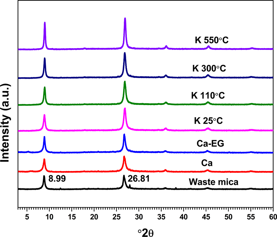
XRD spectra of the waste mica showed well-defined peaks at 8.99 and 26.81°2θ (d values = 0.98 nm and 0.33 nm, respectively) (Fig. 1). The unaltered 8.99°2θ peak under Ca-saturation, Ca-EG, K-saturation and heating confirmed the presence of mica (Fig. 1). Additionally, the XRD patterns remained unchanged after ethylene glycol solvation or heating at 300°C, thereby confirming the absence of expanding clay minerals (e.g. smectite) in the mine waste. The persistence of the 0.98 nm peak after heating at 550°C and the absence of a 1.42 nm peak indicated that no 1:1 or 2:1:1 minerals were present in the mine waste. Examination of the treatment effects (Ca-saturation, Ca-EG, K-saturation and heating) confirmed that micas were the predominant clay minerals in the mine wastes studied and no other distinct mineral phases were detected in appreciable quantities (Patarachao et al., Reference Patarachao, Tyo, Zborowski, Kung, Ng and Mercier2019).
XRD patterns of waste mica: Ca-saturated; Ca-EG-solvated; K 25°C, K 110°C, K 300°C, K 550°C: K-saturated and heated to 25, 110, 300, and 550°C, respectively.

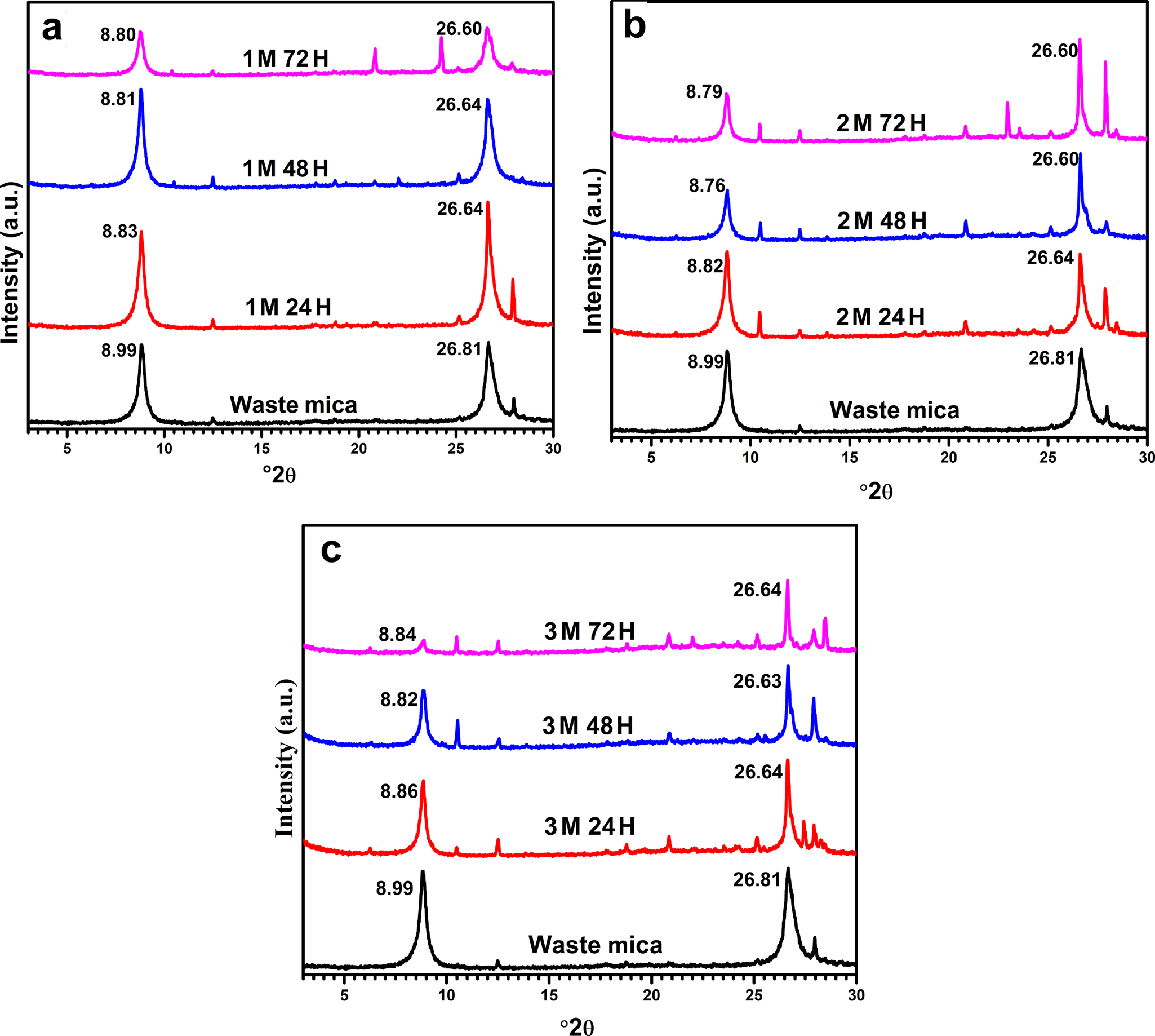
Increments in the time of acid treatment from 24 to 72 h decreased the characteristic diffraction peak intensities of the waste mica progressively, especially at higher acid concentrations. Treatment with 1 M sulfuric acid for 24, 48, and 72 h developed a leftward shift of the characteristic XRD peaks (Fig. 2a). This shift was more pronounced with 2 M sulfuric acid, reaching a maximum after 48 h, but decreased when the concentration was further increased to 3 M. For example, treatment of waste mica with 2 M sulfuric acid for 48 h shifted the characteristic diffraction peaks from 8.99°2θ to 8.76°2θ (d spacing from 0.98 to 1.01 nm) and from 26.81°2θ to 26.60°2θ (d spacing from 0.33 to 0.34 nm) (Fig. 2b). The peak intensities decreased progressively with treatment duration, while additional peaks emerged at greater acid concentrations and longer treatment times, indicating structural changes and possible new phase formation. Sulfuric acid can dissolve mica minerals due to its strong reactivity. Prolonged exposure to this acid leads to partial or complete breakdown of the layered phyllosilicate structure, which in turn could weaken the crystal lattice, enlarge interlayer spaces, and even induce partial amorphization (Fordham, Reference Fordham1990; Jozefaciuk and Bowanko, Reference Jozefaciuk and Bowanko2002). These structural variations scatter X-rays more widely, producing broader and weaker diffraction peaks compared with untreated mica (Jozefaciuk and Bowanko, Reference Jozefaciuk and Bowanko2002). White (Reference White1962) noted that depletion of K in dioctahedral micas (muscovite) increases the (001) reflection intensity more rapidly than the (002), whereas the reverse occurs in trioctahedral micas (biotite). The observed (001)/(002) intensity ratio of 1–3 confirms the predominant trioctahedral biotite nature in the waste mica. The results showed that the interlayer space of waste mica reached its maximum expansion after treatment with 2 M sulfuric acid for 48 h. This behavior can be explained by the gradual removal of interlayer cations and the subsequent protonation of the mineral layers. Sulfuric acid concentration up to 2M and 48 h of treatment facilitated replacement of interlayer cations (e.g. K+, Na+, and Ca2+) with protons, leading to electrostatic repulsion between the layers, hydration of interlayer species, and expansion of the interlayer space (Komadel and Madejová, Reference Komadel and Madejová2006). However, at greater acid concentrations (>2 M), excessive leaching of Al, Fe, and Mg from the octahedral sheets would have degraded structural integrity, causing layer collapse and subsequent reduction in interlayer space. The emergence of new peaks in the XRD patterns following acid treatment indicated structural reorganization and the formation of secondary phases, such as amorphous silica, aluminum oxides, or other residual crystalline structures from partial leaching of octahedral and tetrahedral cations (Krupskaya et al., 2017; Al-Essa, Reference Al-Essa2018). The acid-modified waste mica probably contained a mixture of amorphous material formed due to structural breakdown and some unaltered crystalline mica that remained resistant to acid attack. New diffraction peaks were found at 6.26 and 6.25°2θ (1.41 nm) after 2M 48H and 2M 72H treatments, indicating the formation of expansible phases. Studies have reported that such structures result from depletion of non-exchangeable K in biotite that occur alongside interlamellar expansion (Boyle et al., Reference Boyle, Voigt and Sawhney1967; Fanning et al., Reference Fanning, Keramidas and El-Desoky1989).
XRD patterns of waste micas and acid-treated waste micas for various time periods.

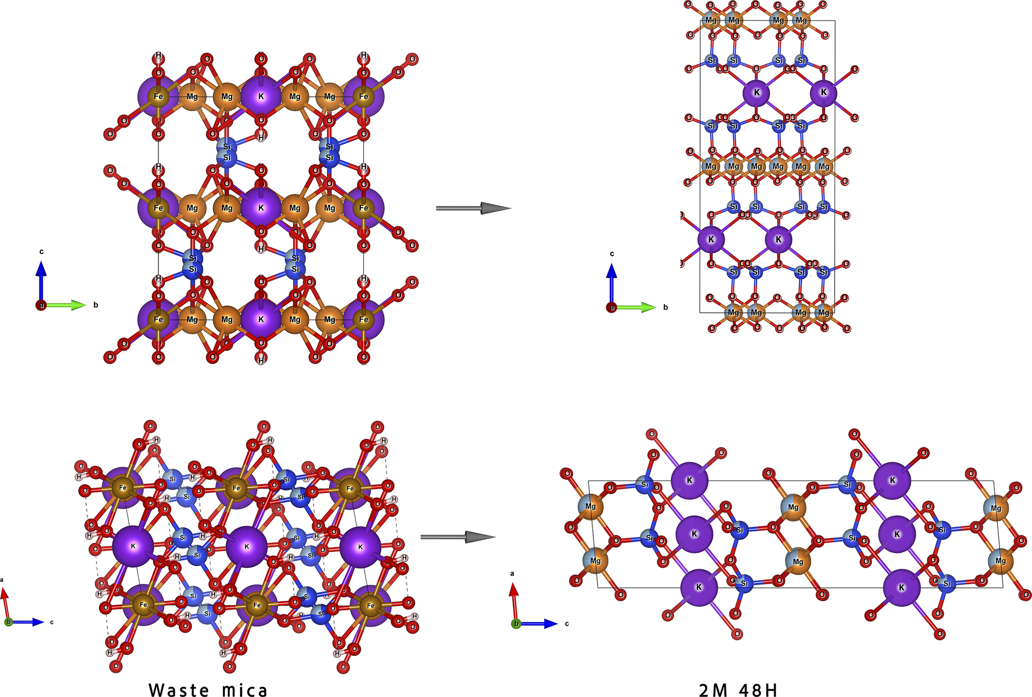
Molecular modeling with VESTA (Momma and Izumi, Reference Momma and Izumi2011), using crystallographic data derived from XRD, was used to visualize the structure of waste micas and acid-treated micas (Fig. 3). The results confirmed potassium release from interlayer spaces after treatment with 2 M sulfuric acid for 48 h, demonstrating the effectiveness of acid activation in promoting cation release and interlayer expansion. Kwon et al. (Reference Kwon, Kim and Roh2021) reported that acid activation of biotite results in the oxidation of Fe(II) to Fe(III) within the octahedral sheets, leading to structural deformation, interlayer wedge expansion, and facilitation of potassium (K) release. The molecular modeling corroborated the findings from elemental and XRD analysis, providing additional support to the conclusions drawn from these analyses.
Changes in the crystal structures of waste mica after treatment with 2 M sulfuric acid for 48 h. The color scheme is: Si (blue), O (red), and K (violet).

FTIR analysis
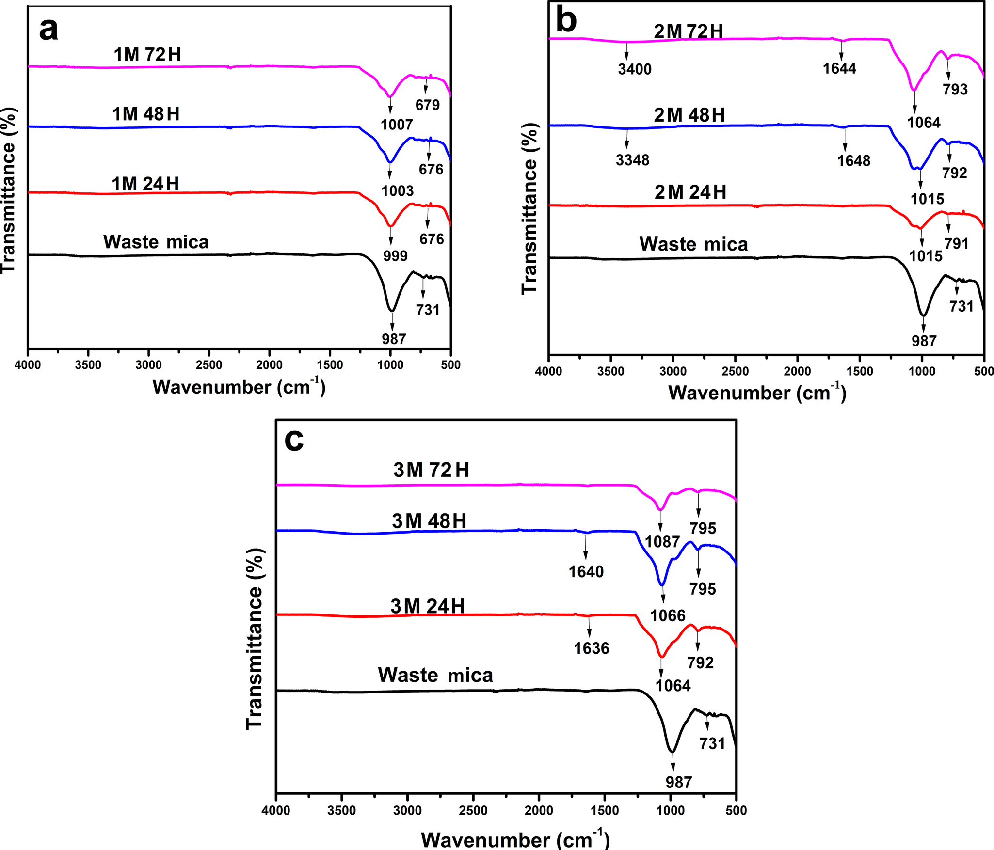
The FTIR spectra of waste mica showed characteristic absorption bands of the tetrahedral and octahedral structural components, intercalated water, and hydroxyl groups. Bands in the 1100–900 cm–1 spectral region correspond to v(Si-O-Si) modes, while those at 900–740 cm–1 are due to octahedral Al-O and Al-O-Al stretching. Waste mica exhibited characteristic bands at 987 cm–1 and 731 cm–1 (Fig. 4) corresponding to Si-O-Si and Al-O-Al vibrations, respectively, of mica (Šontevska et al., Reference Šontevska, Jovanovski, Makreski, Raškovska and Šoptrajanov2008). The low intensity of the band suggests reduced intercalated water content or limited hydration within the mica structure under prolonged acid treatment. The low band intensity also reflects structural disorder in the mica mineral lattice at greater acid concentrations, which consequently alters the intercalated water molecules (Li and Jiang, Reference Li and Jiang2009). The appearance of broader bands at 3400–3200 cm–1, the O-H stretching vibrations, confirmed the presence of intercalated water molecules in the waste mica treated with 2 M sulfuric acid for 48 h and 72 h. The stretching and deformation vibrations of water molecules were observed at 3410 and 1655 cm–1, respectively. The presence of the free amorphous silica phase in the acid-activated mica was confirmed by the prominent shoulders at 1015 cm–1 and increased the intensity of the peak at 792 cm–1 (Balcı, Reference Balcı2018). The FTIR patterns corroborated the recrystallization phases dominated by Si-O bonds and amorphization phases from cation leaching in acid-modified micas. The FTIR spectra confirmed that acid treatments can effectively modify the waste mica.
FTIR spectra of waste micas and acid-treated waste micas for various time periods.

Scanning electron microscopy
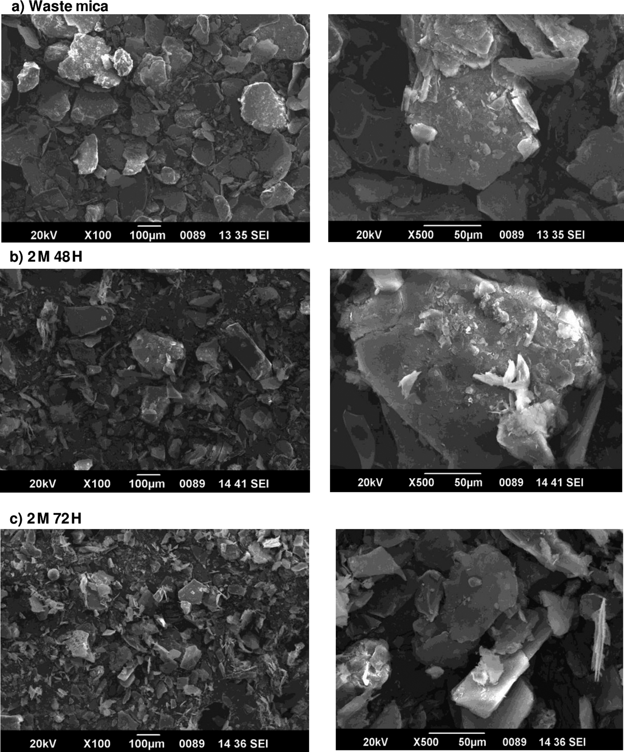
The representative SEM images of the waste mica and acid-treated waste mica (2M 48H and 2M 72H) (Fig. 5) showed that the waste mica was crystalline in a monoclinic form with tabular/prismatic crystals in a pinacoid termination. The morphological features depicted two pinacoid faces and four prism faces forming a pseudo-hexagonal structure of biotite (Fig. 5a). Muscovite, on the other hand, would have shown crystallized structures in a monoclinic system, forming amorphous pseudo-crystals (Rickwood, Reference Rickwood1981; Hosseini, Reference Hosseini2000). The waste mica exhibited slightly bent and fractured mica grains. The SEM images of 2M 48H and 2M 72H indicated a structure disintegration and a reduction in size after acid activation. The SEM images showed that morphological degradation became more pronounced with greater acid concentrations, possibly due to metal leaching from both the tetrahedral and octahedral sheets (described above). Bendou and Amrani (Reference Bendou and Amrani2014) observed similar variations on acid modification of bentonites. Jiang et al. (Reference Jiang, Duanmu, Yang, Gu and Chen2014) reported that acid treatment of palygorskite enhanced disintegration and adsorption performance by generating more active OH- groups and creating additional adsorption sites.
SEM images of raw and acid-treated waste mica.

Zeta potential
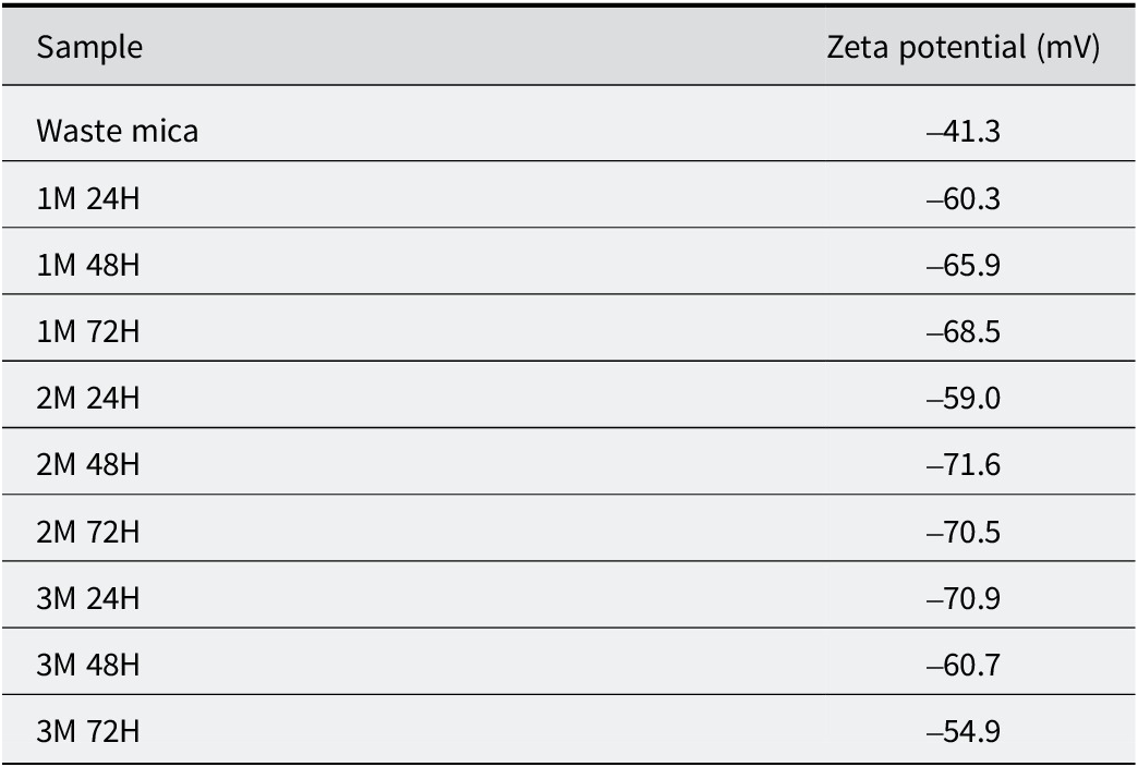
The change in surface charge revealed that acid activation had significant effects at the solid–liquid interface. The waste mica had a zeta potential of –41.3 mV at pH 7 (Table 3). The acid-activated micas exhibited smaller zeta potentials than waste mica, with the minimum observed after treatment with 2 M sulfuric acid for 48 h (–71.6 mV). The lower zeta potential of the modified mica can be attributed to cation leaching from the crystal lattices and further confirmed the structural modifications of waste mica on acid activation (Sawhney, Reference Sawhney1972).
Zeta potential of waste mica and acid-treated waste mica for various time periods

The increase in negative zeta potential after acid modification indicated that the mica suspension had become more stable, thereby providing a greater opportunity for electrostatic interactions between the positively charged ions and the acid-modified mica surfaces (Britz, Reference Britz2018; Gertsen et al., Reference Gertsen, Perelomov, Kharkova, Burachevskaya, Hemalatha and Atroshchenko2024). These findings suggested that mica treated with acid may have a greater capacity for cationic adsorption.
Thermal analysis
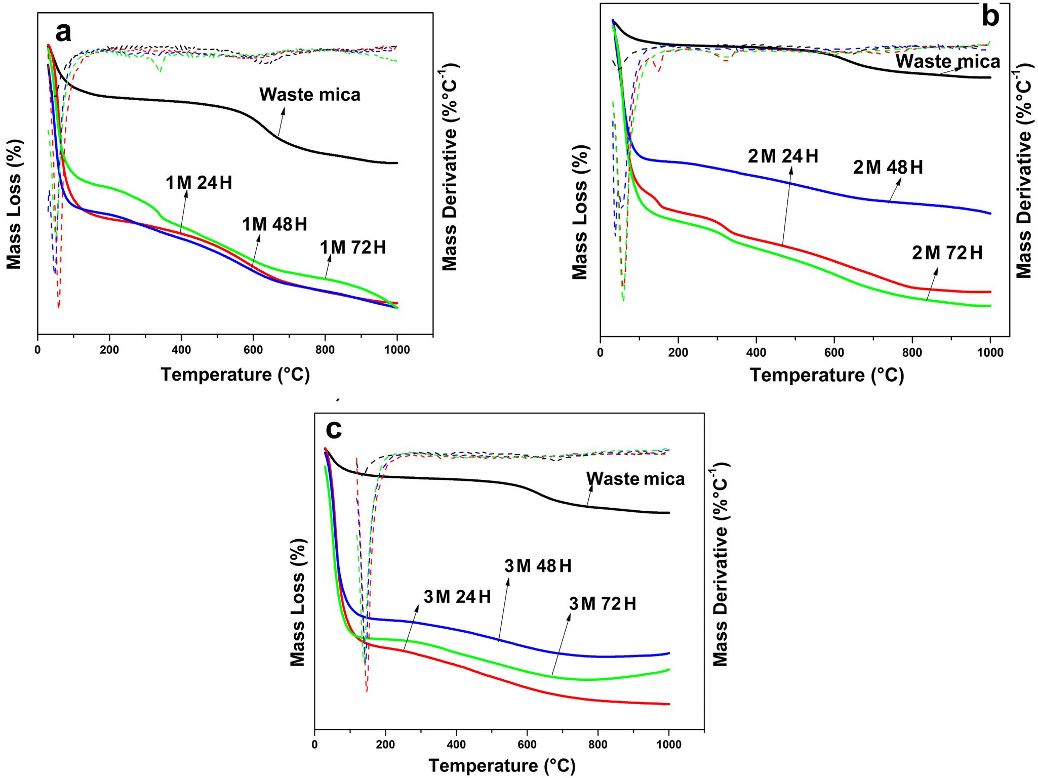
The thermogravimetric analysis (TGA) and derivative thermogravimetry (DTG) patterns (Fig. 6) of the waste mica and acid-modified mica exhibited three distinct mass loss regions: (1) removal of loosely bound physisorbed water (<200°C); (2) dehydration of strongly bound structural and coordinated water (>300°C), and (3) dehydroxylation of structural hydroxyl groups (>500°C). The waste mica exhibited a total mass loss of ~4%, attributed primarily to the loss of physically adsorbed water (~150°C), minor dehydroxylation between 200 and 600°C, and slight structural degradation at higher temperatures. In contrast, the waste mica treated with 2 M sulfuric acid for 48 h showed a greater total mass loss of ~12%, indicating substantial structural and compositional changes due to acid treatment. The TGA curves indicated that the amount of physisorbed water by modified mica had increased with acid concentration. The enhanced surface area and formation of amorphous silica would have promoted water adsorption on the acid-activated mica minerals (Panda et al., Reference Panda, Mishra, Mishra and Singh2010).
TGA and DTG patterns of waste micas and acid-treated waste micas for different time periods.

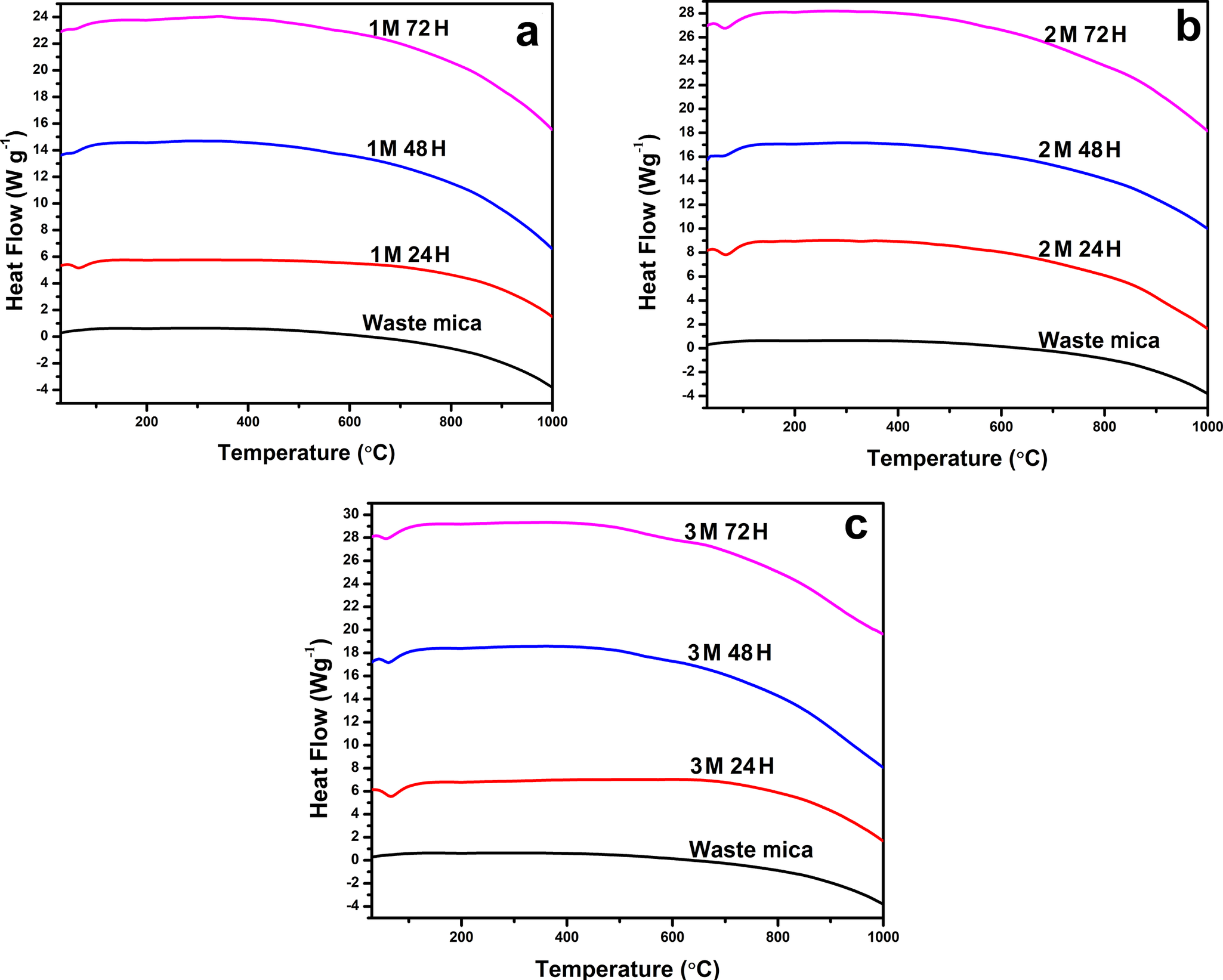
The DSC profiles of the waste and acid-modified mica (Fig. 7) were consistent with the mass loss trends observed in the TGA curves. The DSC curves of both waste mica and acid-treated micas showed endothermic peaks in the region of 800–1000°C. An additional endothermic peak below 200°C was also observed in all the acid-treated samples. The DSC analysis showed a progressive increase in heat flow, indicating a corresponding intensification of thermal events such as dehydroxylation and phase transformations with increasing acid concentration and treatment duration. The waste mica showed an enthalpy change of –412.63 J g–1 and a 4% mass loss, indicating limited thermal activity. After treating the waste mica with 2 M sulfuric acid for 48 h, the enthalpy changes increased to –734.31 J g–1 with a corresponding 12% mass loss, suggesting enhanced breakdown of the mineral structure and removal of volatile components. Treatment of waste mica with 3 M sulfuric acid for 72 h increased the enthalpy change to –1111.27 J g–1, with an increase in mass loss of only 13%, implying that the most decomposable materials had already been removed from the raw mica forms. This trend indicates that acid treatment increases thermal reactivity, but concentration or duration beyond the optimal level leads to diminishing marginal structural changes (i.e. changes per unit of acid added) in the waste mica. The absence of an endothermic peak below 200°C in waste mica is an indication of the well crystallized and non-expandable micas, whereas the appearance of new endothermic peaks in the acid-treated mica at this temperature indicates the formation of expansible 2:1 minerals. The low-temperature endotherm signifies water loss retained on surfaces as well as hydrated interlayer cations (Fanning et al., Reference Fanning, Keramidas and El-Desoky1989). These results support the XRD results and further confirmed that the acid treatment was effective in modifying waste mica. When micas were heated to ~1200°C, the DSC curves showed characteristic peaks that could be attributed to the recrystallization and release of structural -OH groups. As dehydroxylation in micas often happens gradually and does not result in appreciable structural changes, the endothermic peaks related to such reactions were smaller and less pronounced than that found in other expanding clay minerals (Fanning et al., Reference Fanning, Keramidas and El-Desoky1989; Lapides, Reference Lapides1994).
DSC curves of waste micas and acid-treated micas over various time periods.

Conclusions
The present study aimed to investigate the effect of acid activation on waste mica, a material typically discarded, to provide insights into its potential for future environmental remediation efforts. The results showed that acid activation effectively modified the crystal structure of waste mica, with the maximum specific area and reactivity for potential environmental applications obtained upon treatment with 2 M sulfuric acid for 48 h. Acid modification showed a continuous enrichment of silica with a concomitant decrease of all the other elemental oxides with increasing concentrations and treatment time. The elemental composition and molecular formulae showed that with increasing strength of sulfuric acid and duration of treatment, the tetrahedral sheets were transformed progressively to a framework with pockets dominated by Si-O bonds and areas with extensive cation leaching and enriched with amorphization and enhanced surface reactivity. The specific surface area of waste mica (4.32 m2 g–1) increased to its maximum value on activation with 2 M sulfuric acid for 48 h (228.02 m2 g–1). The XRD results and molecular modeling indicated a structural transformation and enhancement of the interlayer spaces of the waste mica with acid concentration and contact time. The FTIR spectra further confirmed that acid treatment was effective in modifying the waste mica. The decrease in zeta potential, after acid activation, indicated enhanced ion release and surface-charge reduction, which could have contributed significantly to improving adsorption efficiency. The TGA and DSC analysis confirmed that acid treatment significantly altered the thermal behavior of the waste mica, increasing the physiosorbed water content and effectively transforming the waste mica into an expansible 2:1 mineral. Increased changes in enthalpy and mass loss indicated that acid treatment improved the thermal reactivity of the waste mica. The appearance of low-temperature endothermic peaks and changes in hydroxyl group decomposition also confirmed enhanced surface area and structural modifications, further verifying the effectiveness of acid activation. The present study demonstrated conclusively that acid treatments are effective in modifying waste mica, providing potential for environmental applications for this hitherto waste material.
Author contributions
Navya M performed the experiments, data analysis, and writing of the original manuscript. Sandeep S conceptualised and supervised the work and contributed to the review and editing of the manuscript.
Acknowledgments
The authors would like to acknowledge the financial grant provided by Kerala State Council for Science, Technology and Environment (KSCSTE) and also thank KSCSTE-Kerala Forest Research Institute, Cochin University of Science and Technology (CUSAT), Centre for Materials for Electronics Technology (C-MET), DST SAIF Cochin, CSIF-University of Calicut, School of Chemical Sciences-Mahatma Gandhi University and Centre for Agricultural Nanotechnology-Tamil Nadu Agricultural University for providing the infrastructural and instrumental facilities required.
Financial support
The Kerala State Council for Science, Technology and Environment (KSCSTE), India, provided the research fellowship for this whole work.
Competing interests
There are no competing interests to declare.
Data availability statement
The data that support the findings of this study are not publicly available because they are part of the associated author’s unpublished PhD work, but they are available upon reasonable request.
Declaration of generative AI and AI-assisted technologies in the writing process
During the preparation of this work the author(s) used AI-assisted technology in order to improve language and readability. After using this tool/service, the authors reviewed and edited the content as needed, and take full responsibility for the content of the publication.