Introduction
Bony inner-ear malformations account for approximately 20 per cent of all cases of congenital sensorineural hearing loss.Reference Jackler, Luxford and House1 Accurate diagnosis and treatment of inner-ear malformations are based on established classification systems commonly used by surgeons, audiologists and radiologists. Jackler et al. published the first classification of inner-ear malformations in 1987 based on polytomographic findings.Reference Jackler, Luxford and House1 According to this classification, the five types of inner-ear malformation are Michel deformity, cochlear aplasia, common cavity, cochlear hypoplasia and incomplete partition.
Recent advances in imaging technology, including high-resolution computed tomography (CT) and high-Tesla magnetic resonance imaging (MRI), are now used to diagnose cochlear anomalies. Magnetic resonance imaging is the method of choice for evaluating the contents of the internal acoustic canal, including the cochlear nerve. Classification of cochlear anomalies is based on the radiological characteristics of the cochlea determined using high-resolution CT and MRI.Reference Sennaroglu2
In 2002, Sennaroğlu and Saatçi classified incomplete partition anomalies as types I and II.Reference Sennaroglu and Saatci3 In 2006, incomplete partition type III, which is associated with an X-linked disease, was added to their classification system.Reference Sennaroglu, Sarac and Ergin4 The cochlear hypoplasia subtypes were classified by Sennaroğlu et al. as subtypes I–III in 2010, with subtype IV added to the classification system in 2016.Reference Sennaroglu2,Reference Sennaroglu, Bajin, Pamuk and Tahir5 According to the classification system, the hypoplastic cochlea has smaller dimensions, and the cochlea and vestibule are considered separate structures.
The four cochlear hypoplasia subtypes (Figure 1a–d) are summarised as follows. In subtype I, the cochlea is a bud-like structure. The cochlea is severely deformed, and there is no modiolus or interscalar septum. In subtype II, the cochlea is smaller than normal, without a modiolus or interscalar septum, but its outer appearance is similar to the normal cochlea. An enlarged vestibular aqueduct and minimally dilated vestibule may accompany subtype II. As there is a wide communication between the cochlea and the internal auditory canal, a ‘gusher’ and/or misplacement of the cochlear implant electrode into the internal auditory canal can occur during implantation. In subtype III, the modiolus is short and the cochlea has fewer than two turns. The internal and external architecture of the cochlea are normal. The vestibule and semicircular canals are usually hypoplastic. In subtype IV, the cochlea is smaller than normal. Whereas the basal turn is normal, the middle and apical turns are hypoplastic.Reference Sennaroglu, Bajin, Pamuk and Tahir5
High-resolution computed tomography axial images, showing cochlear hypoplasia subtypes: (a) subtype I, (b) subtype II, (c) subtype III (arrows depict the cochlear aperture) and (d) subtype IV. AF = anterior/feet; R = right; L = left; PH = posterior/head; A = anterior; P = posterior; AH = anterior/head; RHP = right/head/posterior; LFA = left/feet/anterior; PF = posterior/feet

Cochlear hypoplasia is defined as a smaller than normal cochlea, which occurs because of developmental arrest during the sixth week of gestation, according to GulyaReference Gulya6 and Jackler et al.Reference Jackler, Luxford and House1 In a 2016 histopathological study, Sennaroğlu suggested that the four cochlear hypoplasia subtypes were all associated with developmental arrest that occurs during different periods of gestation.Reference Sennaroglu7
All published cochlear hypoplasia classification systems are based on visual examination of the outer and inner structures of the cochlea, not precise measurement of its structures. Our previous study examined cochlear dimensions in the normal cochlea.Reference Pamuk, Pamuk, Akgöz, Öztürk, Bajin and Sennaroğlu8 However, to the best of our knowledge, the literature does not include any data on hypoplastic cochlear dimensions. Prior to implantation of a cochlear implant, accurate measurement of the cochlear canal length is extremely important for selecting a cochlear implant electrode of optimal length. The present study aimed to radiologically measure hypoplastic cochlear dimensions and determine their effect on cochlear implant selection.
Materials and methods
Participants
The medical records of patients presenting to the Otorhinolaryngology Department at Hacettepe University Hospital, Ankara, Turkey, between 2010 and 2016, who had temporal bone CT images stored in the hospital's picture archiving and communication system were retrospectively reviewed.
The cochlear hypoplasia group consisted of 36 of these patients with sensorineural or mixed hearing loss who were also diagnosed with cochlear hypoplasia according to the Sennaroğlu classification of their CT images. In addition, 24 of the 36 patients also underwent temporal bone MRI. Cochlear hypoplasia patients who underwent CT imaging, follow up and/or treatment at another facility were excluded from the study.
The control group included 40 patients who underwent temporal bone CT because of chronic otitis media with tympanic membrane perforation. None of the controls had sensorineural hearing loss, but all had conductive hearing loss with an air–bone gap of less than 20 dB.
All the participants provided written informed consent, and the study protocol was approved by the Hacettepe University Non-Interventional Clinical Studies Ethics Committee (approval number: GO 15/784-30).
Imaging
High-resolution CT was performed in the axial plane using a four-channel multidetector scanner (Somatom Plus 4 Volume Zoom; Siemens, Erlangen, Germany); all images had 0.5 mm collimation and were 0.5 mm thick. Reformatted axial images parallel to the lateral semicircular canal, and coronal images perpendicular to the lateral semicircular canal, were created. In addition, multiplanar reconstruction reformats were generated in Stenvers view to facilitate visualisation of all cochlear turns in a single plane.
Magnetic resonance imaging was performed using a 3 T scanner (Magnetom Allegra (Siemens) or Ingenia (Philips Healthcare, Amsterdam, Netherlands)) or 1.5 T scanner (Symphony; Siemens) and a standard head coil. The standard temporal bone imaging protocol included axial and sagittal-oblique three-dimensional constructive interference in steady state (Siemens) or ‘Drive’ (Philips) imaging.
Cochlear hypoplasia subtype and side were determined according to the Sennaroğlu classification. Classification was determined at the time of diagnosis by the senior inner-ear specialist (author LS) and retrospectively reviewed for this research. Measurements were made by a radiologist and an ENT specialist.
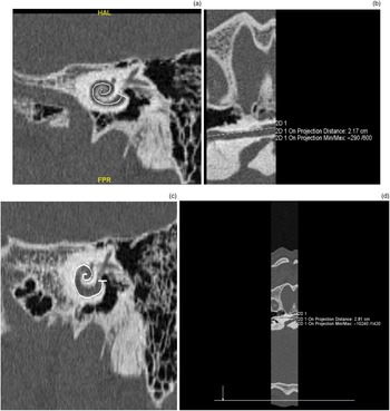
Basal turn height, basal turn length, mid-modiolar height and cochlear aperture were measured using axial reformatted images (Figure 2a–c). Cochlear canal length was measured in a curved multiplanar reconstruction reformat that was created based on the Stenvers plane (Figure 3a–d). Two separate curved multiplanar reconstruction projections were created that extended from the lateral edge of the round window to the helicotrema: one along the midpoint of the cochlear turns, which is referred to as the mid-scalar length of the cochlear canal, and one traced along the lateral wall of the cochlear canal, which is referred to as the cochlear canal length. Additionally, the vestibular system was assessed via both CT and MRI, and the cochlear nerve was evaluated via MRI.
High-resolution computed tomography axial reformatted images, showing the cochlear dimension measurements: (a) basal turn length, (b) basal turn height and (c) mid-modiolar height. 2D = two-dimensional; A = anterior; P = posterior

High-resolution computed tomography images as seen on Stenvers view, showing the cochlear canal measurements: (a & b) mid-scalar cochlear canal length and (c & d) cochlear canal length along the lateral wall. HAL = head/anterior/left; FPR = feet/posterior/right; 2D = two-dimensional

Cochlear aperture width was classified as follows: normal – more than 1.5 mm; stenotic – less than 1.5 mm; and atretic – an undeveloped cochlea. The modiolus was classified as normal, absent, hypoplastic, dysplastic or sclerotic. Cochlear nerve status (normal, aplastic or hypoplastic) was determined by temporal bone MRI. Vestibular structures were defined as normal, dysplastic, hypoplastic or ectatic, and semicircular canals were defined as normal, aplastic, dysplastic or hypoplastic.
Statistical analysis
Statistical analysis was performed using SPSS Statistics for Windows software, version 23.0 (IBM, Armonk, New York, USA) and Microsoft Excel software (Microsoft, Redmond, Washington, USA). Qualitative data are expressed as frequency and percentage values. For quantitative data, range and mean ± standard deviation values were analysed separately for the cochlear hypoplasia and control groups. The Kolmogorov–Smirnov test and the Shapiro–Wilk test were used to evaluate numerical values with a normal distribution. Analysis of variance was used to evaluate numerical parameters that were normally distributed, and the Tamhane test was used for post-hoc corrections. The Kruskal–Wallis and Mann–Whitney U tests were used to analyse numerical parameters that were not normally distributed. The level of statistical significance was set at p < 0.05.
Results
The cochlear hypoplasia group included 36 cochlear hypoplasia patients and the control group included 40 otitis media patients with normal cochleae. In the cochlear hypoplasia group, 47.2 per cent of patients were male (n = 17) and 52.8 per cent were female (n = 19), whereas the control group consisted of 52.5 per cent males (n = 21) and 47.5 per cent females (n = 19); the difference was not significant (p = 0.64). The mean age of patients was 6 ± 6.34 years in the cochlear hypoplasia group versus 30.32 ± 12.7 years in the control group. In total, 28 of the cochlear hypoplasia patients had anomalies in the right ear and 8 had anomalies in the left ear. The control group consisted of 25 right and 15 left ears.
Among the cochlear hypoplasia patients, 31 had profound sensorineural hearing loss and 5 had mixed type hearing loss, of whom 3 had cochlear hypoplasia subtype III and 2 had subtype IV. In all, 25 of the cochlear hypoplasia patients underwent surgery: auditory brainstem implantation (n = 7), cochlear implantation (n = 15), or both auditory brainstem implantation and cochlear implantation (n = 3). Table 1 shows cochlear aperture and modiolus status according to cochlear hypoplasia subtype.
Features of cochlear nerve, SCC, vestibular structures, cochlear aperture and modiolus status according to cochlear hypoplasia subtype

SCC = semicircular canal
Basal turn length (measured in millimetres) was measured on axial sections (Table 2). There was a significant difference in basal turn length between the patients with cochlear hypoplasia subtypes I, II and III and the control group, but not between the cochlear hypoplasia patients with subtypes I–III. There was no significant difference in basal turn length between subtype IV patients and the control group.
Basal turn length, basal turn height and mid-modiolar height according to cochlear hypoplasia subtype

SD = standard deviation
Basal turn maximum height was measured on the axial section passing through the round window. Cochlear dimensions according to cochlear hypoplasia subtype are shown in Table 3. All cochlear dimensions differed significantly between the control group and the cochlear hypoplasia group, regardless of cochlear hypoplasia subtype (p < 0.001). Mean mid-modiolar height (Table 2) was significantly greater in the control group than in the cochlear hypoplasia patients with cochlear hypoplasia subtypes I–III (p < 0.001). Furthermore, there was a significant difference in mean mid-modiolar height between those with subtype I and those with subtypes II–IV (p = 0.01, 0.005 and 0.024, respectively).
Cochlear canal mid-scalar and lateral wall length according to cochlear hypoplasia subtype

SD = standard deviation
Mean mid-scalar length was significantly greater in the control group than in the cochlear hypoplasia patients with subtypes I–III (p < 0.001). Mean mid-scalar length was significantly shorter in the cochlear hypoplasia patients with subtype I than in those with subtypes II–IV (p < 0.001) (Table 3). Mean cochlear duct lateral wall length was significantly shorter in the cochlear hypoplasia group than in the control group (p < 0.001 for subtypes I–III and p = 0.002 for subtype IV) (overall p < 0.001). In addition, mean cochlear duct length (along the lateral wall) was significantly shorter in the cochlear hypoplasia patients with subtype I than in those with subtypes II–IV (p = 0.005, 0.001 and 0.009, respectively), and was significantly shorter in those with subtype II than in those with subtypes III and IV (p = 0.01 and 0.02, respectively) (overall p < 0.001) (Table 3).
Findings related to the cochlear nerve, semicircular canals and vestibular structures, according to cochlear hypoplasia subtype, are shown in Table 1.
Discussion
Inner-ear malformations constitute approximately 20 per cent of all congenital sensorineural hearing loss cases.Reference Sennaroglu, Sarac and Ergin4 Genetic mutations and environmental factors cause inner-ear malformations during inner-ear development. Cochlear development is summarised as follows. At six weeks of gestation, the cochlear canal appears as a diverticulum from the saccular part of the otic vesicle. The cochlear canal completes 2.5 turns between gestational weeks 8 and 10: first the basal turn, and then the middle and apical turns develop. At 20 weeks of gestation, the membranous labyrinth attains its adult configuration. The modiolus is formed via intramembranous ossification, which is completed by gestational week 25. Upon completion of the cochlear canal, the bony labyrinth surrounds the cochlear canal. The cochlea attains its adult size during the 25th week of gestation.
Sennaroğlu reported that cochlear hypoplasia subtypes I–III occur because of arrested development of the membranous labyrinth between gestational weeks six and eight.Reference Sennaroglu7 In cases of subtype IV, the basal turn is normal and the apical turn is hypoplastic, which is caused by arrested development that occurs between the 10th and 20th weeks of gestation.Reference Sennaroglu7 Genetic and vascular factors may play a causative role in fetal developmental arrest. As the blood supply for the developing cochlea is derived primarily from the vascular structures in the internal acoustic canal, in cases of subtype I that lack both a modiolus and an interscalar septum, it is thought that in addition to arrested membranous development, the vascular supply from the cochlear artery is also deficient. In cases of subtype II in which the modiolus is partially defective and the external architecture of the cochlea is normal, subtype II is thought to be the result of a vascular problem associated with the internal radiant arteriole. It is further reported that in cases of subtypes III and IV in which the internal structure is partially defective or normal, cochlear hypoplasia occurs as a result of arrested development of the membranous labyrinth due to genetic abnormalities.Reference Sennaroglu7
The dimensions of the cochlea can be measured via direct, indirect and three-dimensional reconstruction methods. For direct measurement, histological sections are measured microscopically, whereas for indirect measurement serial histological sections or plastic models are measured in two-dimensional graphics.
In 1989, Takagi and SandoReference Takagi and Sando9 measured the dimensions of the cochlea using histological sections and the three-dimensional reconstruction method. In 2014, Würfel et al.Reference Wurfel, Lanfermann, Lenarz and Majdani10 measured the cochlear dimensions of 436 healthy cochleae using cone beam CT, and reported a mean cochlear canal length of 37.9 ± 1.98 mm. Unlike in the present study, they measured cochlear canal length using the lateral wall as the reference point. Meng et al.Reference Meng, Li, Zhang, Li and Qin11 measured canal lateral wall length in 310 cochleae based on temporal bone high-resolution CT and three-dimensional multiplanar reconstruction, and observed a mean cochlear canal length of 35.8 ± 2 mm. Erixon et al.Reference Erixon, Hogstorp, Wadin and Rask-Andersen12 formed a plastic cochlear model on 73 cadavers. After photographing the model, they divided it into quadrants and measured the lateral wall lengths. They reported that mean cochlear canal length was 42 mm (range, 38.6–45.6 mm), mean mid-modiolar height was 3.9 mm (range, 3.3–4.8 mm) and mean basal turn length was 6.8 ± 0.46 mm (range, 5.6–8.2 mm). Because the cochleae were filled with plastic material, mid-scalar measurements could not be obtained.
In the present study, basal turn length was measured in two-dimensional axial sections. Although this method did not yield the actual three-dimensional length of the basal turn, it was used to determine the difference in basal turn length between normal and hypoplastic cochleae, and the measurements yielded significant results. Mean basal turn length in the control group was 8.05 ± 0.77 mm in the present study, versus 9.04 ± 0.31 mm and 6.8 ± 0.46 mm in the studies by Meng et al.Reference Meng, Li, Zhang, Li and Qin11 and Erixon et al.,Reference Erixon, Hogstorp, Wadin and Rask-Andersen12 respectively. In the present study, basal turn length in the cochlear hypoplasia patients with cochlear hypoplasia subtypes I–III was significantly shorter than in the control group, whereas there was no significant difference between subtype IV patients and the control group. Considering that basal turn length is normal in subtype IV, the findings confirm the Sennaroğlu cochlear hypoplasia classification.
The treatment of cochlear hypoplasia varies according to the patient and the cochlear hypoplasia subtype. Stapes fixation can also be seen in patients with subtypes III and IV, and these patients may benefit from stapedotomy.Reference Sennaroglu and Bajin13 In patients with cochlear hypoplasia accompanied by mild to moderate sensorineural hearing loss, normal or near-normal speech development can be achieved with the use of hearing aids. In patients with profound sensorineural hearing loss, the presence or absence of the cochlear nerve should be determined, and treatment should be planned accordingly. In the present study, cochlear nerve aplasia was most common in the patients with subtype I. When cochlear nerve aplasia is detected, the most appropriate treatment is auditory brainstem implantation. When cochlear hypoplasia is diagnosed based on MRI and audiological findings, cochlear implantation can be performed bilaterally if thresholds are present bilaterally. If thresholds can be obtained only unilaterally, a cochlear implantation is performed on the side with the best response and auditory brainstem implantation is performed on the contralateral side with no response; in some cases the two procedures can be performed simultaneously, as was the case in three cochlear hypoplasia patients in the present study.
Choosing the cochlear implant electrode according to cochlear canal length is important for reducing cochlear damage and preserving residual hearing. When the tonotopic organisation of the cochlea is considered, the stimulation of the apical regions matches with low frequencies. In addition, residual hearing may not be preserved as a result of cochlear damage if long electrodes, reaching the apical region, were used during surgery.Reference Adunka and Kiefer14–Reference Skinner, Ketten, Holden, Harding, Smith and Gates17 Therefore, detection of cochlear anomalies before surgery, measurement of cochlear canal length and appropriate cochlear implant electrode selection are the most important factors associated with positive post-operative outcomes. The cochlear implant electrodes used in the present study are divided into three categories ranging in length between 15 mm and 31 mm: standard electrodes (lateral wall), peri-modiolar electrodes and mid-scalar electrodes.
The following cochlear implant electrode recommendations are based on the present study's findings. In cochlear hypoplasia subtype I patients with a cochlear nerve and audiological findings that indicate cochlear implantation, this surgery should be scheduled; the Med-El Compressed cochlear implant electrode (15 mm), or the Med-El Form19™ cochlear implant electrode (19 mm) which has a silicone plug (positioned proximally) to prevent cerebrospinal fluid (CSF) leakage, are recommended in such patients. In patients with cochlear hypoplasia subtypes II and III, the Nucleus CI532 Slim Modiolar (18.4 mm), Nucleus® Contour Advance (19 mm), Med-El Flex20 (20 mm) and Med-El Form19 (19 mm) cochlear implant electrodes are suitable; however, given the risk of CSF leakage, especially in subtype II patients, the Med-El Form19 electrode with silicone plug is most appropriate. In subtype IV patients, all the cochlear implant electrodes can be used except the longest ones, such as the Med-El Standard (31 mm) and FlexSoft (31 mm).
• Basal turn length, mid-modiolar height and mid-scalar length were significantly lower in cochlear hypoplasia subtypes I, II and III
• Cochlear canal lateral wall length was significantly shorter in subtypes I–IV
• Cochlear hypoplasia should be diagnosed when basal turn length is less than 7.5 mm and mid-modiolar height is less than 3.42 mm
• It is important to differentiate subtype II from incomplete partition type I and subtype III from the normal cochlea for optimal cochlear implantation outcomes
• Choosing the cochlear implant electrode according to cochlear canal length is crucial for reducing cochlear damage and preserving residual hearing
In the present study, mean basal turn length was highest (7.5 mm) and mean mid-modiolar height was greatest (3.42 mm) in the cochlear hypoplasia patients with subtype IV. Based on the present findings, we pertain that cochlear hypoplasia be diagnosed when basal turn length is less than 7.5 mm and mid-modiolar height is less than 3.42 mm. Both cochlear dimensions can be easily measured by clinicians on temporal bone CT in the axial section passing through the round window. Following the diagnosis of cochlear hypoplasia, the cochlear hypoplasia subtype should be determined, and the cochlear implant can be selected accordingly.
It is critically important to differentiate cochlear hypoplasia subtype II from incomplete partition type I, and cochlear hypoplasia subtype III from a normal cochlea, to ensure that the most appropriate cochlear implant electrode is chosen, in order to optimise cochlear implantation outcomes and to limit the risk of cochlear damage associated with long cochlear implant electrodes. Cochlear hypoplasia subtype II and incomplete partition type I look similar in appearance, only differing in size. In order to distinguish between cochlear hypoplasia subtype II and incomplete partition type I, the basal turn length must be measured. If the basal turn length is less than 7.5 mm, it should be considered as cochlear hypoplasia subtype II, and if it is more, it should be considered as incomplete partition type I. Particularly in cochlear hypoplasia subtype II patients, given the risk of a ‘gusher’, electrodes with silicone plugs are the most appropriate. Likewise, cochlear hypoplasia subtype III and a normal cochlea look similar, and the same method (basal turn length of less than 7.5 mm categorised as cochlear hypoplasia subtype III, and basal turn length of more than 7.5 mm categorised as normal) can be used to differentiate between them.
The present study measured cochlear dimensions, including cochlear canal length, in patients with cochlear hypoplasia. Based on these measurements, the most appropriate cochlear implant electrode types were determined for cochlear hypoplasia subtypes I–IV. Cochlear implant electrode selection based on radiological measurement of cochlear dimensions will likely optimise cochlear implantation outcomes and limit the occurrence of cochlear damage associated with cochlear implant surgery.
Competing interests
None declared