INTRODUCTION
Schistosomiasis is one of the most prevalent parasitic infections in the world (Steinmann et al. Reference Steinmann, Keiser, Bos, Tanner and Utzinger2006). The three major schistosome species known to infect humans are Schistosoma haematobium, Schistosoma mansoni and Schistosoma japonicum (Ross et al. Reference Ross, Bartley, Sleigh, Olds, Li, Williams and McManus2002). In contrast to S. haematobium, which causes urinary tract morbidity, S. mansoni and S. japonicum contribute to gastrointestinal and liver diseases, especially hepatic fibrosis associated with portal hypertension and splenomegaly (Cheever, Reference Cheever1985). Schistosoma japonicum is a major health risk for more than 50 million Chinese, with approximately 1 million people currently infected (McManus et al. Reference McManus, Gray, Li, Feng, Williams, Stewart, Rey-Ladino and Ross2010). The management of advanced schistosomiasis depends on the degree of hepatic fibrosis and whether or not cirrhosis has developed.
Hepatic fibrogenesis is a complex process that involves activation of hepatic stellate cells (HSCs), accumulation of extracellular matrix (ECM) components and regulation of cytokine networks (Kisseleva and Brenner, Reference Kisseleva and Brenner2007; Friedman, Reference Friedman2008). Transforming growth factor-beta 1 (TGF-β1) has been identified as a pro-fibrogenic master cytokine with multiple effects on inflammation, proliferation, apoptosis, carcinogenesis and ECM turnover (Flisiak and Prokopowicz, Reference Flisiak and Prokopowicz2000; Farrington et al. Reference Farrington, Novak, Liu and Haafiz2010). As a downstream effector of TGF-β1, connective tissue growth factor (CTGF) is a multifunctional heparin-binding glycoprotein that is dramatically enriched in fibrotic liver tissue (Gressner et al. Reference Gressner, Lahme, Demirci, Gressner and Weiskirchen2007; Ding et al. Reference Ding, Jin, Liang, Wang, Chen, Datta, Zhang, Zhang and Chen2013). Accordingly, several in vivo studies demonstrate that increased expression of TGF-β1 and CTGF in the sera and liver tissue is correlated with fibrogenesis (Kanzler et al. Reference Kanzler, Baumann, Schirmacher, Dries, Bayer, Gerken, Dienes and Lohse2001; El-Bassiouni et al. Reference El-Bassiouni, Nosseir, Madkour, Zoheiry, Bekheit, Ibrahim, Ibrahim and El Bassiouny2012; Piao et al. Reference Piao, Brigstock, Zhu, Zhang and Gao2012). Hepatocytes contain substantial amounts of TGF-β1, which is released into the medium if hepatocytes are damaged (Roth et al. Reference Roth, Michel and Gressner1998), suggesting that the elevation of this cytokine in serum may be due to necrosis instead of fibrogenesis. Thus, it is necessary to evaluate the correlation between inflammation and TGF-β1 expression, particularly the gene expression level of TGF-β1 in liver tissue. In addition, CTGF contributes to the recruitment of inflammatory cells and up-regulation of pro-inflammatory cytokines such as tumour necrosis factor-α (TNF-α), interleukin-6 (IL-6), monocyte chemoattractant protein-1 (MCP-1) and interleukin-8 (IL-8) (Sanchez-Lopez et al. Reference Sanchez-Lopez, Rayego, Rodrigues-Diez, Rodriguez, Rodriguez-Vita, Carvajal, Aroeira, Selgas, Mezzano, Ortiz, Egido and Ruiz-Ortega2009). Nevertheless, no significant association has been found between the serum level of CTGF and the grade of inflammation in patients with hepatitis B (Guo-Qiu et al. Reference Guo-Qiu, Nai-Feng, Xiao-Bo, Linxian, Chen, Lixia and Zhao2010). To our knowledge, these associations also remain to be elucidated in schistosomal hepatic fibrosis.
The fibrotic signalling cascade that occurs during chronic inflammation caused by egg granuloma is regulated by various pro-fibrotic mediators (Andrade, Reference Andrade2009). Aside from the mediators described above, heat shock protein 47 (Hsp47) is a collagen-specific molecular chaperone that contributes to molecular maturation of collagen (Tasab et al. Reference Tasab, Batten and Bulleid2000). This protein is highly up-regulated in parallel with collagen type I and III expression during fibrogenesis in the liver, kidney, lung and other tissues (Kawada et al. Reference Kawada, Kuroki, Kobayashi, Inoue, Nakatani, Kaneda and Nagata1996; Moriyama et al. Reference Moriyama, Kawada, Ando, Yamauchi, Horio, Nagata, Imai and Hori1998; Razzaque and Ahmed, Reference Razzaque and Ahmed2002; Ishii et al. Reference Ishii, Mukae, Kakugawa, Iwashita, Kaida, Fujii, Hayashi, Kadota and Kohno2003). Under pathological conditions, Hsp47 expression is higher in human cirrhotic liver associated with alcoholic liver disease, chronic hepatitis C infection and biliary atresia than in normal liver (Brown et al. Reference Brown, Broadhurst, Mathahs, Brunt and Schmidt2005; Deng et al. Reference Deng, Pu, Li, Zhu, Xiang, Zhang and Guo2011). In addition, the mRNA expression of Hsp47 and α B-crystallin initially increases in human liver slices during the first hours of incubation, indicating that both of them may be early markers for HSC activation (van de Bovenkamp et al. Reference van de Bovenkamp, Groothuis, Meijer and Olinga2008). However, the mRNA level of Hsp47 in liver tissue from schistosomal hepatic fibrosis patients remains unknown. Moreover, a large number of immune cytokines are involved in fibrogenesis and previous studies have demonstrated that inflammatory factors such as TGF-β1, interleukin-1 (IL-1) and interleukin-17 (IL-17) are able to promote the expression of Hsp47 in fibroblasts (Sasaki et al. Reference Sasaki, Sato, Yamauchi, Okamoto, Kobayashi, Iyama, Kato, Matsunaga, Takimoto, Takayama, Kogawa, Watanabe and Niitsu2002; Honzawa et al. Reference Honzawa, Nakase, Matsumura, Yamamoto, Uza, Matsuura and Chiba2011). Thus, the correlations between Hsp47 expression and the severity of inflammation and the degree of fibrosis in patients with schistosomiasis remain to be evaluated. To address these gaps in knowledge, on the basis of histology of chronic liver diseases, this study was designed to investigate the expression of Hsp47, TGF-β1 and CTGF and to analyse their correlations with the grade and stage of pathology, and clinical parameters.
PATIENTS AND METHODS
Patients
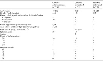
After obtaining approval from the ethical review committee of Tongji Hospital, Tongji Medical College, Huazhong University of Science and Technology, liver biopsy specimens were collected from 58 patients with hepatic fibrosis, including 42 patients with chronic schistosomiasis (CS) and 16 patients with chronic hepatitis B (CHB), and five healthy individuals (HI) from October 2010 to September 2012. All patients with CS had a history of infective water contact and treatment with praziquantel in the past. Patients with CHB were positive for hepatitis B virus surface antigen for at least 6 months. None of the patients exhibited serological evidence of other hepatitis virus infection, autoimmune liver disease, heritable disorder or history of alcoholism or drug abuse. Histological activities of inflammation and fibrosis and clinical symptoms are presented in Table 1. All patients were subjected to laboratory examinations including routine urine, stool and blood tests as well as liver and renal function tests. Morphological parameters such as ultrasonographic analysis of splenomegaly, portal vein width and liver stiffness value were also measured.
Baseline characteristics of the patients and controls

a Data are expressed as mean±s.d.
Liver biopsy specimen collection and pathology
Liver biopsy was performed using a 16-gauge core biopsy needle with a Bard Magnum Biopsy Instrument (Bard Peripheral Vascular, Inc., USA) under the guide of B type ultrasound. Liver biopsy samples that were 1·5–2·2 cm long were fixed immediately in 10% neutral-buffered formalin and processed into paraffin sections. The procedure was repeated once for another specimen that was performed for cryopreservation at −80 °C for future use. Histological sections were stained with haematoxylin and eosin (H&E) and Masson for evaluation. Blinded analysis by two pathologists according to the method of Desmet was used to determine the grade of necroinflammation (G1–G4) and the stage of fibrosis (S0–S4) (Desmet et al. Reference Desmet, Gerber, Hoofnagle, Manns and Scheuer1994; Yuan et al. Reference Yuan, Duan, Wang, Liang and Zhu2002).
Immunohistochemistry (IHC)
For immunohistochemical staining 4-μm thick sections cut from formalin-fixed, paraffin-embedded tissue were used. After deparaffinization in xylene and hydration in ethanol solutions, the sections of liver tissue were subjected to pre-treatment in order to enhance antigen retrieval. Conjugated horseradish peroxidase (HRP) secondary antibodies (Pepro Tech Inc., USA) were used for immunostaining with the following primary antibodies: rabbit polyclonal antibody to Hsp47 (1:150), rabbit polyclonal antibody to TGF-β1 (1:200) and goat polyclonal antibody to CTGF (1:200) (Santa Cruz, CA, USA). The immunostaining was performed according to the manufacturer's instructions. As a final step, counterstaining with haematoxylin was performed. Positive signals were seen as yellow, brown or tan staining. The immunostained sections were examined using Olympus TH4-200 (Olympus, Japan). The extent of the immunoreactivity in parenchymal liver cells, defined as the mean percentile value of immunoreactive cells, independently obtained by two pathologists, was semiquantitatively scored as follows (Turato et al. Reference Turato, Calabrese, Biasiolo, Quarta, Ruvoletto, Tono, Paccagnella, Fassina, Merkel, Harrison, Gatta and Pontisso2010): 0 = absence of immunoreactive cells; 1 = 1–10% immunoreactive cells; 2 = 11–50% immunoreactive cells; 3 = >50% immunoreactive cells. Intra- and inter-observer differences were <5% and discordant cases were re-evaluated by the two observers.
RNA isolation and real-time PCR
Total RNA was extracted using Trizol Reagent (Invitrogen, USA) according to the manufacturer's instructions and quantified using a spectrophotometer (Beckman Coulter, USA) for nucleic acid analyses. Total RNA (up to 1 μg) was reverse transcribed to cDNA using the FAQ-201 RT reagent kit (Toyobo Bio Inc., Japan) according to the manufacturer's protocol. All PCR reactions were performed in the C1000 Thermal Cycler System (Bio-rad CFX96, USA) using QPK-101 SYBR Green Real-time PCR Master Mix (Toyobo Bio Inc., Japan). The primers were designed following standard procedures (http://www.ncbi.nlm.nih.gov/tools/primer-blast/) and were as follows:
-
human HSP47 forward: 5′-CTTCGCTGATGACTTCGTGC-3′,
-
reverse: 5′-ATGAAGCCACGGTTGTCCAC-3′;
-
TGF-β1 forward: 5′-TGGCGATACCTCAGCAACC-3′,
-
reverse: 5′-CTAAGGCGAAAGCCCTCAAT-3′;
-
CTGF forward: 5′-GAGGAGTGGGTGTGTGTGACGA-3′,
-
reverse: 5′-GGACCAGGCAGTTGGCTCTA-3′;
-
Beta-Actin forward: 5′-AACTGGAACGGTGAAGGTGAC-3′,
-
reverse: 5′-TGTGGACTTGGGAGAGGACTG-3′.
Cycling conditions were 40 cycles of denaturation at 95 °C for 30 s, annealing at 58 °C for 15 s, and extension at 72 °C for 30 s. Real-time fluorescence measurements was recorded and the threshold cycle (Ct) value for each sample calculated by the above sequence detector (Heid et al. Reference Heid, Stevens, Livak and Williams1996). The amount of mRNA was calculated using the comparative C t method and was expressed as 2−ΔΔCt . The 2−ΔΔCt method presented the data as fold change in mRNA expression of the target gene, normalized to the mRNA expression of the housekeeping gene.
The specificity of each amplicon was determined by melt curve analysis and 1% agarose gel electrophoresis. Each biopsy sample obtained from the same patient was tested in duplicate, and the average of two Ct values was used in this study.
Statistical analysis
Continuous normally distributed variables were analysed by one-way analysis of variance with post Tukey's HSD multiple comparison test, and non-normal variables were analysed for different groups (non-paired analysis) by a non-parametric Kruskal–Wallis with post Dunn's multiple comparison test. Student t-test for parametric samples was used for two-group comparisons. Associations between the mRNA levels of Hsp47, TGF-β1 and CTGF with the histological features (stage of fibrosis, grade of inflammation) were analysed using the Jonckheere–Terpstra test. Correlations between clinical data and mRNA levels of these molecules were performed by Spearman rank correlation test. All statistical analyses were performed using the SPSS program, version 12.0 (SPSS Inc., Chicago, USA). Statistical significance was accepted at P<0·05.
Ethical aspects
Informed consent was obtained from all patients. This study was designed and conducted according to the principles of the Declaration of Helsinki and was approved by the Ethics Committee of Tongji Hospital.
RESULTS
Forty-two cases of CS, 16 cases of CHB and five HI were enrolled in the study. No complications of bleeding, infection and pneumothorax were found immediately after liver biopsy. In the CS group, 18 patients had 1–3 courses for antischistosomiasis treatment, and 24 patients had 4–6 courses. One patient had a history of splenectomy in stages 2 and 4.
Morphological changes in biopsy
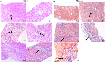
Pathological lesions caused by S. japonicum infection were observed in the livers of CS patients. The schistosome eggs were randomly lodged in the liver, although clustering of eggs was sometimes seen. Schistosoma japonicum eggs were found in liver specimen sections of 20 patients with CS. In the portal areas, capsule and parenchyma of the liver, large numbers of infiltrating eosinophils were seen in the specimens, by H&E staining. In addition, Masson staining showed that collagen fibres were deposited at the periphery of the eosinophilic granuloma. A comparison of typical H&E stain and Masson stain images of liver sections is shown in Fig. 1.
Histopathological classification of liver specimens. (A) Evaluation of inflammatory grade in liver biopsy specimens by H&E staining. G1, little inflammation around the portal vein; G2, local inflammatory cell infiltration around the granulomas; G3, extensive inflammatory cell infiltration with multiple foci of periportal interface inflammation. Arrows indicate regions of egg deposition; (B) Evaluation of fibrotic stage in liver biopsy specimens by Masson staining. S0, no fibrotic areas, normal amount of connective tissue around the portal vein; S1, mild fibrosis in the periportal area restricted to perisinuous lobules; S2, periportal fibrosis evident with fibrous septa formation; S3, fibrosis and fibrous septaformation accompanying disorder of lobular structures; S4, widespread fibrotic proliferation and enlarged fibrous septa. Arrows show dense collagen deposits in liver tissue.

Based on standard guidelines for fibrotic stage classification (Desmet et al. Reference Desmet, Gerber, Hoofnagle, Manns and Scheuer1994; Yuan et al. Reference Yuan, Duan, Wang, Liang and Zhu2002), 15 (36%) cases of CS patients were in stage 1; 10 (24%) cases were in stage 2; 11 (26%) cases were in stage 3; and 6 (14%) cases were in stage 4. Based on classification of inflammatory grade, 26 (62%) cases of CS patients were in grade 1; 11 (26%) cases were in grade 2; and 5 (12%) cases were in grade 3 (Table 1).
Comparison of the protein levels of Hsp47, TGF-β1 and CTGF in CS, CHB and HI
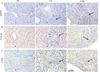
The protein levels of Hsp47, TGF-β1 and CTGF were evaluated by IHC. Low expression of Hsp47, TGF-β1 and CTGF was observed in liver specimens from HI. However, the protein levels of Hsp47, TGF-β1 and CTGF were elevated in patients with chronic hepatic fibrosis. Representative photomicrographs showed Hsp47-positive cells were localized mainly in areas of inflammation and fibrosis (Fig. 2A). In liver specimens of patients with chronic hepatic fibrosis, TGF-β1 was observed in hepatocytes at the border between the parenchyma and connective tissue and entrapped in fibrotic bands surrounding the pseudolobules (Fig. 2B). CTGF was detected in portal tracts and fibrous septa predominantly in the fibrogenesis area, at the interface between the fibrous septa and liver parenchyma (Fig. 2C).
Immunohistochemical evaluation of the protein levels of HSP47, TGF-β1 and CTGF in the liver. (A) Immunostaining for Hsp47 in HI and patients with CS and CHB. (B) Immunostaining for TGF-β1 in HI and patients with CS and CHB. (C) Immunostaining for CTGF in HI and patients with CS and CHB. Regions of fibrosis displaying positive cytochemistry reactions are indicated by arrows.

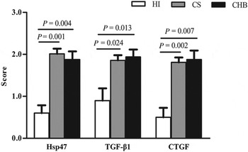
Statistical analysis of the IHC scores indicated that Hsp47 expression in patients with CS and CHB was significantly higher than in those with HI (P = 0·001 and P = 0·004, respectively). Semi-quantitative scoring of Hsp47 immunostaining in CS group was 2·0 (1·00–2·62) (median with interquartile range), CHB group was 2·0 (1·12–2·50) and HI group was 0·5 (0·25–1·00). The expression of TGF-β1 was higher in the CS and CHB groups compared with HI (P = 0·024 and P = 0·013, respectively). The immunoreactive score for TGF-β1 in CS group was 2·0 (1·00–2·50), in CHB group was 2·0 (1·50–2·50) and HI group was 1·0 (0·25–1·50). The expression of CTGF was higher in CS and CHB groups compared with HI group (P = 0·002 and P = 0·007, respectively). The staining intensity of CTGF in CS group was 2·0 (1·00–2·50), in CHB group was 2·0 (1·00–2·50) and HI group was 0·5 (0·00–1·00) (Fig. 3). However, there were no statistically significant differences between CS and CHB groups in regard to the expression of these molecules (P = 0·564, P = 0·756 and P = 0·765, respectively).
Semi-quantitative scoring of Hsp47, TGF-β1 and CTGF expression in the liver. The semiquantitative method was used to score the immunoreactivity of parenchymal liver cells as follows: score of 0, absence of positively stained cells; score of 1, fewer than 10% positively stained cells; score of 2, 11–50% positively stained cells; score of 3, more than 50% positively stained cells. Intra- and inter-observer differences were <5% and discordant cases were re-evaluated by the two observers.

Comparison of the mRNA levels of Hsp47, TGF-β1 and CTGF in CS patients and HI
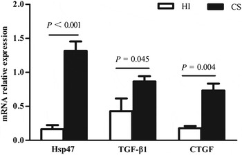
The mRNA expression levels of Hsp47, TGF-β1 and CTGF were detected in 42 specimens from CS patients and five HI donors. The mRNA levels of Hsp47, TGF-β1 and CTGF were remarkably higher in CS patients than those in HI (P<0·001, P = 0·045, P = 0·004, respectively) (Fig. 4). Compared with HI group, an 8-fold increase in the expression of Hsp47 was noted in the liver tissues obtained from CS patients. The expression of TGF-β1 and CTGF was more than 2-fold and 4-fold higher in CS patients than in HI group, respectively.
Relative expression of Hsp47-mRNA, TGF-β1-mRNA and CTGF-mRNA in liver tissues from HI and CS patients. Expression levels of the target gene were relative to the mRNA expression of Beta-Actin.

Correlations with the progression of hepatic fibrosis
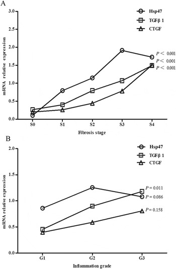
To better define the correlation of the gene expression of Hsp47, TGF-β1 and CTGF with the progression of hepatic fibrosis, results were evaluated according to the extent of fibrosis and the grade of inflammation. As shown in Fig. 5A, the expression of Hsp47, TGF-β1 and CTGF mRNA was positively associated with the fibrosis stages (P<0·001 in all cases). The mRNA level of Hsp47 was significantly increased in CS patients with stage 2 fibrosis, and reached a maximum in patients with stage 3 fibrosis compared with stage 0 (P = 0·038, P<0·001, respectively). Although a slight decline occurred in stage 4, Hsp47 mRNA expression in these patients was significantly increased in comparison to stage 0 (P<0·001). In contrast, the expression of TGF-β1 and CTGF was significantly elevated in stage 3 fibrosis (P<0·001, P = 0·011, respectively), later increases than shown by Hsp47.
The dynamic changes of Hsp47, TGF-β1 and CTGF mRNA expression in the progression of schistosomiasis hepatic fibrosis. (A) The mRNA levels of Hsp47, TGF-β1 and CTGF with respect to fibrosis stage; (B) The mRNA levels of Hsp47, TGF-β1 and CTGF with respect to inflammation grade. Expression levels of the target gene were relative to the mRNA expression of Beta-Actin. Different icons represent median values.

Based on inflammation grade classification guidelines, the mRNA level of TGF-β1 was correlated with grade of inflammation (P = 0·011), while the expression of Hsp47 and CTGF was not (P = 0·086, P = 0·158, respectively) (Fig. 5B).
Correlations with clinical parameters
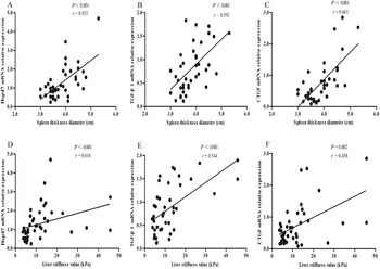
In order to further evaluate the role of Hsp47, TGF-β1 and CTGF in inflammatory and fibrotic progression, we compared the mRNA levels of these molecules in CS patients with some clinical parameters, such as laboratory examinations (e.g. alanine aminotransferase (ALT), aspartate aminotransferase (AST), ratio of AST/ALT (AARI), g-glutamyl transpeptidase (GGT), alkaline phosphatase (ALP), total bilirubin and total albumin) and imaging tests (e.g. spleen thickness diameter, portal vein width and liver stiffness value). The mRNA levels of Hsp47 and CTGF did not correlate with biochemical liver parameters, except for serum GGT level (r = 0·378, r = 0·300; P = 0·014, P = 0·046, respectively) (in supplementary data). In contrast, the mRNA level of TGF-β1 was correlated with serum levels of AST, ALP and GGT (r = 0·359, r = 0·330, r = 0·470; P = 0·02, P = 0·033, P = 0·002, respectively) (in supplementary data), but not with serum levels of ALT, AARI, albumin, total bilirubin (P = 0·051, P = 0·831, P = 0·641, P = 0·138, respectively) (data about ALT shown in supplementary data; the others not shown). Non-invasive methods, namely, ultrasonography and transient elastography, were utilized to determine spleen thickness diameter, portal vein width and liver stiffness value. The mRNA levels of Hsp47, TGF-β1 and CTGF were positively correlated with spleen thickness diameter (n = 40; r = 0·553, r = 0·593, r = 0·661, respectively; P<0·001 in all cases) (Fig. 6A–C) and liver stiffness value (r = 0·616, r = 0·544, r = 0·458 and P<0·001, P<0·001, P = 0·002, respectively) (Fig. 6D–F), but not with portal vein width (P = 0·57, P = 0·53, P = 0·775, respectively) (data not shown).
Correlations between mRNA levels of Hsp47, TGF-β1 and CTGF with spleen thickness diameter or liver stiffness value. (A–C) Correlations between mRNA levels of Hsp47, TGF-β1 and CTGF with spleen thickness diameter (n = 40); (D–F) Correlations between mRNA levels of Hsp47, TGF-β1 and CTGF with liver stiffness value (n = 42). Expression levels of the target gene were relative to the mRNA expression of Beta-Actin.

DISCUSSION
In the present study, we examined the expression of Hsp47, TGF-β1 and CTGF in liver biopsy specimens from patients with CS and CHB, and from HI. Our results showed that not only were the expression levels of Hsp47, TGF-β1 and CTGF significantly higher in patients with hepatic fibrosis than in HI, their gene expression also appeared to be positively correlated with the stage of fibrosis, which provided evidence of their involvement in the progression of schistosomal hepatic fibrosis. Moreover, TGF-β1 gene expression increased parallel to the grade of inflammation. As particular attention was paid to the non-invasive diagnostic parameters, we broadened the focus of our study by providing indirect evidence of the major roles the three molecules play by analysing the correlations between their mRNA expression and certain parameters. The mRNA levels of Hsp47 and CTGF had a relationship with serum level of GGT, spleen thickness diameter and liver stiffness value, which were increased in parallel with the extent of hepatic fibrosis (Lu et al. Reference Lu, Zeng, Wan, Li, Mao, Li, Qiu, Cao, Ye, Cai, Chen, Wang, Wu, Zhu and Zhou2003; Morais et al. Reference Morais, Carvalho Bde, Melo, Melo, Lopes, Domingues, Juca, Martins, Diniz and Montenegro2010; El-Bassiouni et al. Reference El-Bassiouni, Nosseir, Madkour, Zoheiry, Bekheit, Ibrahim, Ibrahim and El Bassiouny2012). Interestingly, the mRNA expression of TGF-β1 correlated not only with these parameters, but also with serum levels of AST, ALP and GGT, which represented the severity of inflammation (Lu et al. Reference Lu, Zeng, Wan, Li, Mao, Li, Qiu, Cao, Ye, Cai, Chen, Wang, Wu, Zhu and Zhou2003). On the basis of histology of chronic liver diseases, the early elevation of Hsp47 gene expression was found at stage 2 fibrosis, while increased expression of TGF-β1 and CTGF was detected at stage 3 fibrosis. Thus, it is suggested that TGF-β1 is involved in the progression of inflammation, and that Hsp47 might be a potential biomarker of early onset of hepatic fibrosis in human S. japonicum infection.
There is little information in the medical literature on detecting the gene expression of pro-fibrotic molecules in liver biopsy as percutaneous liver biopsy is invasive. Although Brown et al. investigated Hsp47 expression in human liver obtained from patients undergoing liver transplantation, they divided groups according to the aetiology of cirrhosis instead of the stage of fibrosis, and did not illustrate how many patients were collected in the study (Brown et al. Reference Brown, Broadhurst, Mathahs, Brunt and Schmidt2005). Their results showed that immunoreactive Hsp47 increased substantially, and Hsp47 transcripts tended to be more abundant in cirrhotic livers compared with controls, but the differences were not statistically significant. The authors raised the possibility that Hsp47 gene expression was constitutive in activated human HSC and levels of Hsp47 transcripts increased during periods of active fibrogenesis. Nevertheless, this possibility might be verified by a study examining levels of Hsp47 mRNA in liver biopsies at various stages of fibrosis. In addition, a few studies utilized IHC and ELISA (enzyme-linked immunosorbent assay), and revealed that TGF-β1 and CTGF were up-regulated in hepatic fibrosis induced by hepatitis B virus infection, hepatitis C virus infection and non-alcoholic fatty liver (Kanzler et al. Reference Kanzler, Baumann, Schirmacher, Dries, Bayer, Gerken, Dienes and Lohse2001; Lu et al. Reference Lu, Zeng, Wan, Li, Mao, Li, Qiu, Cao, Ye, Cai, Chen, Wang, Wu, Zhu and Zhou2003; Colak et al. Reference Colak, Senates, Coskunpinar, Oltulu, Zemheri, Ozturk, Doganay, Mesci, Yilmaz, Enc, Kiziltas, Ulasoglu and Tuncer2012). In our opinion, quantitative evaluation of mRNA levels in liver specimens was helpful to investigate their correlation with the progression of CS. Although the number of patients in the present study was sufficient, the size of sub-groups of patients with grade 3 or 4 inflammation, as well as that of patients with stage 4 fibrosis, was limited. In such patients, liver biopsies were performed rarely, and consequently we had a small number of liver biopsy specimens for such patients.
Previous studies confirmed the identity of Hsp47-positive cells as HSC based on co-localization of Hsp47 expression with synaptophysin, an established marker of HSCs, in human liver (Brown et al. Reference Brown, Broadhurst, Mathahs, Brunt and Schmidt2005; Sato et al. Reference Sato, Murase, Kato, Kobune, Sato, Kawano, Takimoto, Takada, Miyanishi, Matsunaga, Takayama and Niitsu2008). Consistent with these results, we observed that immunoreactive Hsp47 obviously increased in patients with CS and CHB compared with HI. Although CHB patients had lower Hsp47 expression than CS patients, no statistically significant difference was found between the two groups. It is demonstrated that the observed up-regulation of Hsp47 is related to an increase in collagen deposition, irrespective of the aetiology of the liver disease (Brown et al. Reference Brown, Broadhurst, Mathahs, Brunt and Schmidt2005).
Results of a study performed by Sato et al. (Reference Sato, Murase, Kato, Kobune, Sato, Kawano, Takimoto, Takada, Miyanishi, Matsunaga, Takayama and Niitsu2008) indicated that Hsp47 was involved in the pathogenesis of chronic hepatic fibrosis and might be a potential target for antifibrotic therapy. We previously reported similar results when using Hsp47-targeted shRNA in mouse hepatic fibrosis associated with S. japonicum (Huang et al. Reference Huang, Tao, Li, Ma, Xu, Ai, Fan, Jiao and Ning2014). On the other hand, it was reported that Hsp47 expression could be induced by many inflammatory factors, such as TGF-β1, IL-1 and IL-17 (Sasaki et al. Reference Sasaki, Sato, Yamauchi, Okamoto, Kobayashi, Iyama, Kato, Matsunaga, Takimoto, Takayama, Kogawa, Watanabe and Niitsu2002; Honzawa et al. Reference Honzawa, Nakase, Matsumura, Yamamoto, Uza, Matsuura and Chiba2011). Consequently, we paid attention to the relationship between expression of Hsp47 and progression of fibrosis and inflammation. In our study sample, composed of 42 CS patients, the mRNA level of Hsp47 increased significantly with aggravation of hepatic fibrosis, initially increasing at stage 2, peaking at stage 3 and then declining slightly at stage 4. Observations made using in situ hybridization in carbon tetrachloride (CCl4)-treated rat liver were also relevant to this question. While the abundance of Hsp47-expressing cells increased throughout the period of CCl4 administration, Hsp47-expressing cells decreased slightly in areas of ‘completed’ fibrosis at 12 weeks of CCl4 treatment (Masuda et al. Reference Masuda, Fukumoto, Hirayoshi and Nagata1994). This decline in Hsp47 expression might be attributed to the fact that Hsp47 is related not only to secretion in all activated HSCs, but also to the number of HSCs (Krizhanovsky et al. Reference Krizhanovsky, Yon, Dickins, Hearn, Simon, Miething, Yee, Zender and Lowe2008; Nishikawa et al. Reference Nishikawa, Takahara, Asada, Shigenaga, Otaka, Kitagawa and Koide2010). In addition, liver compensatory function decreased during cirrhosis (stage 4) which limited the proliferation of HSCs and thereby lowered Hsp47 expression. Although a slight decline of Hsp47 expression at stage 4 fibrosis had no effect on its correlation with the extent of fibrosis, the level of Hsp47 might underestimate advanced hepatic fibrosis in CS patients. In contrast to the results of previous in vitro experiments (Sasaki et al. Reference Sasaki, Sato, Yamauchi, Okamoto, Kobayashi, Iyama, Kato, Matsunaga, Takimoto, Takayama, Kogawa, Watanabe and Niitsu2002; Honzawa et al. Reference Honzawa, Nakase, Matsumura, Yamamoto, Uza, Matsuura and Chiba2011), increased mRNA level of Hsp47 did not correlate with the degree of inflammation in CS patients. Therefore, the biological relevance of Hsp47 expression with inflammatory grade and fibrotic stage merits further epidemiological study.
TGF-β1, the key mediator of fibrogenesis, can regulate the activation and transformation of HSCs, and the accumulation of ECM in hepatic fibrosis. Located downstream of TGF-β1, CTGF is vital in the formation of connective tissues and is over-expressed in CS (Huang et al. Reference Huang, Zhu, Li, Jiao, Xu, Tao, Fan, Ma, Guo and Ning2012; Hao et al. Reference Hao, Xie, Peng, Ma, Zhou, Zhang, Kang, Wang, Bai, Wang and Jia2013). Similar to the results of previous studies, we found that mRNA levels of TGF-β1 and CTGF were significantly higher in CS patients than in HI. The results of published studies agree that the expression of TGF-β1 and CTGF was related to the stage of fibrosis (Abou-Shady et al. Reference Abou-Shady, Friess, Zimmermann, di Mola, Guo, Baer and Buchler2000; Kanzler et al. Reference Kanzler, Baumann, Schirmacher, Dries, Bayer, Gerken, Dienes and Lohse2001; Piekarska et al. Reference Piekarska, Piekarski, Omulecka, Szymczak and Kubiak2006). In the present study, the mRNA levels of TGF-β1 and CTGF increased along with the severity of hepatic fibrosis due to S. japonicum infection. Interestingly, the grade of inflammation was correlated with the mRNA level of TGF-β1 in CS patients, but not with that of CTGF. The results of a few published studies are contradictory. Castilla et al. and Malizia et al. performed studies on TGF-β1 at the molecular level. They found a significant correlation between the presence of mRNA TGF-β1 and sub-indexes of inflammation and fibrosis (Castilla et al. Reference Castilla, Prieto and Fausto1991; Malizia et al. Reference Malizia, Brunt, Peters, Rizzo, Broekelmann and McDonald1995). However, a few studies reported that the expression of TGF-β1 was increased in foci of piecemeal necrosis, but failed to find a significant correlation between TGF-β1 expression and inflammation or fibrosis (Banner et al. Reference Banner, Allan, Savas, Baker, Barnard and Bonkovsky1997; Piekarska et al. Reference Piekarska, Piekarski, Omulecka, Szymczak and Kubiak2006). It is hard to establish whether the high expression of TGF-β1 is a result of chronic damage of cells or is the cause of their damage. The answer requires further study. Notably, we could only conclude that TGF-β1 was involved in the progression of inflammation since there were no patients with grade 4 inflammation. Furthermore, a previous study involving patients infected with hepatitis B virus (Guo-Qiu et al. Reference Guo-Qiu, Nai-Feng, Xiao-Bo, Linxian, Chen, Lixia and Zhao2010) showed no significant association between the serum levels of CTGF and the grade of inflammation. This result is consistent with the findings of our study.
In the clinical setting, non-invasive tests are increasingly precise in identifying the cause of liver disease and even the amount of hepatic fibrosis, since liver biopsy is invasive and is associated with rare but serious complications (Castera and Pinzani, Reference Castera and Pinzani2010; Ding et al. Reference Ding, Wu, Ma, Wang, Wu, Guo, Qi and Ning2012). To further investigate the role of Hsp47, TGF-β1 and CTGF in pathogenesis of schistosomal hepatic fibrosis, we examined correlations of their mRNA levels with clinical parameters. Consistent with our histological results, mRNA expression of TGF-β1 was correlated with serum levels of AST, ALP and GGT, all of which were closely related to inflammatory activity (Lu et al. Reference Lu, Zeng, Wan, Li, Mao, Li, Qiu, Cao, Ye, Cai, Chen, Wang, Wu, Zhu and Zhou2003). The mRNA level of TGF-β1 exhibited strong correlation with serum level of GGT, while the mRNA levels of Hsp47 and CTGF were only weakly correlated with it. Our study also confirmed that the mRNA levels of the three molecules correlated with spleen thickness diameter and liver stiffness value, both of which, to some extent, represented the severity of fibrosis (Li et al. Reference Li, Chen, Ross, Burke, Yu, Li, Zhou and McManus2011). Notably, the mRNA level of Hsp47 exhibited the best correlation with liver stiffness values, while that of CTGF was highly correlated with spleen thickness diameter. These results suggest that Hsp47 and CTGF may contribute more direct effort to the progression of fibrosis than TGF-β1. The mRNA levels of Hsp47, TGF-β1 and CTGF were not significantly correlated with portal vein width, possibly because the width is less sensitive to portal hypertension than spleen thickness diameter (De Cock, Reference De Cock1986). However, this negative result should be interpreted with caution because of the sample size used in the present study. Larger sample sizes and more variants should be used in future studies to confirm this result.
In summary, the results obtained in this study suggest that Hsp47, TGF-β1 and CTGF are involved in the pathogenesis of schistosomal hepatic fibrosis. Two novel observations were made in this study: first, the expression of Hsp47 mRNA was found to increase, starting at stage 2, with aggravation of hepatic fibrosis induced by S. japonicum infection. This indicates that Hsp47 may be a suitable maker for the early stage of schistosomal hepatic fibrosis. Second, the mRNA levels of Hsp47 and CTGF were found to exhibit strong correlations with liver stiffness value and spleen thickness diameter, respectively. Overall, our results identify the roles of pro-fibrosis factors in human schistosomal hepatic fibrosis, and may aid in choosing therapeutic targets for early stage of fibrosis.
SUPPLEMENTARY MATERIAL
To view supplementary material for this article, please visit http://dx.doi.org/10.1017/S0031182014001115.
ACKNOWLEDGEMENTS
We are sincerely grateful to PhD candidate Hongyan Wang for proofreading this article. The authors declare that no conflicts of interest exist.
FINANCIAL SUPPORT
This work was supported by grants from the National Natural Science Foundation of China [81171558, 81100282], National twelfth ‘five years’ project in Science and Technology [2012ZX10002-002-004] and Changjiang Scholars and Innovation Research Team in University [IRT1131].