INTRODUCTION
While reviewing literature on sarcocystosis in animals, we found considerable confusion concerning the Sarcocystis species in camels (Dubey et al. Reference Dubey, Calero-Bernal, Rosenthal, Speer and Fayer2015). The morphological descriptions were often vague, and there are no archived specimens for verification. Here, we have summarized available reports on Sarcocystis infection in camels and provided redescription of sarcocysts of two species, Sarcocystis cameli and Sarcocystis ippeni.
REVIEW OF LITERATURE
Species names
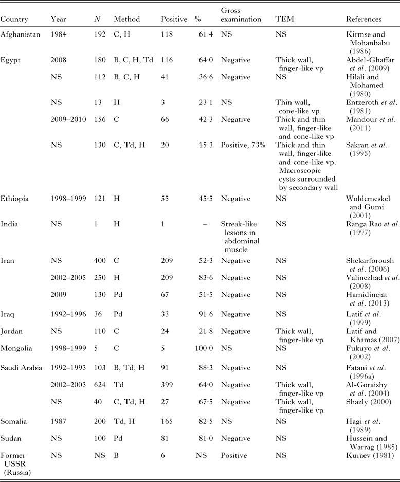
Mason (Reference Mason1910) first reported sarcocysts in muscles of camels slaughtered for food in Cairo, Egypt. All old and emaciated camels had numerous sarcocysts that were found in virtually all muscles, including the heart, but the numbers of camels infected or examined were not provided. Sarcocysts were up to 12 mm long and less than 1 mm wide, appearing as white lines, with thin or thick cyst walls, but no measurements of the thickness of the wall was given; the parasite was named as S. cameli (Mason, Reference Mason1910). Dubey et al. (Reference Dubey, Speer and Fayer1989) arbitrarily termed the so called thick-walled sarcocyst of Mason (Reference Mason1910) S. cameli, but did not name the thin-walled species which was subsequently called S. ippeni by Odening (Reference Odening1997). Neither Dubey et al. (Reference Dubey, Speer and Fayer1989) nor Odening (Reference Odening1997) examined specimens reported by Mason (Reference Mason1910) or sarcocysts from other camels. The presence of thick- and thin-walled sarcocysts was confirmed in camels from Saudi Arabia (Fatani et al. Reference Fatani, Hilali, Al-Atiya and Al-Shami1996a) and Somalia (Hagi et al. Reference Hagi, Hassan and Di Sacco1989) (Table 1). Abdel-Ghaffar et al. (Reference Abdel-Ghaffar, Mehlhorn, Bashtar, Al-Rasheid, Sakran and El-Fayoumi2009) studied the prevalence of Sarcocystis infection in camels in Cairo, Egypt. Microscopic sarcocysts, found in 116 of 180 camels from an abattoir, were 120–170 × 50–100 µ m in size and of one morphological type. Dogs that were fed heavily infected camel meat excreted Sarcocystis sporocysts (Table 2). The parasite studied was called Sarcocystis camelicanis without elaborating on the new name.
Prevalence of Sarcocystis sarcocysts in camels

NS, Not stated; B, bioassay in dog; C, compression/muscle squash; G, gross examination; H, histology; Pd, pepsin digestion; Td, trypsin digestion; TEM, transmission electron microscopy; vp, villar protrusions.
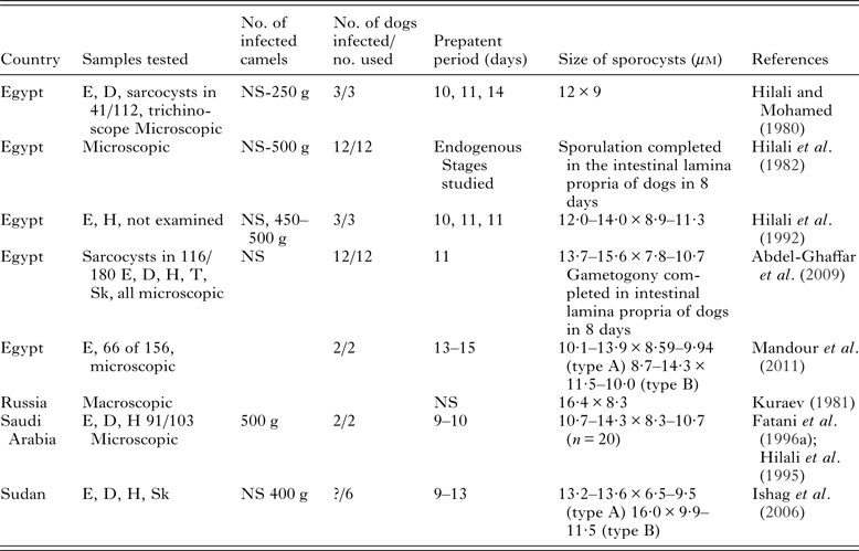
Excretion of Sarcocystis sporocysts in feces of dogs fed camel meat.

NS, not stated; D, diaphragm; E, esophagus; H, heart; Sk, skeletal muscle; T, tongue.
Ishag et al. (Reference Ishag, El Amin and Osman2001, Reference Ishag, Majid and Magzoub2006) in Sudan studied transmission of Sarcocystis between camels and dogs. They found two types of sarcocysts, thick- and thin-walled, in a camel fed sporocysts from dogs (Ishag et al. Reference Ishag, El Amin and Osman2001) and two different sized sporocysts (Table 2, 13·2–13·6 × 6·5–9·5 and 16·0 × 9·9–11·5 µ m) in dogs that were fed camel meat (Ishag et al. Reference Ishag, Majid and Magzoub2006). They named the larger sporocyst in the dog that was fed camel meat as a new species, Sarcocystis camelocanis, but gave no description of the sarcocyst.
To add to this confusion, another new species from the camel was named, Sarcocystis miescheri, based on finding oocysts in feces of dogs fed naturally infected camel meat (Mandour et al. Reference Mandour, Rabie, Mohammed and Hussein2011). Illustrations provided by the authors resemble Cystoisospora ohioensis oocysts measuring 20·8–26·7 × 18·5–20·7 µ m with a thick wall and containing two sporoblasts, and bearing no resemblance to other species of Sarcocystis. The bradyzoites, measuring 21·5–32·8 × 7·7–17·7 µ m, appeared to be artefacts misidentified as bradyzoites (Dubey et al. Reference Dubey, Calero-Bernal, Rosenthal, Speer and Fayer2015).
There are therefore currently five named Sarcocystis species in camels, namely S. cameli, S. ippeni, S. camelicanis, S. camelocanis and S. miescheri.
Sarcocyst size
There is considerable confusion concerning the size of sarcocysts. As stated earlier, Mason (Reference Mason1910) found sarcocysts in camels that were up to 12 mm long. Kuraev (Reference Kuraev1981) in Russia reported macroscopic sarcocysts in the oesophagi of six camels. Thick- and thin-walled sarcocysts between 6 and 15 mm long with a variety of shapes including oval, spindle and cylindrical, were present. Dogs fed infected camel tissues excreted 16·4 × 8·3 µ m sized sporocysts; no details of the experiment were provided. This report needs confirmation and is mentioned only in the context of a complete review of Sarcocystis infection in camels. Sakran et al. (Reference Sakran, Abdel-Aziz and Abdel-Ghaffar1995) reported macroscopic sarcocysts in 95 of 130 oesophagi and 25 of 50 diaphragms of camels from Cairo, Egypt. The results of this investigation are difficult to reconcile with their subsequent paper where they did not find macroscopic sarcocysts in camels from Cairo, Egypt (Abdel-Ghaffar et al. Reference Abdel-Ghaffar, Mehlhorn, Bashtar, Al-Rasheid, Sakran and El-Fayoumi2009). Sarcocysts were found in tissue sections of 116 of 180 camels; in 60% of oesophagi, 50% of diaphragms, 40% of tongues and 10% of hearts (Abdel-Ghaffar et al. Reference Abdel-Ghaffar, Mehlhorn, Bashtar, Al-Rasheid, Sakran and El-Fayoumi2009). Sarcocysts were 120–170 × 50–100 µ m in size, and only one morphologic type of sarcocyst was found. Both reports are by the same group of scientists (Sakran et al. Reference Sakran, Abdel-Aziz and Abdel-Ghaffar1995; Abdel-Ghaffar et al. Reference Abdel-Ghaffar, Mehlhorn, Bashtar, Al-Rasheid, Sakran and El-Fayoumi2009). There is speculation whether the epidemiology of sarcocystosis in camels has changed drastically between 1995 when the Sakran et al. (Reference Sakran, Abdel-Aziz and Abdel-Ghaffar1995) study was published vs the recent study (Abdel-Ghaffar et al. Reference Abdel-Ghaffar, Mehlhorn, Bashtar, Al-Rasheid, Sakran and El-Fayoumi2009). The point is raised because of the condemnation of meat with grossly visible sarcocysts.
Prevalence of sarcocysts
Sarcocysts or Sarcocystis-like bradyzoites have been reported in up to 91% of one-humped camels from several countries (Table 1) but the species of Sarcocystis were not determined.
Life cycle studies and excretion of sporocysts by dogs
Dogs fed naturally infected camel meat containing microscopic sarcocysts in Egypt and Saudi Arabia excreted sporocysts, and gametogonic stages were found in small intestines of dogs (Table 2). Because camel meat fed to dogs was not examined microscopically in each instance, it is uncertain if the dogs were hosts for one or both microscopic sarcocyst species.
Ultrastructural studies
Two types of sarcocysts have been described from camels. Sarcocysts with finger-like villar protrusions (vp) (variety A) and conical projections (variety B), but they have not been assigned to specific species.
Variety A. Abdel-Ghaffar et al. (Reference Abdel Ghaffar, Entzeroth, Chobotar and Scholtyseck1979) first reported ultrastructure of sarcocysts from camel in Egypt. Microscopic sarcocysts (130–180 × 60–110 µ m) were found in oesophagi and diaphragms (number of infected was not stated) of 44 camels examined. Sarcocysts had smooth wall by light microscopy (LM) (Abdel-Ghaffar et al. Reference Abdel Ghaffar, Entzeroth, Chobotar and Scholtyseck1979). Ultrastructurally, the cyst wall had 1·2–1·6 µ m long vp with a maximum width of 0·5 µ m. Bradyzoites were 8–12 × 2·5–3·8 µ m in size. Only one morphologic type was described; the parasite was not named. Abdel-Ghaffar et al. (Reference Abdel-Ghaffar, Mehlhorn, Bashtar, Al-Rasheid, Sakran and El-Fayoumi2009) in Cairo, Egypt added further to the description of this type of sarcocyst in camels in Cairo, Egypt. They reported 16–18 knob-like structures on each vp. As stated earlier they called this parasite S. camelicanis. Similar sarcocyst type was reported in camels from Iran (Motamedi et al. Reference Motamedi, Dalimi, Nouri and Aghaeipour2011), Jordan (Latif and Khamas, Reference Latif and Khamas2007) and Saudi Arabia (Al-Goraishy et al. Reference Al-Goraishy, Bashtar, Al-Rasheid and Abdel-Ghaffar2004).
Variety B. Entzeroth et al. (Reference Entzeroth, Abdel Ghaffar, Chobotar and Scholtyseck1981) found this parasite in three of 13 camels from Cairo, Egypt. Sarcocysts were 120–150 × 50–80 µ m in size. Cyst wall was not described by LM. Ultrastructurally, cyst wall had knob-like elevations on the surface. The cone-like vp were 0·5–1·4 µ m long. Bradyzoites were 10–12 × 2·5–4·0 µ m in size. Only one morphologic type was described.
Clinical sarcocystosis
In two separate experiments, young camels orally inoculated with Sarcocystis spp. sporocysts from dogs became ill. In the first experiment, two 6-month old camels in Saudi Arabia were inoculated orally with 250 000 or 750 000 sporocysts from experimentally infected dogs (Fatani et al. Reference Fatani, Hilali, Al-Atiya and Al-Shami1996a, Reference Fatani, El-Sebaie and Hilalib). Both camels became anorectic, developed pyrexia, became restless and anemic 29 days post inoculation (p.i.). One camel was euthanized 34 days p.i. and the second died day 41 p.i.; hemorrhages were found in viscera and muscles. Histopathological findings were not reported.
In the second experiment, two 1-month old camels in Sudan were inoculated orally with 1 000 000 sporocysts from feces of experimentally infected dogs (Ishag et al. Reference Ishag, El Amin and Osman2001). Both camels became anorectic, lethargic and anemic, beginning 20 day p.i. Camel 1 died 26 day p.i.; post mortem examination revealed hemorrhages in several organs and immature cysts containing metrocytes in the brain. The second camel was given food medicated with Amprolium® (100 mg kg−1 body weight), starting the day of sporocyst inoculation and continuing for 30 days. This camel remained asymptomatic and mature sarcocysts were found in muscles at necropsy on 110 p.i.
The objective of the present paper is to provide proper description of two types of sarcocysts by LM and transmission electron microscopy (TEM) and assign them to specific species.
MATERIAL AND METHODS
Naturally infected camels
Oesophageal tissues were collected from two adult camels (Camelus dromedarius) (nos. 4 and 5) on 15 January 2015 from an abattoir in Giza, Egypt. Tissues were fixed in glutaraldehyde (GF) or formalin (FF). The FF tissues were processed for paraffin embedding. The paraffin blocks and the GF samples were transported to the Faculty of Veterinary Science, University of Pretoria, Onderstepoort, Republic of South Africa for light and electron microscopic examinations. For LM, paraffin-embedded sections were cut at 5 µ m thick and examined after staining with hematoxylin and eosin (H and E). For TEM, GF tissue from camel no. 5 (cysts #1, 6, 7, 8), were processed using standard techniques. Briefly, the samples were post-fixed in 1% osmium tetroxide in Millonig's buffer (pH 7·4), dehydrated through a series of graded ethanols, infiltrated with an epoxy resin/propylene oxide mixture before being embedded in absolute resin and polymerized at 60 °C overnight. A further four tissue cysts, located in paraffin blocks (by matching with H and E sections) from camel no. 4 (cysts # 2, 3, 4, 5), were deparaffinized (Van den Berg Weermans and Dingemans, Reference Van den Berg Weermans and Dingemans1984). Toluidine blue-stained resin sections of all eight microcysts were photographed with an Olympus BX63 compound microscope (Olympus, Wirsam, South Africa). Ultrathin resin sections were contrasted with uranyl acetate and lead citrate and examined in a Philips CM10 TEM (FEI, Eindhoven, The Netherlands) operated at 80 kV. Digital images were captured with a Megaview III side-mounted digital camera and iTEM software (Olympus Soft Imaging Solutions GmbH, Münster, Germany).
RESULTS
Macromorphology and LM
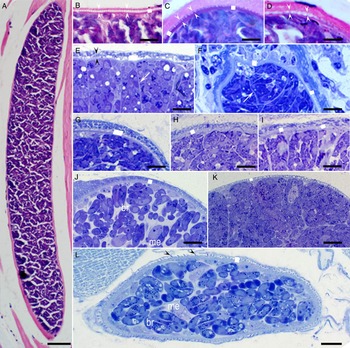
Twenty-two sarcocysts were found in H and E stained sections. All were mature and microscopic. The largest sarcocyst was 700 × 100 µ m (Fig. 1A and B). Eight sarcocysts (#1–8) were located in 1 µ m Toluidine blue-stained sections; they were 150 × 60 µ m (cyst #1), 270 × 45 µ m (cyst #2), 120 × 100 µ m (cyst #3), 120 × 50 µ m (cyst #4), 110 × 65 µ m (cyst #5), 226 × 80 (cyst #6), 47 × 38 (cyst #7) and 93 × 30 (cyst #8). The description is correlated between sections stained by Toluidine blue and by TEM but not with H and E stained sections.
Sarcocysts from camels from Egypt. Figures (C) and (E) are from camel no. 5, the remainders are from camel no. 4. (A–D), 5 mm sections stained with H and E, (E–L), Toluidine blue. Scale bar applies to all figures; 50 μm in (A), 10 μm in (B–I) and 5 μm in (L). The opposing arrowheads point to vp. The white squares point to thickness of the sarcocyst wall. The species of Sarcocystis was not identified in H and E stained sections. Based on TEM, sarcocysts in (E–I) are S. cameli and (J–L), S. ippeni. It is difficult to speciate these sarcocysts based on LM. (A, B) The largest sarcocyst found, probably S. cameli sarcocyst. The vp are very thin and barely visible and whitish areas are probably degenerated host tissue between vp. (C) Probably S. ippeni based on triangular vp. (D) Probably S. cameli. The sarcocyst wall on the right side appears different than on the left side. (E) Note indistinct cyst wall divided by septa. (F) S. cameli. Note prominent cyst wall. (G–K) Sarcocysts with prominent septa. (L) Sarcocystis ippeni sarcocyst with conical projection (arrowheads). Note pale me and banana shaped br. Abbreviations: vp, villar protrusions; TEM, transmission electron microscopy; LM, light microscopy; me, metrocytes; br, bradyzoites.

In H and E stained sections, all sarcocysts appeared to be thin walled (<2 μ m). All sarcocysts were mature. Representative images are shown in Fig. 1B–D. In some sarcocyst, conical projections could be seen on the sarcocyst wall (Fig. 1C). In 1 µ m Toluidine blue-stained sections, the structure of the sarcocyst wall was not clear, even at 1000X magnification (Fig. 1E–K). However, in one cyst photographed at higher magnification, conical projection was visible (Fig. 1L). In Toluidine blue-stained sections, metrocytes were stained faintly and appeared of different shapes. The bradyzoites were banana-shaped and 10–12 µ m.
TEM
Two structurally distinct sarcocysts were recognized by TEM, varieties A and B in both camels.
Variety A sarcocyst (S. cameli)
Three sarcocysts were studied, two from camel no. 4 and one from camel no. 5. Sarcocyst #1, 6 were GF cysts. Sarcocyst #2 was from camel no. 4 and was deparaffinized. The sarcocyst wall consisted of an outermost parasitophorous vacuolar membrane (pvm) that was lined by an electron dense layer (edl) that was up to 50 nm thick (Fig. 2E and H). The pvm had numerous vp at regular intervals (Fig. 2A–D). The host myocyte was degenerated along the vp to a varying degree, giving the impression that vp were apart (Fig. 1A–D). The vp were slender, with a maximum length of 3 µ m from the base to the tip, and approximately 0·5 µ m width (Fig. 1E). Several microtubules were present from the tip of the villus to the middle of ground substance (gs) layer; the tubules were smooth, were without granules and had fine cross-striations on the surfaces of the tubules. On each villus, there were several rows (16 or more) of knob-like projections (pr) of medium electron density. In one cross-section of a vp, 11 pr up to 100 nm long, were visible at regular intervals (Fig. 2H). The pr seems to be interconnected (Fig. 2D and G). Electron dense, evenly distributed hair-like structures were seen on vp tips, both in GF and the FF vp (Fig. 2E and F). The gs was 0·5–1·0 µ m thick (Fig. 2A). The deeper part (juxtaposed with bradyzoites) of the gs was smooth and more electron dense than the outer part towards the vp. The microtubules of the vp originated from the outer part of gs; and the base of these tubules was electron lucent. The gs continued in to the interior of sarcocyst as septa and thus the gs at the origin of septa appeared thicker than in other areas.
TEM of S. cameli sarcocyst walls. Note pvm lined by edl, vp, gs layers (gs1, gs2), pr, mt, hair-like structures at vp tips (double arrowheads) and hc. (A) The vp are interspersed with vacuolated (degenerated) hc. GF, cyst #1. (B) The vp are at regular intervals. FF, cyst #2. (C) Note vp cut at an angle, and me. GF, cyst #6. (D) Note projections (arrow) from vp. GF, cyst #6. (E) Slender vp with thick edl and electron-lucent pr along the villar length. GF, cyst #1 (F) Note hair-like structures at the villar tips (arrowheads) and prominent mt at the base of the vp. FF, cyst #2. (G) The vp at the edge of cyst interconnected pr. FF, cyst #2. (H) Cross-section of vp showing 11 pr at the periphery at regular intervals, and numerous internal mt with electron lucent centers. GF, cyst #1. Abbreviations: TEM, transmission electron microscopy; pvm, parasitophorous vacuolar membrane; edl, electron dense layer; vp, villar protrusions; gs, ground substance; pr, protrusions; mt, microtubules; hc, host cell; FF, formalin fixed; GF, glutaraldehyde fixed; me, metrocytes.

Only a few metrocytes were seen. They were globular to oblong in shape and 6–10 µ m long (Fig. 3A and B). They contained 1 or 2 nuclei (nu), endoplasmic reticulum, a few to several amylopectin granules, few dense granules but no rhoptries (Fig. 3A). Bradyzoites were 12–14 × 2·5–4·0 µ m in size. It was difficult to find longitudinally cut bradyzoites (showing the conoid and the posterior end with nucleus) because of their compactness in the sarcocyst (Fig. 3 B and C). The bradyzoites had a double-membraned plasmalemma consisting of an outer membrane (om) and an inner membrane (im), a conoid (co), micronemes (mn), rhoptries (rh), amylopectin granules (am), dense granules (dg), micropore (mp), a mitochondrion (mc) and a terminal to subterminally located nucleus (Figs 3 and 4). The papillary co was truncated. Thickening of the plasmalemma was seen in some bradyzoites at the conoidal end (Fig. 4B). A mp was seen, 3 µ m from the conoidal end (Fig. 4B). Electron dense granular material and few secretory droplets were seen below the mp (Fig. 4C). Micronemes were numerous and were dispersed throughout the anterior one-third part of the bradyzoite (Fig. 4). Micronemes were approximately 250 × 50 nm in size with tapering or round ends. Most mn were arranged in rows, but some were haphazardly arranged at the conoidal end (Fig. 4). Some mn were present in the co (Fig. 4A). Only two rh were seen in any one plane of section; the blind bulbous end extended up to conoidal third of bradyzoite. Amylopectin granules were numerous and dispersed in throughout the bradyzoite (Fig. 3). The single mc was convoluted (Fig. 3B). The dg were 50–125 nm in diameter and located mostly in the middle part of bradyzoites (Fig. 3C).
TEM of S. cameli me and br. Note co, numerous mn, several dg of different sizes concentrated in the middle part of the br, a nu and rh with long slender neck. (A) An electron lucent me showing two nuclei, a few am granules, three dg and several mn that are indistinct. GF, cyst #1. (B) A longitudinally cut br and a me dividing nu and formation apical end of a zoite (arrow). GF, cyst #6. (C) Two longitudinally cut br with their conoidal ends at opposing ends. GF, cyst #6. Abbreviations: me, metrocytes; br, bradyzoites; co, conoid; mn, micronemes; dg, dense granules; nu, nucleus; rh, rhoptries; am, amylopectin; GF, glutaraldehyde fixed; mc, mitochondrion.

TEM of conoidal parts of br of S. cameli. GF, cyst #1. (A) Longitudinal section of conoidal part of br #1. Note co with two droplets of sc at the conoidal tip, two rh (rh1, rh2) with bulbous posterior blind ends. Note differences in electron density of dg and rh contents. The mn are arranged in rows. Bradyzoite #2 conoidal part is cut obliquely. Note cr and st. (B) Conoidal part of a br. Note double-membraned plasmalemma (om, im), and an extra layer towards the co (arrowheads). Note a mp (arrow) and a dg. The mn are arranged haphazardly towards the mp. (C) Details of pellicle with om and im at the micropore (mpc) junction. The im is interrupted at the mp opening and collar/rim-like (white arrowheads) structure is present at the opening (mpc). Electron dense secretory material and two droplets surrounds the mp. (D) Cross/oblique section through the co. Note 22 subpellicular tubules (arrows) originating from the polar ring. Abbreviations: TEM, transmission electron microscopy; br, bradyzoites; GF, glutaraldehyde fixed; co, conoid; sc, secretions; rh, rhoptries; dg, dense granules; mn, micronemes; cr, conoidal ring; st, subpellicular microtubules; mp, micropore; om, outer plasmalemma membrane; im, inner membrane; mp, micropore.

Variety B (S. ippeni)
Five sarcocysts were studied from both camels (Figs 5–7). Sarcocysts were 110–120 × 50–100 µm in size. The sarcocyst wall had vp that were often conical in shape (Figs 5 and 6). The gs was approximately 1 µ m thick and smooth. The vp were at regular intervals. The vp were approximately 1·0–1·2 µ m wide at the base, approximately 1 µ m long with a blunt tip. The distal 0·25 µ m tip was electron dense. Each villus had microtubules that originated mid of the gs layer. The mt were smooth and some were criss-crossed at the base (Fig. 6). The total width of the cyst wall from the tip of the vp to the base of gs was 2·3–3·0 µ m. The gs towards the bradyzoites was more electron dense than the gs towards the vp (Fig. 5). Within the same sarcocyst, some vp were not conical and more finger-like and some were stubby (Fig. 5). Some vp also had hair-like structures at the tips and sides (Fig. 6B). Cross-section of vp showed tubules with an electron dense core. Oval to spindle-shaped metrocytes were nucleated and contained very few organelles (Fig. 5). Bradyzoites were 12–13·5 × 2·0–3·0 µ m in size (Fig. 7). They contained two rh (Fig. 7B), numerous mn, one long mc and subterminal nucleus (Fig. 7). The mn were up to 300 nm long and located in the conoidal third part of bradyzoites. The mp was 300 × 540 nm in size and surrounded by electron-dense material (Fig. 7C). Numerous am granules were concentrated in the posterior half of the bradyzoite (Fig. 7A).
TEM of S. ippeni sarcocyst walls. GF, cyst #7. Note the vp are cut at different angles. The gs layer is mostly electron lucent and not well demarcated. The mt in vp are more electron dense towards the villar tips. (A) Note vp cut at different angles. (B) A metrocyte below indistinct gs layer. Abbreviations: TEM, transmission electron microscopy; GF, glutaraldehyde fixed;gs, ground substance; vp, villar protrusions; mt, microtubules; hc, host cell; me, metrocyte; nu, nucleus.

Details of conical vp from two sarcocysts of S. ippeni. (A) Note criss-crossing mt and knob-like thickening of the vp. FF, cyst #3 (B) Details of part of the vp with a blunt tip. Arrowheads point to hair-like structures on the villar tip and sides. FF, cyst #3. (C) Note variable thickness of the edl. The edl is thicker at the villar tips and thinned at the base of villi (arrowheads). The microtules are of various densities, smooth and without granules. GF, cyst #7. Abbreviations: vp, villar protrusions; mt, microtubules; FF, formalin fixed; edl, electron dense layer.

TEM of br of S. ippeni. GF, cyst #7. Note co, numerous mn, two rh (rh1, rh2), a convoluted mc, granules and a nu. (A) Longitudinally cut br with elongated nu. (B) Coinoidal part. Note electron dense contents of rh, and ds. (C) Conoidal part of a br showing two rh opening in co. Also note mp of another br. Dense flocular material surrounds the mp. Abbreviations: TEM, transmission electron microscopy; GF, glutaraldehyde fixed; co, conoid; mn, micronemes; rh, rhoptries; mt, mitochondrion; am, amylopectin; nu, nucleus; ds, dense granules; mp, micropore.

Specimens deposited
Voucher specimens of histological sections stained with Toluidine blue and H and E from camels 4 and 5 are deposited in the United States National Parasite Collection in the Division of Invertebrate Zoology and National Museum of Natural History, Smithsonian Institution, Washington, DC under USNM number 1283485.
DISCUSSION
From the review of literature and the findings presented here, it is clear that there are two structurally distinct Sarcocystis species in the one-humped camel. Before the discovery of the life cycle of Sarcocystis in 1972, Sarcocystis species were often named for the host species and often only one species was thought to parasitize a given host. Heydorn et al. (Reference Heydorn, Gestrich, Mehlhorn and Rommel1975) conclusively showed that more than one structurally distinct species may exist in each host. They proposed new names for Sarcocystis species based on the intermediate host and the definitive host (e.g. Sarcocystis bovicanis for the species with cattle and dog cycle). They suggested to replace old names with new names because the original descriptions were inadequate, and no type specimens were available (Dubey et al. Reference Dubey, Speer and Fayer1989). Their application to the International Code of Zoological Nomenclature was rejected and with a view ‘A name is or remains available even though it is found that the original description relates to more than one taxonomic unit. The species must be simply redescribed’ (Levine, Reference Levine1977).
This scenario is now applicable to Sarcocystis species in camel. There are no type specimens deposited for any Sarcocystis species in camel. Mason (Reference Mason1910) who first reported Sarcocystis in camel did not describe the parasite adequately and the name S. cameli that he proposed was only briefly mentioned in the discussion. This name was largely ignored until Dubey et al. (Reference Dubey, Speer and Fayer1989) arbitrarily assigned one sarcocyst species to be named S. cameli; Abdel-Ghaffar et al. (Reference Abdel Ghaffar, Entzeroth, Chobotar and Scholtyseck1979) had reported unique structure of this parasite but they did not name it. Odening (Reference Odening1997) proposed a new name, S. ippeni, for the parasite that Entzeroth et al. (Reference Entzeroth, Abdel Ghaffar, Chobotar and Scholtyseck1981) had described. Abdel-Ghaffar et al. (Reference Abdel-Ghaffar, Mehlhorn, Bashtar, Al-Rasheid, Sakran and El-Fayoumi2009) ignored all previously assigned names and called the parasite they studied as S. camelicanis, continuing with the earlier philosophy of Heydorn et al. (Reference Heydorn, Gestrich, Mehlhorn and Rommel1975). An additional problem with the description of the sarcocysts was that there was no correlation of description by LM and TEM, and specimens are not available for verification. We have now filled this vacuum and properly described the two Sarcocystis species, and deposited specimens in a museum available to all scientists.
Taxonomic summary
In the present study, S. camelicanis is synonymized with S. cameli. The names S. camelocanis and S. miescheri are declared invalid because of the inadequate description or erroneous identification of sporocysts, and without description of sarcocysts. Two species S. cameli and S. ippeni are redescribed.
The taxonomical position is summarized below:
Sarcocystis cameli (Mason, Reference Mason1910) amended Dubey, Hilali, Van Wilpe, Calero-Bernal, Verma and Abbas (Syn. S. camelicanis Abdel-Ghaffar et al. (Reference Abdel-Ghaffar, Mehlhorn, Bashtar, Al-Rasheid, Sakran and El-Fayoumi2009)).
Diagnosis. Sarcocysts microscopic, appear thin-walled by LM. By TEM, sarcocyst wall has unique vp, type 9j (Dubey et al. Reference Dubey, Calero-Bernal, Rosenthal, Speer and Fayer2015), these are upright, slender, up to 3·0 µ m long and 0·5 µ m wide, with knob-like protrusions that appeared to be interconnected in a mesh-like structure, microtubules in vp are smooth, originate at midpoint of the gs and continue up to the tip. Total thickness of the sarcocyst wall with gs layer was 3·5 µ m. Bradyzoites were approximately 14–15 × 3–4 µ m in size. Dog is most likely definitive host.
Sarcocystis ippeni (Odening, Reference Odening1997) amended Dubey, Hilali, Van Wilpe, Calero-Bernal, Verma and Abbas.
Diagnosis. Sarcocysts microscopic, appearing thin walled by LM. By TEM, sarcocyst wall has unique type 32 (Dubey et al. Reference Dubey, Calero-Bernal, Rosenthal, Speer and Fayer2015) conical vp with an electron dense knob. The vp approximately 1·0 µ m long, 1·2 µ m wide at the base and 0·25 µ m at the tip, microtubules in vp originate at midpoint of gs and continue up to tip, criss-crossed, smooth and without granules or dense areas. The total thickness of the sarcocyst wall (from the base of gs to vp tip) was 2·3–3·0 µ m. Bradyzoites 12·0–13·5 × 2·0–3·0 µ m in size.
The status of thick-walled and macroscopic sarcocysts in camels needs further investigation. Nothing is known of the Sarcocystis infection in bactrian camel (Camelus bactrianus).
FINANCIAL SUPPORT
R. Calero-Bernal is a postdoctoral fellow (ref. PO12010) funded by the Department of Employment and Innovation of the Regional Government of Extremadura (Spain) and the European Social Fund.