Cardiac strangulation is a potentially life-threatening complication of epicardial pacemaker insertion. Gradual constriction of the cardiac mass occurs when the pacemaker leads become adherent to the heart’s surface during somatic growth. Up until recent years, it has been rarely reported and may not be recognised as a cause of sudden death in pacemaker patients without a careful post-mortem. More worryingly, presenting symptoms can be relatively non-specific and a high index of suspicion is required. We review the case of a young boy presenting with cardiac strangulation at our institution 7 years after pacemaker insertion and evaluate the available literature.
Clinical summary
A 7-year-old boy presented with a history of tiredness and episodes of non-specific chest pain. He had a diagnosis of 2:1 AV block with intermittent complete heart block and a structurally normal heart. An epicardial DDD pacemaker was sited at 6 months of age. Bipolar leads (CapSure Epi Medtronic, Minneapolis, Minnesota, United States of America) were placed on the right atrial appendage and on the free wall of the right ventricle. The generator was replaced at age 5 years keeping the original leads in situ.
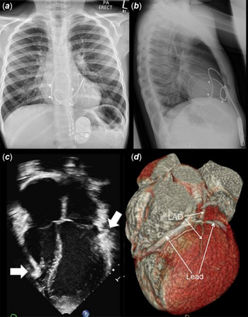
The chest X-ray suggested that the epicardial leads were likely to be encircling the heart (Fig 1a and b). The echocardiogram demonstrated indentation of the right ventricular free wall with an adjacent bright area towards the apex and an indentation in the basal anterolateral wall of the left ventricle (Fig 1c). There was mildly impaired function and clear septal dyssynchrony. A gated cardiac CT scan demonstrated an indentation of the right ventricular free wall present in both systole and diastole. The two leads passed over the anterior wall of the left ventricle, the more lateral of which compressed the mid LAD and first diagonal branch and indented the basal LV wall, just below the mitral annulus (Fig 1d). His pacemaker was functioning normally and the ECG showed atrial sensing with ventricular pacing at a rate of 88 bpm.
(a and b) AP and lateral chest X-ray demonstrating characteristic looping of epicardial leads suggestive of cardiac strangulation. Posterior looping of leads best seen on the lateral projection. (c) Transthoracic echocardiogram four-chamber view demonstrating indentation of the inferior right ventricular free wall and basal anterolateral wall of the left ventricle by the pacing leads (white arrows). (d) Reconstruction from gated cardiac CT demonstrating pacing lead compressing the left anterior descending (LAD) coronary artery.

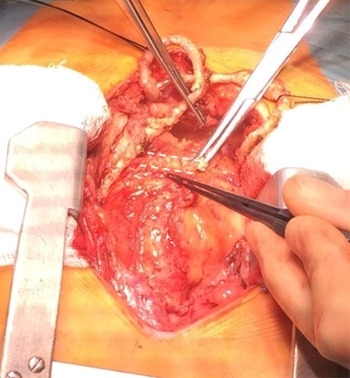
Urgent surgical revision of his system was arranged. The surgical findings showed epicardial leads running circumferentially around the heart. The ventricular leads were running inferiorly and looping behind the heart and then anteriorly onto the free wall of the right ventricle. The atrial leads were running leftwards crossing the heart anteriorly and looping around the left ventricle posteriorly, slightly lower than the left AV groove. The leads were heavily calcified and embedded (Fig 2). They were successfully removed and replaced with new leads. The ventricular lead was placed on the apex of the left ventricle to improve function and the atrial lead on the right atrium; care was taken to minimise the amount of lead slack in the pericardium. He made a rapid recovery and no longer complains of chest pain.
Heavily calcified and embedded leads removed from the anterior surface of the right ventricle. Lead bed outlined with dotted lines.

Discussion
Cardiac strangulation following implantation of epicardial pacemaker leads is a life threatening and rarely reported complication. It occurs when the pacemaker leads are adherent to the heart’s surface and become embedded around it. With time and somatic growth, there is a gradual constriction of the cardiac mass. Depending on the point of maximum compression, there may be impaired filling of the heart, valvar insufficiency or arrhythmia. Leads may impinge on the coronary arteries causing myocardial ischaemia and sudden death. Reference Eyskens, Mertens and Moerman1–Reference Mah, Prakash and Porras5
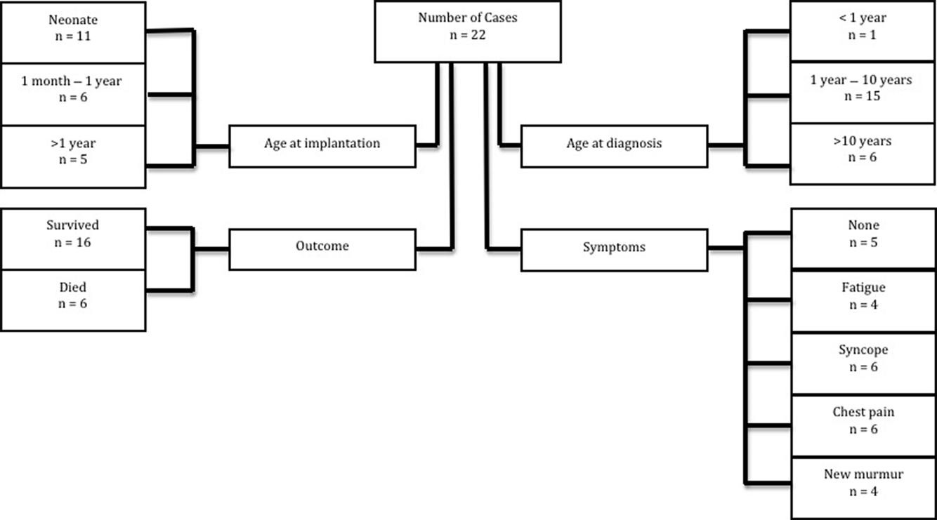
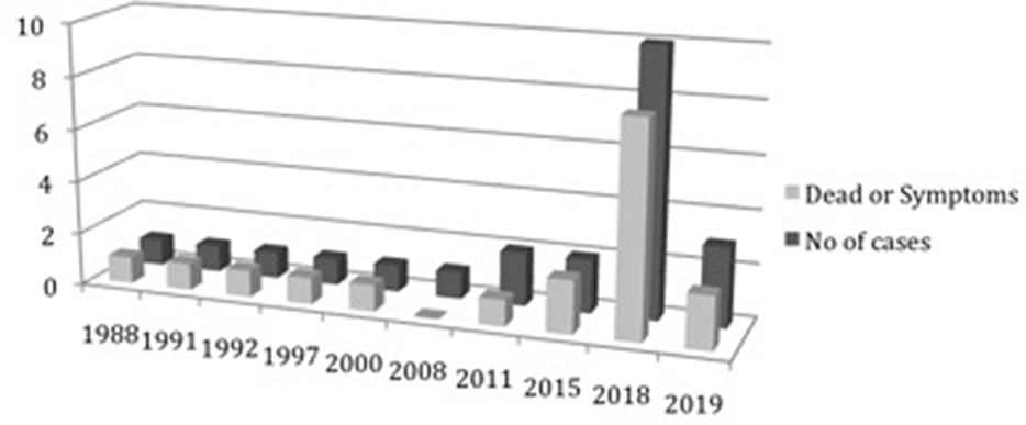
The published incidence of cardiac strangulation is estimated to be 0.016% worldwide. Reference Carerras, Duncan and Djurdjev3 There has been an increasing awareness in recent years with two institutions taking the initiative to systematically review their patient cohorts following the death of an index patient. Reference Carerras, Duncan and Djurdjev3,Reference Mah, Prakash and Porras5 Unsurprisingly, the number of local cases found and associated mortality was higher than the reported averages. A literature search of MEDLINE and PubMed revealed 20 reports of cardiac strangulation in North America, Europe and Japan – these cases are summarised in Fig 3 together with our case and Breatnach et al in this issue. Six cases had a fatal outcome. The seemingly exponential rise in reported cases over the past 3 years may be due to cases identified by prospective surveillance. Reference Carerras, Duncan and Djurdjev3,Reference Mah, Prakash and Porras5 It is likely that there is under-reporting as sudden death in pacemaker patients may not be recognised to be due to strangulation without a careful post-mortem examination. Reference Janik, Hejna and Straka4 Figure 4 demonstrates the increase in number of reports over recent years.
Flowchart summarising presentation and outcome of cases.

Number of cardiac strangulation cases reported per year, including those deceased.

The main characteristic of patients presenting with cardiac strangulation is lead implantation at a very young age. Among the cases reported in the literature, 10 were implanted in the newborn period and 16 within the first year of life. Age less than 6 months, weight less than 7.3 kg, placement of a dual-chamber system, evidence of postpericardiotomy syndrome or post-operative infection all increase the risk of cardiac strangulation. Reference Carerras, Duncan and Djurdjev3 Both excess pericardial lead looping around the heart and somatic growth of the patient have a role to play. Reference Eyskens, Mertens and Moerman1,Reference Janik, Hejna and Straka4 The presence of minimal adipose tissue in paediatric patients makes the coronary arteries on the surface of the heart more prone to compression by an adherent lead and myocardial ischaemia may present many years later. The narrower space between the heart surface and sternum in children may predispose to adherence and compression of the left anterior descending artery. Reference Takeuchi and Tomizawa6 Mah et al report the highest incidence of cardiac strangulation (5.5%) and recognise that embedded leads may compress the coronary artery without actual strangulation of the heart. Reference Mah, Prakash and Porras5
Cardiac strangulation can occur with any type of epicardial lead. Reference Takeuchi and Tomizawa6 Symptoms include chest pain or syncope related to ischaemia from coronary artery compression. Reference AlHuzaimi, Roy and Duncan7 A new onset murmur may indicate valvar dysfunction. Five patients presented with the collapse that was a terminal event. Worryingly, several authors report vague/general symptoms such as tiredness highlighting the need for a high index of suspicion and the need for a reliable screening tool. Reference Mah, Prakash and Porras5,Reference Brenner, Gaines and Cordier8
Carerras et al suggest 3-yearly PA and lateral chest X-ray monitoring during the child’s growth and describe the classic appearance of posterior looping of the leads (Fig 1a and b). They discontinue such screening in patients who have attained their full adult size assuming the lead pattern on X-ray is not suggestive. Reference Carerras, Duncan and Djurdjev3 Isolated chest X-ray has been shown to have modest sensitivity, but high specificity for detecting coronary artery compression and any patient with suspicious symptoms should proceed to evaluation by CT or coronary angiography. Reference Carerras, Duncan and Djurdjev3,Reference Mah, Prakash and Porras5 Regular echocardiography as part of a pacing check to assess function may also alert to potential strangulation by identifying indentation (as in this case), new valvar dysfunction or exaggerated dyssynchrony. It is important to remember that abandoned leads also pose a risk and the possibility of cardiac strangulation in such patients should be considered even when they have replacement endocardial systems if they present with non-specific symptoms. Reference Janik, Hejna and Straka4,Reference Takeuchi and Tomizawa6 Worryingly, the strangulation and coronary compression from epicardial leads may only present many years after they have been implanted. Reference Janik, Hejna and Straka4 We propose that serial chest X-ray and echocardiography should form part of the pacemaker check/patient review, with further coronary imaging reserved for those patients who have suspicious findings on the investigation or worrying symptoms.
Surgical removal of the leads can be tedious and risks damaging the coronary arteries Reference Janik, Hejna and Straka4 as long-standing leads become calcified with encapsulating fibrous tissue adherent to the heart (Fig 2). Replacement leads need to be carefully sited avoiding multiple loops, and minimising excess lead left within the pericardium, it was not always clear in the cases reviewed where leads placed. Placement of excess lead within the pacemaker pocket, pleural space or fixed to the diaphragm may prevent future compression.
This case highlights a potentially deadly complication of epicardial pacing in young patients that is likely to be under-reported. Surgical technique should minimise redundant lead within the pericardium. Serial assessment is required, initially with AP and lateral chest X-ray and then regular echocardiography, at least until full adult growth is attained. A low threshold for coronary artery imaging with CT or angiography is necessary for unexplained symptoms. If cardiac strangulation is diagnosed prompt surgery is needed.
Acknowledgements
None.
Financial support
This research received no specific grant from any funding agency, commercial or not-for-profit sectors.
Author contributions
MLM collected data, researched cases, prepared and revised manuscript. SK collected data, SS revised manuscript, AJS revised manuscript, ER concept and revised manuscript.
Conflicts of interest
None.