Introduction
The study of the ontogeny of a species in fish farms is essential for adequate reproductive management and to obtain repopulation or commercial objectives. To this end, diverse studies have been performed in Coho salmon (Oncorhynchus kisutch) that have considered aspects of their ontogeny (Phillips & Campbell, Reference Phillips and Campbell1962; Shumway et al., Reference Shumway, Warren and Doudoroff1964; Ostrander et al., Reference Ostrander, Landolt and Kocan1988, Reference Ostrander, Landolt and Kocan1989; Piferrer & Donaldson, Reference Piferrer and Donaldson1989). However, there are no studies of early embryogenesis that specifically cover the morphometric characterization of the first blastomere stage and that are associated with embryo quality. The morphology of the first blastomeres (symmetrical or normal, and asymmetrical or abnormal) has been studied as a predictive tool for early embryonic quality in different species (Kjørsvik et al., Reference Kjørsvik, Mangor-Jensen and Holmefjord1990, Reference Kjørsvik, Hoehne-Reitan and Reitan2003; Bromage et al., Reference Bromage, Bruce, Basavaraja and Rana1994; Shields et al., Reference Shields, Brown and Bromage1997; Rideout et al., Reference Rideout, Trippel and Litvak2004; Penney et al., Reference Penney, Lush, Wade, Brown, Parrish and Burton2006; Pavlov & Emel'yanova, Reference Pavlov and Emel'yanova2008; Avery et al., Reference Avery, Killen and Hollinger2009; Bobe & Labbé, Reference Bobe and Labbé2010; Vásquez et al., Reference Vásquez, Llanos-Rivera and Castro2010). The information obtained would be important for: (1) early determination of the fertility rates; (2) standardization of tools that allow early diagnosis of embryonic quality in salmonids, to eliminate eggs with a less probability of success during hatching or that produce unviable larvae (to lower operational costs) (Rideout et al., Reference Rideout, Trippel and Litvak2004; Penny et al., Reference Penney, Lush, Wade, Brown, Parrish and Burton2006; Pavlov & Emel'yanova, Reference Pavlov and Emel'yanova2008); and/or (3) standardization of protocols to procure tetraploid examples with the recognition and/or characterization of the first cleavage (Hershberger & Hostuttler, Reference Hershberger and Hostuttler2005) and in this way to optimize activities for repopulation and/or commercial production with sterile specimens (interploid tetraploid).
For the early evaluation of the quality of embryos in salmonid fish, overripeness of the eggs (Barnes et al., Reference Barnes, Hanten, Sayler and Cordes2000; Lahnsteiner, Reference Lahnsteiner2000; Aegerter & Jalabert, Reference Aegerter and Jalabert2004; Samarin et al., Reference Samarin, Ahmadi, Azuma, Rafiee, Amiri and Naghavi2008), increase in size of the eggs during the hydration period that follows fertilization (Lahnsteiner & Patzner, Reference Lahnsteiner and Patzner2002), and the physical-chemical parameters of the coelomic fluid and of the eggs (Lahnsteiner & Patzner, Reference Lahnsteiner, Weismann and Patzner1999; Lahnsteiner, Reference Lahnsteiner2000), were used as indicators.
To date, the symmetry of the first blastomeres of fish embryos has only been considered from a qualitative point of view. Valdebenito et al. (Reference Valdebenito, Sánchez, Effer and Ubilla2012) and Effer et al. (data unpublished), detailed morphometric aspects for the first time to evaluate this mentioned symmetry, of the first blastomeres of Atlantic salmon (S. salar) and their relationship with the quality of embryos. In this investigation, the objective was to characterize morphometrically the first blastomeres of Coho salmon eggs. This preliminary information would allow the creation of a protocol to evaluate the quality of salmonid species gametes and/or embryos.
Materials and methods
Procurement of the gametes and artificial fertilization
The oocytes were collected in June 2010 from two female Coho salmon (O. kisutch) of 2 years of age; these females were maintained initially in seawater and then transported to freshwater in January 2010 at the Belén del Sur fish farm, Araucanía Región-Chile. The female and male gametes were extracted by abdominal incision and massage, respectively.
Fertilization was performed separately with semen from 11 males and a pool of eggs, divided into 33 homogenous aliquots (three replicas of approximately 100 oocytes per each male). Each replicate was fertilized with 10 μl of semen from one male using the standard reproduction methods of the fish farm. After fertilization, each aliquot of eggs was introduced into a micro-incubator of hydrophobic material, with a tag that had colour and numerical codes to identify the male and replicate used. Eggs were then grouped into a Zoug jar type incubator at 5°C in dark.
Procurement and processing of embryonic discs
Eggs were incubated for 19 h at 5°C (95°C h of accumulated heat), then approximately 50 eggs of each replicate were fixed in Stockard solution and stored in tagged containers.
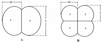
The chorion of the eggs was removed and the blastodisc was extracted by tweezers under a stereoscopic Olympus SZ60 (JAPAN) magnifying lens equipped with a Micropublisher 3.3 RTV digital camera (CANADA). In total, 660 microphotographs were taken of the embryonic discs at a magnification of ×56 (60 microphotographs per group including the replicates), which were divided into three categories: (1) unfertilized blastodiscs; (2) blastodiscs with two blastomeres (after the first cleavage); and (3) blastodiscs with four blastomeres (after the second cleavage); then all were characterized morphometrically with the software Q Capture Pro 5.0 (CANADA), in which the diameter of the unfertilized blastodisc, and the length and width of each blastomere were measured (Fig. 1).
Criteria used to determine the length (L) and width (W) of the blastomeres and the designation of number of each blastomere in embryos with two (A) or four blastomeres (B).

For morphological characterization of the four blastomere blastodiscs, 100 photographs were selected randomly and classified into categories according to their morphology and frequency of appearance following the criteria proposed by Shields et al. (Reference Shields, Brown and Bromage1997). For the morphometric description, we proceeded in the same way for the each blastodisc, and evaluated symmetry by a comparison of the proportions of the lengths (L1/L2, L1/L3, L1/L4, L2/L3, L2/L4, L3/L4) and widths (W1/W2, W1/W3, W1/W4, W2/W3, W2/W4, W3/W4) of all the blastomeres (of the same blastodisc) (Fig. 1B), using the highest value as the denominator of both the proportion analyses.
For 20 blastodiscs (control blastodiscs) with four blastomeres, symmetrical cleavages and normal appearance were selected visually following the criteria proposed by Shields et al. (Reference Shields, Brown and Bromage1997). Then the morphometric symmetry was determined, as described in the previous paragraph, to compare the blastodiscs characterized visually as abnormal or asymmetrical.
Statistical analysis
Values for proportions of length and width of the symmetrical or normal blastomeres and for the asymmetrical or abnormal blastomeres, were analyzed with the statistical software GraphPad Prism® version 5.0 (GraphPad Software, San Diego CA). A one-way analysis of variance (ANOVA) test was used for the non-parametric samples. Additionally, and in a parallel manner, a Dunnet's multiple comparisons test was done to evaluate any statistically significant differences between the proportions of length and width of the blastodiscs sampled from the control group. The level of significance was fixed at a P-value < 0.05 (n = 10 replicates).
Results
Morphological characterization
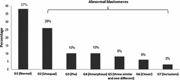
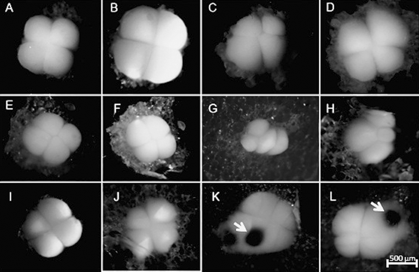
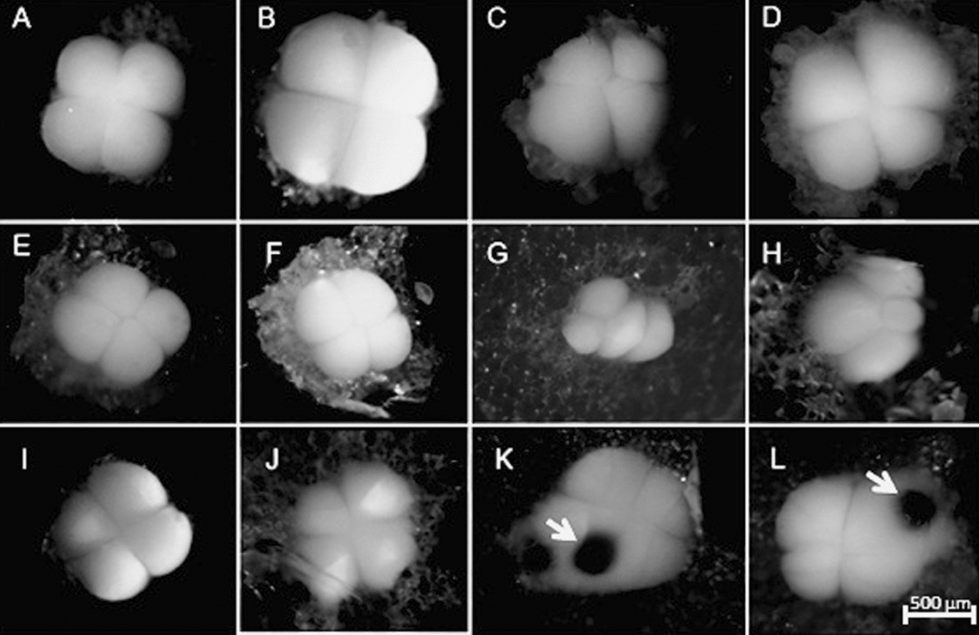
Figure 2 shows the seven groups of blastomeres classified by morphology and frequency of appearance in embryos of Coho salmon, from which six groups were considered to be abnormal morphologically. Around 37% of the analyzed embryos presented normal blastomeres (category 1: G1) (Fig. 3A,B); 26% with unequal blastomeres (G2) (Fig. 3C,D); 10% had ‘pie-shaped’ blastomeres (G3) (Fig. 3E,F); 10% amorphous blastomeres (G4) (Figure 3G,H); 8% with three equal and one unequal blastomeres (G5) (Fig. 3I); 6% with ‘clover’ shaped blastomeres (G6) (Fig. J), and 3% blastomeres with inclusions (G7) (Fig. 3K,L).
Morphological categories and percentage of appearance of the observed groups of blastomeres in Coho salmon eggs (O. kisutch). n = 100.

Microphotographs of normal and abnormal blastomeres of Coho salmon (S. kisutch). (A, B) Normal (G1). (C, D) Unequal (G2). (E, F) ‘Pie-shaped’ (G3). (G, H) Amorphous (G4). (I) Three equal and one unequal blastomeres (G5). (J) ‘Clover-shaped’ blastomeres (G6). (K, L) Blastomeres with inclusions (G7) (arrows). Magnification ×56.
Morphometric characterization
A normal embryonic disc from a Coho salmon has a discoid shape with a mean diameter of 985.37 ± 93.66 μm. When the first segmentation occurred, the mean length and width of the blastomeres was 823.81 ± 82.53 μm and 492.12 ± 48.04 μm, respectively. When the second segmentation occurred, the blastomeres had a mean length and width of 443.73 ± 46.98 μm and 431.85 ± 45.85 μm, respectively.
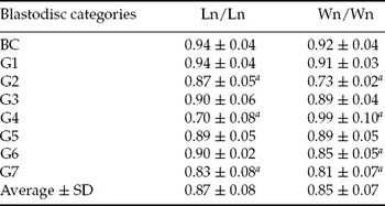
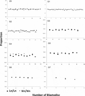
The mean value of the proportions of length and width of the blastomeres in the eight categories that were analyzed morphologically was 0.87 ± 0.08 and 0.85 ± 0.07, respectively (Table 1, Fig. 4).
Mean values of the proportions of lengths and width of blastomeres of O. kisutch in blastodiscs

Control blastodiscs (CB), normal (G1), unequal (G2), ‘pie-shaped’ (G3), amorphous (G4), three equal and one unequal blastomeres (G5), ‘clover-shaped’ (G6) and with inclusions (G7).
aSignificant difference within the control group (BC). SD, standard deviation.
Mean values of the proportions of lengths and widths of blastomeres of O. kisutch of control blastodiscs (CB) (n = 20), normal (G1) (n = 38), unequal (G2) (n = 26), ‘pie-shaped’ (G3) (n = 10), amorphous (G4) (n = 10), three equal and one unequal (G5) (n = 8), ‘clover-shaped’ (G6) (n = 6) and with inclusions (G7) (n = 3). Ln/Ln and An/An: proportions of lengths and widths of blastomeres of a same blastodisc, where ‘n’ is the random number of a blastomere.

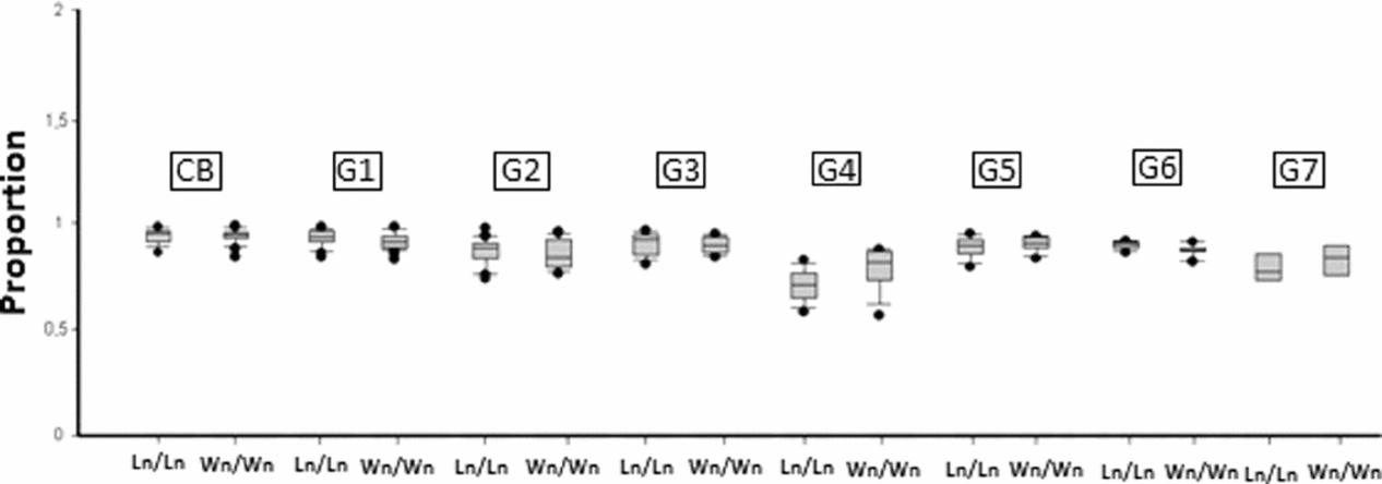
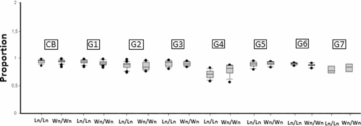
When comparing the mean values of the proportions of lengths (0.94 ± 0.04) of control blastodiscs (CB) regarding the other sampled groups, statistically significant differences were found with the proportions of blastodiscs G2: unequal (0.87 ± 0.05), G4: amorphous (0.7 ± 0.07) and G7: with inclusions (0.83 ± 0.08) (P < 0.05). When considering the mean values of the proportions of widths (0.93±0.04) of control blastodiscs (CB) regarding the other sampled groups, statistically significant differences were found with the proportions of blastodiscs G2: unequal (0.73 ± 0.02), G4: amorphous (0.79 ± 0.1), G6: ‘clover-shaped’ blastomeres (0.85 ± 0.05) and G7: with inclusions (0.81 ± 0.07) (P < 0.05) (Table 1 and Fig. 5).
Mean values of the proportions of length and width of the blastomeres of O. kisutch embryos: control (CB) (n = 20), normal (G1) (n = 38), unequal (G2) (n = 26), ‘pie-shaped’ (G3) (n = 10), amorphous (G4) (n = 10), three equal and one unequal blastomeres (G5) (n = 8), ‘clover-shaped’ (G6) (n = 6) and with inclusions (G7) (n = 3). Ln/Ln and Wn/Wn: proportions of lengths and widths of blastomeres of a same blastodisc, where ‘n’ is the random number of a blastomere.
Discussion
The mean diameter of the blastodiscs of Coho salmon found in this study was 985.37 ± 93.66 μm, similar to that reported for other species of salmonids, such as Oncorhynchus mykiss: 941.44 μm (Valdebenito et al., Reference Valdebenito, Sánchez, Effer and Ubilla2012), 1000 μm (Ballard, Reference Ballard1973) and Salvelinus fontinalis: 880 μm (Ballard, Reference Ballard1973), Salmo salar: 950 μm (Gorodilov, Reference Gorodilov1996) and 1,015.58 μm (Effer et al., data unpublished).
Seven categories or groups of blastodiscs were found based on morphology of the blastomeres, from which six presented blastomeres that had abnormal cleavage patterns. These patterns can be generated by different factors such as overmaturity or ageing of the eggs (Valdebenito et al., unpublished data), elevated temperatures during the incubation phase (Pavlov & Moksness, Reference Pavlov and Moksness1994), and/or triploidy induction (Rani, Reference Rani2005; Valdebenito et al., unpublished data). The abnormal pattern found in the highest percentage (26%) was G2 (unequal blastomeres, Fig. 3C,D) followed by G3 (‘pie-shaped’ blastomeres, Fig. 3E,F) with a 10% appearance, values that are reported in the characterization of the first blastomeres of S. salar by Effer et al. (data unpublished) and in Gadus morhua by Avery et al. (Reference Avery, Killen and Hollinger2009). This last author determined, for this species, that the mortalities generated by these anomalies did not generate statistically significant differences regarding normal blastodiscs and, on the contrary, these could produce normal specimens. This finding contrasts that for G7 (blastomeres with inclusions, Fig. 3K,L), found in Hippoglossus hippoglossus by Shields et al. (Reference Shields, Brown and Bromage1997); this author determined that there was a correlation of higher presence of blastomeres with inclusions in this species with a lower hatching percentage. The previous finding suggests that not all abnormal cleavage patterns are severe and can still result in viable larvae, and that other abnormal patterns do not correct themselves during embryonic development, causing higher detriment to the embryos and/or the final larvae (mortality, increase in larval teratogeny, lower percentage of hatching, etc.). It would be useful, therefore, to be able to relate these abnormalities with parameters of embryonic quality during the later stages of embryogenesis, such as survivability at 150 accumulated thermal units (ATU). Thus we aimed to establish if a relationship exists between these anomalies and embryonic quality, and to produce an evaluation tool for early embryonic quality in salmonids.
In contrast with the morphological criteria for the evaluation of symmetry of the first blastomeres in fish adopted by several groups (Kjorsvik et al., Reference Kjørsvik, Mangor-Jensen and Holmefjord1990; Shield et al., Reference Shields, Brown and Bromage1997; Kjorsvik et al., Reference Kjørsvik, Hoehne-Reitan and Reitan2003; Rideout et al., Reference Rideout, Trippel and Litvak2004; Avery & Brown, Reference Avery and Brown2005; Penney et al., Reference Penney, Lush, Wade, Brown, Parrish and Burton2006; Moran et al., Reference Moran, Smith, Gara and Poortenaar2007; Pavlov & Emel'yanova, Reference Pavlov and Emel'yanova2008; Avery et al., Reference Avery, Killen and Hollinger2009; Hamoutene et al., Reference Hamoutene, Lush, Drover and Walsh2009), in this study the symmetry of the embryos of two and four blastomeres was considered to determine the proportions of lengths and width of all of the blastomeres (from the same blastodisc). This approach was an alternative to that considered in S. salar by Effer et al. (data unpublished), in which the symmetry of the morphometric form is compared, but only between two blastomeres (homologous blastomeres, in embryos at the second cleavage stage).
Statistical analysis detected significant differences in at least three of the six groups of blastodiscs that presented visually abnormal cleavage patterns, both for the proportions of lengths and widths (Table 1). These findings could indicate: (1) the need to make the evaluation technique of the symmetry of the morphometric form more precise, for which consideration of volumetric measurements of the blastomeres (proportions of volume between blastomeres of a same blastodisc) is proposed, instead of linear measurements; and (2) that there exists an evident difference between the cleavage patterns in blastodiscs of Coho salmon. This difference could be expressed in advanced stages of embryogenesis and translate into either good or bad quality embryos. Cleavage patterns must be correlated with indicators of quality in the later stage of embryogenesis of this species to clarify effects on the progeny, and in this manner to establish a solid base for the creation of an early evaluation tool for the quality of embryos in salmonids.
Acknowledgements
The investigation was financed by the FONDEF D06I1020 project. We also wish to acknowledge the Belén del Sur, (Chile) fish farm for providing the gametes for the experiment.