Introduction
Following more than a century of sometimes controversial work, it is now well established that descending fibers from the brain terminate in the retinas of all classes of vertebrate. This descending input has evolved on at least six separate occasions within the vertebrate lineage, with different nuclei providing the origin of these pathways that very likely serve a number of different functions (Reperant et al., Reference Reperant, Ward, Miceli, Rio, Medina, Kenigfest and Vesselkin2006, Reference Reperant, Medina, Ward, Miceli, Kenigfest, Rio and Vesselkin2007). While the mammalian retina possesses only a few descending fibers (Gastinger et al., Reference Gastinger, Tian, Horvath and Marshak2006), it is in the bird retina, fortuitously the retina in which Cajal first described an input from the brain (Ramón y Cajal, Reference Ramón y Cajal1889), that descending input is most highly elaborated and has been most extensively studied. The central unanswered question clearly concerns the function of this input. Not only is this an interesting question in its own right, possibly requiring that we adjust our ideas of what the retina does, but it might perhaps illuminate the more general questions about the function of descending input throughout the visual system and elsewhere in the brain.
Not long after Cajal described fibers from the brain terminating in the bird retina, Wallenberg (Reference Wallenberg1898) found the nucleus from which these fibers originate. This small nucleus, located in the caudal midbrain underneath the cerebellum, is now called the isthmo-optic nucleus (ION; Fig. 1). This was confirmed much later (Cowan & Powell, Reference Cowan and Powell1963) when it was shown that this nucleus contains roughly 10,000 neurons (Cowan & Powell, Reference Cowan and Powell1963; Wolf-Oberhollenzer, Reference Wolf-Oberhollenzer1987), giving rise to a bundle of fibers that form the isthmo-optic tract before merging with the optic nerve and crossing to the contralateral eye. These fibers entering the retina have been given several names, including centrifugal fibers or retinopetal fibers, but in this review we will refer to them as efferent fibers. We will refer to the entire loop from retina to brain and back to the retina as the centrifugal visual system (CVS). Chicken, pigeon, and quail have provided the three experimental subjects for virtually all investigations of the CVS, and though minor differences distinguish these three species (see, e.g., Crossland et al., Reference Crossland, Cowan, Rogers and Kelly1974), in general, results are interchangeable between all three and we consider them together.
Dorsal surface of a chicken brain with fluorescent staining resulting from the injection of red and green Alexa-conjugated cholera toxin subunit B into the right and left eyes, respectively. The cerebral hemispheres are visible in the upper half of the image and the midbrain has been exposed by removing the cerebellum. The left (red) and right (green) optic tecta are visible on the left and right margins of the image. Between the two tecta, on the dorsal surface of the midbrain, are two ovoid structures measuring approximately 1 mm across (arrows). These are the left and right IONs located roughly 2 mm from the midline. Anterior is up. Scale bar is 1 mm. From Lindstrom et al., (Reference Lindstrom, Nacsa, Blankenship, Fitzgerald, Weller, Vaney and Wilson2009).

Our intention in this review is to bring together results gathered over several decades of work on the CVS of the bird. Some excellent anatomical and physiological studies of this system lay a foundation for theories of its function. A determination of function however requires more than this and a truly convincing case would describe the forces that drove evolution to develop and maintain this system in birds, though apparently not in mammals. Regrettably, there is a scarcity of studies that could help in this area. The ideas we develop in this review could benefit from field studies of predation, detailing vulnerability to, and escape from, different modes of attack under different conditions, but these data are similarly not available. Accepting that these limitations imply that no argument can be more than plausible, we show that with the addition of a few new results, a novel hypothesis about the function of this system emerges.
Lesion studies
Though Cajal considered his discovery of brain input to the retina to be important, he offered no definite opinion on the function of these fibers. He seems to have favored the idea, proposed by others, that fibers from the brain were “conductors of expectant attention” whose role, as we would now say, is to increase the gain in a local region of retina (Ramón y Cajal, Reference Ramón y Cajal1952). A number of hypotheses have subsequently been put forward and are summarized in Table 1, but before considering these further, we will look at a potential shortcut to understanding function.
Suggested functions for the bird CVS

The shortcut to which we refer is the classic approach of observing the behavioral deficits following lesion or ablation of a neural structure. This inductive approach has yielded extraordinarily valuable insights into the function of many brain structures and has been applied to the ION in four independent studies (Rogers & Miles, Reference Rogers and Miles1972; Shortess & Klose, Reference Shortess and Klose1977; Knipling, Reference Knipling1978; Hahmann & Gunturkun, Reference Hahmann and Gunturkun1992).
There is general agreement from these studies that ION lesions have little or no effect on either visual acuity or pattern discrimination. Indeed, a main conclusion of these studies, each employing a few visual tasks, is that lesion of the ION has little apparent effect on visual performance. Three studies support the finding that lesioned birds have some impaired discrimination of grit from grain, at least under dim lighting conditions (Rogers & Miles, Reference Rogers and Miles1972; Shortess & Klose, Reference Shortess and Klose1977; Hahmann & Gunturkun, Reference Hahmann and Gunturkun1992), and of these, one study reports that lesioned animals are also less responsive to novel objects introduced into the lateral visual field (Rogers & Miles, Reference Rogers and Miles1972). This study by Rogers and Miles also reported that lesioned animals perseverate at pecking and are unable to pick up grain as a consequence of failing to open their beaks. This abnormality, not reported in the other studies, suggests that structures other than the ION were damaged during lesioning. Behavioral deficits from unintended damage seem likely for a number of reasons. The ION is a small nucleus, of somewhat variable shape (Holden & Powell, Reference Holden and Powell1972), completely covered by the cerebellum. In addition, it lies within the tectal commisure and the mesencephalic root of the trigeminal nerve passes through the middle of the ION. Damage to any of these other structures might well degrade performance on the behavioral tests employed in these studies.
A more subtle difficulty follows from the fact that the ION is surrounded by a loosely defined halo of anatomically distinct cells, called ectopic neurons (Clarke & Cowan, Reference Clarke and Cowan1976; Cowan & Clarke, Reference Cowan and Clarke1976; Hayes & Webster, Reference Hayes and Webster1981; Fritzsch et al., Reference Fritzsch, Crapon de Caprona and Clarke1990; Woodson et al., Reference Woodson, Shimizu, Wild, Schimke, Cox and Karten1995; Li & Wang, Reference Li and Wang1999). These ectopic neurons also project efferent fibers into the contralateral retina, but their terminations are strikingly different from those originating in the ION. Fibers originating in the ION are called “restricted” efferent fibers because their terminals are compact, typically covering 0.003 mm2 of retina, whereas those originating from ectopic neurons are called “widespread” efferent fibers since their terminals typically cover 0.091 mm2 (Woodson et al., Reference Woodson, Shimizu, Wild, Schimke, Cox and Karten1995). These differences between widespread and restricted efferent fibers suggest they serve different functions and, largely because much less is known about the widespread efferent fiber pathway, we will consider only the restricted efferent fiber pathway. Except where distinctions between the two efferent pathways are clarified, by “efferent” we mean the restricted efferent pathway.
Taken as a whole, lesion studies have not provided any unambiguous clues about function and we suspect that at least some of the results are due to collateral damage. An intrinsic limitation of lesion studies is, of course, that the experimenter can only find behavioral deficits that are assayed for, and it is not clear that the appropriate behavior has yet been monitored. Regrettably, in the case of the CVS, the shortcut offered by lesioning combined with behavioral studies has thus far been a dead end. To understand the function of the CVS, we must instead rely on the slower, step-by-step process of examining the anatomy and physiology of the components of this system.
Anatomy and physiology of the CVS
The ION and optic tectum
The structure of the ION is, at first sight, confusing but can best be understood as a collapsed hollow ball that has subsequently been folded (McGill et al., Reference McGill, Powell and Cowan1966; Holden & Powell, Reference Holden and Powell1972; Fig. 2a–2d). Neurons are arranged in two adjacent layers, each one neuron thick, that constitute the walls of the collapsed ball. The ION comprises two cell types: local GABAergic interneurons and the output neurons (typically referred to as IO neurons). The approximately 180 GABAergic interneurons in the nucleus have very broad dendritic fields and are thought to be inhibitory (Miceli et al., Reference Miceli, Reperant, Rio and Medina1995). Each IO neuron gives rise to an axon that runs in the isthmo-optic tract to the contralateral eye and terminates in a single efferent terminal. Neurons of the ION are driven chiefly by visual input from the contralateral eye, and consistent with this, anatomical studies have shown that input to the ION comes from the ipsilateral optic tectum (Crossland & Hughes, Reference Crossland and Hughes1978; Woodson et al., Reference Woodson, Reiner, Anderson and Karten1991; Miceli et al., Reference Miceli, Reperant, Marchand and Rio1993; Uchiyama et al., Reference Uchiyama, Yamamoto and Ito1996) after minimal processing in that nucleus (Holden, Reference Holden1968; Holden & Powell, Reference Holden and Powell1972).
Organization of the pigeon ION. (a) and (b) are anterior and posterior views of the whole nucleus showing its folding. (d) shows six horizontal sections through the structure corresponding to the numbers in (a) and (b). In (c) are shown diagrammatic views of sagittal sections of the nucleus at the relevant positions marked by arrows in the reconstruction shown in (a). Section c1 is drawn as seen from the medial aspect looking laterally and c2 from the lateral aspect. (e) Receptive fields of IO neurons plotted on the anterior inferior quadrant of the visual field. Each of the eight series of receptive fields, 38–46, represents sequentially encountered neurons in eight horizontal penetrations of a microelectrode through the isthmo-optic nucleus traveling from lateral to medial. Track 45, for example, corresponds roughly to the horizontal plane marked 5 in panel (b). The map of visual space is precisely retinotopic with posterior visual space represented at the lateral margin of the nucleus and anterior visual space in the medial margin. Panels (a) through (d) taken from McGill et al. (Reference McGill, Powell and Cowan1966), panel (e) taken from Holden and Powell (Reference Holden and Powell1972), both with permission.

The optic tectum is the major recipient of retinal output in the avian visual system, unlike the mammalian visual system where the lateral geniculate nucleus of the thalamus is the primary recipient of retinal input. Tectal input to the ION is mediated by a specific group of cells in lamina h, called tecto-IO neurons. There is evidence that tecto-IO neurons make one-to-one connections with IO neurons (Woodson et al., Reference Woodson, Reiner, Anderson and Karten1991; Uchiyama et al., Reference Uchiyama, Yamamoto and Ito1996; Li et al., Reference Li, Hu and Wang1999). In addition to tectal input, the ION also receives input from the brain stem, including the reticular formation and the raphe nucleus (Miceli et al., Reference Miceli, Reperant, Bavikati, Rio and Volle1997, Reference Miceli, Reperant, Rio, Hains and Medina2002; Medina et al., Reference Medina, Reperant, Miceli, Bertrand and Bennis1998). Stimulation of the visual Wulst, a region analogous to the mammalian visual cortex, is also effective in driving IO neurons (Uchiyama et al., Reference Uchiyama, Matsutani and Watanabe1987), but this probably comes via the optic tectum, which itself is known to have Wulst input (Leresche et al., Reference Leresche, Hardy and Jassik-Gerschenfeld1983).
There is strong evidence from both anatomy and physiology, at least in the pigeon, suggesting that the input to the ION is derived chiefly from the dorsal retina. The anatomical evidence comes from the finding that tecto-IO neurons are predominantly located in the ventral tectum which itself receives input from the dorsal retina (Woodson et al., Reference Woodson, Reiner, Anderson and Karten1991). The physiological evidence comes from a very careful and detailed study in pigeon by Holden and Powell (Reference Holden and Powell1972) in which they mapped the receptive fields of IO neurons and carefully reconstructed the electrode tracks through the nucleus. Their results convincingly show that input to the ION is largely from dorsal retina. A similar study in chicken (Miles, Reference Miles1972c) implied that input is from the entire retina, and indeed the gross structure of the pigeon and chicken ION differ in a way that would be consistent with a greater dorsal input in the pigeon (Miles, Reference Miles1972c; Crossland et al., Reference Crossland, Cowan, Rogers and Kelly1974), but this apparent difference needs to be more closely examined.
Notwithstanding this bias for input from the dorsal retina, the ION contains an orderly retinotopic representation of the visual world (Holden & Powell, Reference Holden and Powell1972, Fig. 2e). IO neurons are strongly and phasically driven by moving targets positioned within their central receptive fields (Holden & Powell, Reference Holden and Powell1972; Miles, Reference Miles1972c; Uchiyama et al., Reference Uchiyama, Nakamura and Imazono1998). Some disagreement exists on the size of receptive fields. Holden and Powell report central receptive fields of about 5 deg (1972), whereas Miles (Reference Miles1972c) and Li et al. (Reference Li, Xiao, Fu and Wang1998) report at least double that. Differences in method may underlie these discrepancies. Optimal stimuli move at rates between 5 and 30 deg/s, though IO neurons respond to movement over a huge range of velocities (Miles, Reference Miles1972c). A majority of IO neurons are either directionally selective, mostly preferring targets moving anteriorward, or are relatively nonselective, except for having a minimum response to posteriorward movement. Although IO neurons respond to the onset as well as the offset of light for stationary spots, they greatly prefer dark moving edges to light edges, especially for large stimuli (Miles, Reference Miles1972c).
Outside the central excitatory receptive field lies a very broad and powerful suppressive surround that covers virtually the whole of the rest of the visual field. Even a small spot of 2–5 deg diameter at a remote location in the retina is sufficient to suppress responses in the receptive field center (Uchiyama et al., Reference Uchiyama, Nakamura and Imazono1998). This powerful inhibition is not a feature of tectal responses (Hughes & Pearlman, Reference Hughes and Pearlman1974) and is presumably mediated by the large-field GABAergic interneurons in the ION. These very large suppressive surrounds have led to the interpretation that IO neurons compete in a winner-take-all system that selects one region of the retina on the basis that it responds most strongly (Uchiyama et al., Reference Uchiyama, Nakamura and Imazono1998; Uchiyama, Reference Uchiyama1999).
The efferent fibers
The fibers leaving the ION include some of the largest diameter fibers found in the optic nerve and they are fully myelinated, remaining so even within the retina (Dowling & Cowan, Reference Dowling and Cowan1966; Cowan, Reference Cowan1970; Nickla et al., Reference Nickla, Gottlieb, Marin, Rojas, Britto and Wallman1994). Since the number of efferent fibers seen in the retina is roughly similar to the number leaving the ION, axons are thought not to branch. Once in the retina, efferent fibers fan out in the fiber layer then cross the inner plexiform layer (IPL) before synapsing with neurons in the inner nuclear layer (INL). Efferent fibers, we now know, synapse exclusively in the ventral retina (Hayes & Holden, Reference Hayes and Holden1983a; Uchiyama et al., Reference Uchiyama, Ito and Tauchi1995; Fischer & Stell, Reference Fischer and Stell1999; Lindstrom et al., Reference Lindstrom, Nacsa, Blankenship, Fitzgerald, Weller, Vaney and Wilson2009).
The synapses, formed by efferent fibers of the restricted type, are most unusual in several respects. First, the presynaptic boutons, of which there are between 5 and 25 in the chicken, are large, about 2 μm in diameter, and are pressed against the soma of a particular type of amacrine cell. This cluster of large boutons has a very distinctive appearance, resembling a bunch of grapes or, as Cajal characterized it, a pericellular nest. Second, in the chicken, and apparently quail as well (Uchiyama & Ito, Reference Uchiyama and Ito1993), each efferent fiber forms this unusual synapse with only a single postsynaptic partner (Fig. 3), though in the pigeon, efferent fibers sometimes have two or three postsynaptic partners (Wolf-Oberhollenzer, Reference Wolf-Oberhollenzer1987; Woodson et al., Reference Woodson, Shimizu, Wild, Schimke, Cox and Karten1995). Third, each synaptic bouton is loaded with vesicles and there are multiple active zones for each bouton, suggesting that synaptic input to the soma is massive (Lindstrom et al., Reference Lindstrom, Nacsa, Blankenship, Fitzgerald, Weller, Vaney and Wilson2009). The amacrine cells that are targeted by efferent fibers are themselves very unusual in that they have no proper dendrites but instead have a tight basketwork of anastomosing processes that envelop the large boutons of the efferent terminal. Remarkably, these synapses are confined to the cellular layer (the INL) rather than the synaptic layer (the IPL).
A collapsed Z-stack image from a retinal flat mount showing FluoroRuby back-labeled efferent terminals (red) and parvalbumin immunofluorescence (green). Target cells are clearly distinguishable from the other parvalbumin-positive amacrine cells based on their larger size and more intense staining. Every target cell is postsynaptic to a pericellular nest formed by a single efferent fiber. Scale bar is 50 μm. From Lindstrom et al. (Reference Lindstrom, Nacsa, Blankenship, Fitzgerald, Weller, Vaney and Wilson2009).

The retinal targets of efferent fibers
Some studies have claimed that the postsynaptic partners of efferent fibers are in fact displaced ganglion cells (Maturana & Frenk, Reference Maturana and Frenk1965; Woodson et al., Reference Woodson, Shimizu, Wild, Schimke, Cox and Karten1995). The distribution of displaced ganglion cells and efferent terminals is indeed similar (Hayes & Holden, Reference Hayes and Holden1983a,b), but not identical, and several studies make it clear that this claim is erroneous in all three species examined (Dowling & Cowan, Reference Dowling and Cowan1966; Hayes & Holden, Reference Hayes and Holden1983a; Fritzsch et al., Reference Fritzsch, Crapon de Caprona and Clarke1990; Uchiyama & Ito, Reference Uchiyama and Ito1993; Nickla et al., Reference Nickla, Gottlieb, Marin, Rojas, Britto and Wallman1994; Lindstrom et al., Reference Lindstrom, Nacsa, Blankenship, Fitzgerald, Weller, Vaney and Wilson2009). In our studies of chicken in which we recorded from the postsynaptic partners of pericellular nests, only in one instance out of several hundred was a displaced ganglion cell seen to be the postsynaptic partner of the efferent fiber (Lindstrom et al., Reference Lindstrom, Azizi, Weller and Wilson2010).
While the major synapse made by the efferent fiber is undoubtedly between the pericellular nest and a single type of amacrine cell, efferent terminals have two additional presynaptic structures. The first type is commonly observed and consists of a few extremely narrow fibers, or tendrils, that branch off from the pericellular nest and travel a few tens of micrometers, each terminating in a single very small synaptic bouton on unknown postsynaptic partners (Dogiel, Reference Dogiel1895; Chmielewski et al., Reference Chmielewski, Dorado, Quesada, Geniz-Galvez and Prada1988; Lindstrom et al., Reference Lindstrom, Nacsa, Blankenship, Fitzgerald, Weller, Vaney and Wilson2009). The second kind of presumed presynaptic structure is tethered to the pericellular nest by a fiber approximately 1 μm in diameter. This odd structure is spherical with a diameter of about 5 μm, giving it the appearance of a ball attached to the efferent terminal by a long chain. This ball-and-chain structure is only observed on a subset of efferent terminals, and there is seldom more than one per terminal. We presume this is a specialized presynaptic terminal though, once again, its postsynaptic partner is unknown. We should also point out that all three synaptic elements of the efferent terminal (the pericellular nest, tendrils, and ball and chain), as well as the soma of the postsynaptic amacrine cell, are strongly positive for nitric oxide synthase (Morgan et al., Reference Morgan, Miethke and Li1994; Goureau et al., Reference Goureau, Regnier-Ricard, Jonet, Jeanny, Courtois and Chany-Fournier1997; Fischer & Stell, Reference Fischer and Stell1999; Posada & Clarke, Reference Posada and Clarke1999; Cellerino et al., Reference Cellerino, Novelli and Galli-Resta2000; Rios et al., Reference Rios, Lopez-Costa, Fosser, Brusco and Saavedra2000; Lindstrom et al., Reference Lindstrom, Nacsa, Blankenship, Fitzgerald, Weller, Vaney and Wilson2009). The significance of this is presently obscure, but it raises the possibility that nitric oxide could diffuse locally and affect nearby neurons.
Unlike canonical amacrine cells, the amacrine cells postsynaptic to the pericellular nest have an axon that can extend many millimeters to distant regions in the retina. Recognizing the unusual anatomy of this neuron, Cajal (Ramón y Cajal, Reference Ramón y Cajal1896) called these cells, “association amacrine cells” (reviewed in Uchiyama & Stell, Reference Uchiyama and Stell2005), though they are now commonly known as efferent target neurons, IO target cells, target amacrine cells, or simply, target cells. For the most part, target cell axons head dorsally, though a few extend laterally and a very few short axons may run ventrally. Axons characteristically make abrupt turns before terminating in a field of small branches bearing boutons. These termination fields, located in the uppermost stratum of the IPL, subtend an angle of roughly 1–3 deg of visual space (Uchiyama et al., Reference Uchiyama, Aoki, Yonezawa, Arimura and Ohno2004). The identity of the recipients of these boutons is unknown but both Cajal and Uchiyama et al. describe them as amacrine cells (Fig. 4; Ramón y Cajal, Reference Ramón y Cajal1952; Uchiyama et al., Reference Uchiyama, Aoki, Yonezawa, Arimura and Ohno2004).
Cajal’s scheme for signal flow through the bird retina. Signals flow through bipolar cells (g) to ganglion cells (stippled) and exit the retina via ganglion cell axons. The efferent pathway brings signals back into the retina via efferent fibers (a), which make synapses with the somata of association amacrine cells (b). The axons (c) of these association, or target, amacrine cells terminate some distance away where they contact “ordinary” amacrine cells (d). These ordinary amacrine cells then synapse with ganglion cells (e). Modified from Cajal (Ramón y Cajal, Reference Ramón y Cajal1972) with permission.

The CVS is a positive feedback loop with homotopic organization
The details of the way in which target cells affect their postsynaptic partners are unclear; however, it is clear that the net effect on nearby ganglion cells is excitatory. Two complementary electrophysiological experiments support this. In one kind of experiment, the output of the ION was reversibly inhibited by cold block. The cold block experiments of Pearlman and Hughes (Reference Pearlman and Hughes1976) found that 77 ganglion cells, out of a population of 107, showed diminished response to visual stimuli while the ION was cooled. Although the effect seems real, it is a small one and a similar study by Miles (Reference Miles1972d) was unable, perhaps for technical reasons, to demonstrate any consistent effect of cooling.
The complementary experiment, recordings of ganglion cell activity during stimulation of efferent fibers, has also been reported. These experiments show that visual stimulation produces increased ganglion cell activity during efferent fiber stimulation (Galifret et al., Reference Galifret, Condé-Courtine, Repérant and Serviere1971; Miles, Reference Miles1972a; Pearlman & Hughes, Reference Pearlman and Hughes1976; Uchiyama & Barlow, Reference Uchiyama and Barlow1994). The effects are, however, rather small and it is uncertain whether the increase in activity is due to a suppression of the inhibitory surround of ganglion cells, as argued by Miles (Reference Miles1972a) and Pearlman and Hughes (Reference Pearlman and Hughes1976), or an increase in sensitivity without a change in spatial properties (Uchiyama & Barlow, Reference Uchiyama and Barlow1994). In all of these experiments, it is inevitable that, in addition to the restricted efferent fibers, the widespread efferent fibers must also have been affected and it is not clear therefore to which type of efferent fiber any effects are attributable. The anatomy of the restricted efferent pathway, in particular the huge synapse between efferent terminals and target cells, suggests that efferent input should exert a powerful effect on the retina, but where effects have been seen in these experiments, it is surprising how small they are.
Several of the ambiguities and uncertainties attending the above experiments were circumvented in a clever study by Li et al. (Reference Li, Xiao, Fu and Wang1998). In this study, anesthetized pigeons were given visual stimulation in the form of moving spots on a tangent screen while simultaneous recordings were made from the optic tectum and the ION. The electrode used to record from the ION was double barreled, allowing lidocaine, a Na+ channel blocker, to be injected at the recording site (Fig. 5). When the receptive field of the tectal recording overlapped with the receptive field of the ION recording, local injection of lidocaine into the ION strongly reduced the visual responses of the tectal units. Crucially, when the receptive fields of tectal units and ION units did not overlap, injection of lidocaine into the ION did not significantly reduce tectal visual responses. Because there is no direct connection from the ION to the tectum, the influence of the ION must be operating through its effect on the retina. Like the experiments with cold block and those using stimulation of the efferent fibers, this experiment argues that efferent input to the retina conveys positive feedback. Unlike those experiments though, this one is unambiguously an effect of the restricted efferent pathway. It also shows that efferent input exerts a strong influence on retinal signaling since lidocaine reduced tectal responses by an average of 59%. The most important result of this study, however, is that it establishes that the CVS forms a homotopic loop. In other words, the same ganglion cells in the dorsal retina that provide input to the ION via the tectum, receive feedback from those same IO neurons via the intermediation of target cells.
Schematic illustration of the recording arrangement used by Li et al. (Reference Li, Xiao, Fu and Wang1998). Simultaneous recordings were obtained from the optic tectum (e) and the ION (E). Recordings from the ION were made with double-barreled pipets that allowed small local injections of lidocaine (L) that temporarily quieted those neurons. The relative position of the cerebellum (Cb) and other nuclei are indicated, as are the positions of some recording sites in the ION. Scale bar is 1 mm. Modified from Li et al. (Reference Li, Xiao, Fu and Wang1998) with permission.

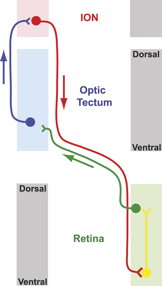
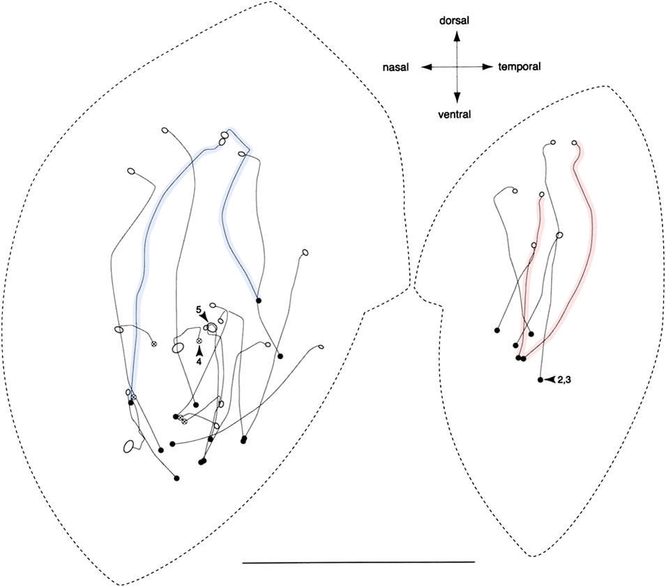
From the evidence presented thus far, the organization of the CVS would seem to be straightforward. It consists of a homotopic loop in which ganglion cells, mostly in the dorsal retina, project to the tectum, which projects to the ION, which then projects to the ventral retina, with the loop being completed by target cells in the retina (Fig. 6). Target cells are excitatory to ganglion cells, perhaps through disinhibition, and in fact, with the exception of the wide-field inhibition in the ION, all connections in the CVS are effectively excitatory so that the system is one of positive feedback. Up to the projection of the ION onto the ventral retina, the system is organized in a familiar retinotopic manner; however, a discovery by Uchiyama et al (Reference Uchiyama, Aoki, Yonezawa, Arimura and Ohno2004) shows that in the ventral retina this retinotopy is broken in an unexpected way. By labeling individual target cells in the retina, their study showed that target cell somata lying next to each other in the ventral retina can nevertheless send their axons to terminate far apart from each other. Conversely, nearby target cell terminations can be provided by target cell somata quite distant from each other (Fig. 7). In short, the wiring from the ventral retina to target cell terminations, mostly in the dorsal retina, appears chaotic. What does this mean?
The general organization of the CVS of the bird. Ganglion cells (green), typically in the dorsal retina, project to the contralateral optic tectum. After minimal processing by the optic tectum, tecto-isthmo-optic fibers (blue) project to the isthmo-optic nucleus where they each make a one-to-one connection to a neuron in this nucleus. These IO neurons (red) in turn project back to the contralateral retina where they terminate in the ventral half making one-to-one synapses with association, or target, amacrine cells (yellow). These target cells project to the dorsal retina where they complete the loop.

Superimposed tracings of fluorescently labeled target cell axons in the flat-mounted retina of quail. Target cell somata are indicated by dark circles, while the open circles show the terminal field of each axon. Highlighted in blue are two target cells with distant somata but adjacent terminal fields. Highlighted in pink are two adjacent somata whose axon terminations are far apart. Scale bar is 5 mm. Modified from Uchiyama et al. (Reference Uchiyama, Aoki, Yonezawa, Arimura and Ohno2004) with permission.

The seemingly random relationship between the positions of a target cell soma and its terminal field
One interpretation of the chaotic relationship between target cell somata and terminations rests heavily on the idea that target cells are slave neurons driven exclusively by efferent fiber input (Uchiyama & Stell, Reference Uchiyama and Stell2005). The argument is this. If efferent fibers provide the only input to target cell somata, then the position of these somata in the ventral retina is of no consequence; they can settle anywhere. The only consideration that matters is that each target cell’s axon is connected to the correct location (as defined by the receptive field of its efferent input), usually in the dorsal retina. And clearly, alignment of the target cell axon terminal with the IO-neuron receptive field is achieved by some means since the entire system is a homotopic loop.
Two recent results make this interpretation seem unlikely. The first is that target cell somata and their presynaptic partners, the efferent fibers, are very tightly confined to the ventral retina. In one chicken retina examined, of the 7193 efferent terminals found, only one was in the dorsal retina (Lindstrom et al., Reference Lindstrom, Nacsa, Blankenship, Fitzgerald, Weller, Vaney and Wilson2009). This precision does not fit well with the idea that target cell soma position is inconsequential. The second is that electron microscopy has shown that woven in between the large boutons of the efferent terminals are much smaller processes that also synapse upon the target cell (Fig. 8A). From antibody staining, it appears that efferent fibers are glutamatergic, while at least some of the small processes are GABAergic. The vesicular glutamate transporter, vGlut2, is found in the efferent terminals (Fig. 8B), and GAD, the synthetic enzyme for GABA, is found in some of the fine processes (Fig. 8C). Patch clamp recordings and pharmacology experiments support the presence of both glutamatergic and GABAergic input to the target cell, though surprisingly, both are excitatory (Lindstrom et al., Reference Lindstrom, Azizi, Weller and Wilson2010). Almost certainly these fine processes providing excitatory GABAergic input come from amacrine cells. The identity of these amacrine cells is unknown, and they might be narrow-field or wide-field amacrine cells but whatever kind they are, they surely carry information specific to a more or less local region of the ventral retina.
Organization of the synapse between the efferent fiber and the target cell. (A) A transmission electron microscopy image of a target cell at the level of its synapse. Individual processes belonging to the target cell are colored green, while efferent terminals are colored red, with processes that could not be unambiguously identified left uncolored. The large efferent terminal boutons can be seen pressed into the target cell soma and wrapped by target cell processes. Some of the unidentified processes likely belong to the target cell or efferent terminal, but others (colored yellow) are different, having very light cytoplasm and a low density of small pleomorphic synaptic vesicles. One process of this type is seen presynaptic to one of the target cell dendrites (blue circle). The immunohistochemistry images in (B) demonstrate that efferent terminals (identified by FluoroRuby labeling following injection of this tracer into the ION, red in a and c) likely release glutamate as they colocalize (yellow in c) with antibody staining for vGlut2, a vesicular glutamate transporter (green in b and c). For comparison in (C), the synthetic enzyme for GABA, GAD65/67 (green in b and c), is found in small puncta scattered throughout the dendritic region of the target cell (identified here by its intense labeling with antibodies to parvalbumin, red in a and c). The size and distribution of these GAD+ puncta resembles that of the small light processes seen in EM (those colored yellow in A). Scale bar in (A) is 2 μm. Scale bars in (B) and (C) are 10 μm. A from Lindstrom et al. (Reference Lindstrom, Nacsa, Blankenship, Fitzgerald, Weller, Vaney and Wilson2009), B and C from Lindstrom et al. (Reference Lindstrom, Azizi, Weller and Wilson2010).

These new results would seem to make the system less intelligible, implying as they do, that patches of ventral retina are connected, seemingly at random, with other patches mostly in the dorsal retina. We believe, in fact, that the resolution of this paradox is the key to understanding the function of the system.
Suggested functions for the CVS
In evaluating competing hypotheses about CVS function, we hold two general considerations to be paramount. First, the CVS is clearly designed to work rapidly. This conclusion is supported by many pieces of evidence such as the large diameter of the myelinated efferent fibers (Cowan, Reference Cowan1970), the minimal number of neurons in the CVS loop, and the large size of the synapses from efferent fibers to target cells. Holden & Powell (Reference Holden and Powell1972) estimated that the delay between the visual stimulus and return of the descending signal to the retina is only 75 ms, just a few milliseconds longer than the first appearance of a signal in the optic tectum. Whatever function is served by the CVS must place a premium on speed.
The second paramount consideration is that the size and complexity of the ION in different species is more closely linked to way of life rather than phylogeny. Although only a tiny sample of the 10,000 species of birds have been examined, the evidence to date suggests that ground feeding birds such as the passerines, sparrow and greenfinch; galliforms, including chicken and quail; and columbiforms, including pigeon (reviewed in Reperant et al., Reference Reperant, Miceli, Vesselkin and Molotchnikoff1989) have the most highly developed ION (8,000–10,000 neurons in a highly differentiated nucleus). In contrast Anseriforms, ducks and geese, have a smaller (∼3500 neurons) loosely differentiated ION (Sohal & Narayanan, Reference Sohal and Narayanan1974) and birds of prey, at least falconiforms and strigiforms, have a much reduced ION (900–1400 neurons in a poorly differentiated nucleus, Weidner et al., Reference Weidner, Reperant, Desroches, Miceli and Vesselkin1987). As shown by the positioning of these different groups of birds on the phylogenetic tree shown in Fig. 9, the size of the ION is not well predicted by phylogeny. A relationship to feeding style is apparent however and is sharply illustrated in a study comparing the ION of one apodiform, the common swift, with those of three passerines, the barn swallow, blackbird, and hawfinch (Feyerabend et al., Reference Feyerabend, Malz and Meyer1994). Compared to the ground feeding blackbird and hawfinch with 8000–12,000 neurons per ION, the ION of the swallow is much reduced and more closely resembles that of the swift (1900–3000 neurons), rather than its passerine relatives. The similarity of ION size in swift and swallow is most easily accounted for by their shared lifestyle characterized by feeding on the wing.
A phylogeny of birds showing those orders with known CVS anatomy. The phylogenetic tree with major clades labeled (left) is modified from Hackett et al. (Reference Hackett, Kimball, Reddy, Bowie, Braun, Braun, Chojnowski, Cox, Han, Harshman, Huddleston, Marks, Miglia, Moore, Sheldon, Steadman, Witt and Yuri2008). Major clades in which the CVS has not been quantitatively examined are italicized. Orders for which the CVS has been studied in at least one species are listed to the right along with the number of cells found in the ION. Subscripted numbers refer to the following references: 1, Feyerabend et al. (Reference Feyerabend, Malz and Meyer1994); 2, Weidner et al. (Reference Weidner, Reperant, Desroches, Miceli and Vesselkin1987); 3, Wolf-Oberhollenzer (Reference Wolf-Oberhollenzer1987); 4, Lindstrom et al. (Reference Lindstrom, Nacsa, Blankenship, Fitzgerald, Weller, Vaney and Wilson2009); 5, Sohal & Narayanan (Reference Sohal and Narayanan1974); 6, Reperant et al. (Reference Reperant, Miceli, Vesselkin and Molotchnikoff1989).

Previous Theories
As laid out in Table 1, a number of ideas have been advanced for the function of the CVS. These ideas range from the vaguest of suggestions to specific hypotheses with supporting evidence, but all can be classified into three broad and nonexclusive categories, “motor guidance,” “adaptation” and “attention,” with a fourth category of “other” to accommodate the remainder.
The category of motor guidance includes two appealing hypotheses. The first is that the CVS helps stabilize the retinal image through an involvement of the accessory optic system (AOS) and subsequently, head and eye movement. By increasing the gain of the AOS, Woodson et al (Reference Woodson, Shimizu, Wild, Schimke, Cox and Karten1995) suggest that this system improves the accuracy of pecking. This hypothesis rests on the supposition that efferent fibers synapse with displaced ganglion cells, which are known to project to a nucleus called nBOR, a part of the AOS and a nucleus concerned with eye movements. As already described however it is target cells, rather than displaced ganglion cells, that are the primary recipient of restricted efferent input. The possibility exists that minor synapses made by efferent fibers (such as the tendril synapses) are on displaced ganglion cells, but the major role of the efferent pathway is tied to the target cells. A further indication that eye stabilization is unlikely to be the function of the CVS lies in the observation that nBOR neurons have very large receptive fields and prefer backward moving stimuli (Wylie & Frost, Reference Wylie and Frost1990), whereas ION cells have much smaller fields and generally have a response minimum for backward motion (Holden, Reference Holden1970; Miles, Reference Miles1972c).
The second hypothesis is that the CVS is somehow involved directly in pecking behavior. This is superficially attractive as it potentially explains why this system is highly developed in ground feeding birds but reduced in others. It also seems plausible since pecking is a rapid behavior and might require rapid feedback to ensure successful targeting. Unfortunately, the details of pecking behavior, which has been carefully examined in pigeons, are inconsistent with this. The peck itself lasts only 60 ms (Goodale, Reference Goodale1983) and is likely too brief to allow any visually guided corrections; moreover, the eyes are closed approximately 35 ms after peck initiation. However, this does not preclude the possibility that the CVS might play a role in peck initiation.
During peck initiation, the object to be pecked is aligned in the small frontal area of the visual field in which there is binocular overlap. Since the head remains stationary about 56 mm from the ground during this period, the image of the object to be pecked falls on the dorsal temporal retina in the region called the red field. Efferent fibers do not project to this region (Woodson et al., Reference Woodson, Shimizu, Wild, Schimke, Cox and Karten1995); moreover, the small amount of data we presently possess about the projection of target cells does not indicate any preferential innervation of the red field either (Uchiyama et al., Reference Uchiyama, Aoki, Yonezawa, Arimura and Ohno2004). This would seem to make it unlikely that the CVS is concerned with adjusting the initial head trajectory. The final hypothesis concerning the influence of the CVS on pecking behavior suggests that the CVS is designed to enhance discrimination of grain in the lateral visual field. Again, this suggestion is poorly matched to the topology of the system, and it has no exceptional requirement for speed. Furthermore, the strong preference of IO neurons for moving objects is inconsistent with this hypothesis.
The general idea that the CVS is involved in adaptation, either dark adaptation or “dynamic adaptation,” has some supporters but also some substantial difficulties. It is unclear why a mechanism of this sort should be most developed in ground feeding birds, and unclear also why efferent input should be entirely to the ventral retina.
Lastly, the general idea that the CVS is somehow concerned with attention has a lot to recommend it and has been elaborated in several different directions. The idea that the CVS is involved with switching attention, either between the two eyes (Holden, Reference Holden1966), which in ground feeding birds have almost completely different visual fields, or from dorsal to ventral retina (Clarke et al., Reference Clarke, Gyger and Catsicas1996), both have the problem that the CVS seems unnecessarily complex to function simply as a binary switch. Furthermore, the idea that the CVS suppresses activity in one eye or one half of a retina is inconsistent with the overall positive feedback of the CVS. A different kind of attentional hypothesis derives from Uchiyama’s insight that the wide-field inhibitory interneurons of the ION allow for global competition between visual stimuli, thereby “highlighting” retinal regions of interest (Uchiyama et al., Reference Uchiyama, Nakamura and Imazono1998; Uchiyama, Reference Uchiyama1999). This suggestion is consistent with the strongly suppressive effects of small stimuli distant from the classical receptive field of IO neurons, and it receives support from computational simulations (Uchiyama et al., Reference Uchiyama, Nakamura and Imazono1998; Uchiyama, Reference Uchiyama1999). Because of its firm grounding, the idea of global competition within the ION has to be incorporated into any wider theory of CVS function.
A new hypothesis
We begin by suggesting that the natural stimuli that drive IO neurons are moving shadows cast on the ground or on objects in the environment. This is consistent with the preference of these neurons for dark moving edges and also with ION input, at least in pigeon, being chiefly from the dorsal retina, which looks at the ground. Ground feeding birds like the pigeon and chicken have panoramic vision in which the two eyes see virtually the whole visual world with only a 22-deg area of binocular overlap anteriorly, and a 44-deg blind region behind them (Martin, Reference Martin, Zeigler and Bischof1993). As Uchiyama has pointed out, the ION is a nucleus in which a winner-take-all competition allows the selection of the most salient stimulus (Uchiyama et al., Reference Uchiyama, Nakamura and Imazono1998; Uchiyama, Reference Uchiyama1999). Considering a moving shadow then, ganglion cells in the retina detect the image of the shadow and transmit a signal through the optic tectum to the ION (Fig. 10a and 10b). We suggest that the ION selects this shadow and produces an output in a group of efferent fibers projecting to target cells in the ventral retina (Fig. 10c and 10d). This output from the ION to the ventral retina is in essence a prediction about the existence and possible location of an object in the sky. That object is potentially an aerial predator. If the image of an aerial predator excites amacrine cells near a target cell receiving efferent input, these amacrine cells will provide excitatory GABAergic input to the target cell (Fig. 10e) and together these two inputs, from the efferent fiber and a local amacrine cell, would be expected to drive the target cell strongly. In turn, because the system is homotopic, this target cell will further excite ganglion cells in the shadowed patch of dorsal retina (Fig. 10f). Because of the extra input from the ventral retina, the ION will select this stimulus as the most salient and will lock-on, temporarily binding these two stimuli together and continuing to track them as signals pass iteratively around the CVS loop.
A model of CVS function. In this sequential diagram a shadow falling on the ground excites a group of ganglion cells in the dorsal retina (a). This pattern of excitation is transmitted to the optic tectum and from there to the ION via tecto-isthmo-optic neurons (b). Wide-field GABAergic interneurons in the ION create a winner-take-all competition in which the most salient inputs are selected (c) and activate efferent fibers running to the ventral retina (d). These efferent fibers are widely dispersed within the ventral retina and constitute a prediction or guess about the likely position of an object in the dorsal visual field —in other words, the sky. A target cell driven by efferent input that also receives local input corresponding to an object in the sky (e) will further excite ganglion cells under the shadow (f). The enhanced responses of these ganglion cells created by this association of the two stimuli will ensure their selection in the next round of winner-take-all competition in the ION and the positive feedback of the system means it will continue to track the shadow and aerial object together as they move.

This hypothesis incorporates the suggestions of others, for example, that the CVS performs “a sort of scanning mechanism acting without any movement of the eye” (Galifret et al., Reference Galifret, Condé-Courtine, Repérant and Serviere1971), or “covert attentional scanning” (Ohno & Uchiyama, Reference Ohno and Uchiyama2009), and is similar to the suggestion by Holden (Reference Holden1990) that when searching for food a “sky watch” system is turned on. It resolves the paradox that target cells connect seemingly at random to other regions of retina. We would argue that connections are not random at all but instead represent two conditional probability distributions. The wiring from the ION to the ventral retina represents the likelihood of finding an aerial predator in a particular patch of sky, given the position of its shadow. Conversely, the projection of target cell axons from the ventral to the dorsal retina reflects the likelihood of a shadow’s location, given the position of an object in the sky.
Many factors must affect the geometry of the relationship between object and shadow. The angle of the sun, the head angle of the bird, and the topography upon which the shadow is cast are all important factors. Evolution, we argue, has fashioned statistical best guesses based on these factors weighted by ecological realities such as the time of day, and hence sun angle, at which most predation takes place. The fact that target cells and efferent terminals are confined to the ventral retina is because aerial predators are confined to the sky; moreover, the details of the distribution of efferent fiber terminations in the ventral retina (Lindstrom et al., Reference Lindstrom, Nacsa, Blankenship, Fitzgerald, Weller, Vaney and Wilson2009) are significant. The extreme ventral retina has only a low density of terminations, suggesting that predators directly overhead are uncommon. The densest region of terminations lies a little below the equator of the eye and is chiefly nasal rather than temporal, suggesting that the system is tuned for predators at low angles approaching from behind. Target cells project mostly to the dorsal retina because, typically, shadows fall on the ground, but they can also fall on trees and other objects that are imaged in the ventral retina. In this regard, we would expect that differences in the visual environment of different species could produce different projection patterns. We would also suggest that the preference of IO neurons for anteriorward movement is because the blind region behind the bird represents the direction from which it is most vulnerable to surprise attack.
Plenty of anecdotal material going back to classical times suggests that shadows induce fear in ground feeding birds (McCartney, Reference McCartney1947) but surprisingly, very little behavioral work has been done on the responses of birds to shadows. An exception is a study in which chickens were subject to models moved overhead (Hirsch et al., Reference Hirsch, Lindley and Tolman1955). The work was undertaken to reexamine the claim that silhouettes of hawks induced fearful behavior more than the silhouettes of geese. In this study, no difference could be found between the two types of silhouette. However, the authors did make an unexpected discovery that fear behavior was very significantly increased if the model cast a shadow.
While behavioral data to support this hypothesis are virtually absent, it is clear that for ground feeding birds, aerial predation is a major cause of mortality. In a large study of Bobwhite quail using telemetric tagging to determine the fate of some 2600 individuals, it was found that these birds have an extraordinarily high (63%) annual probability of mortality from aerial predators (Cox et al., Reference Cox, Peoples, DeMaso, Lusk and Guthery2004). Given this, there must be a huge selection pressure for the detection of, and escape from, aerial predators, a conclusion that is supported by many aspects of bird behavior and morphology that are seen as adaptations to avoid, detect, and escape from predators (Caro, Reference Caro2005).There would, of course, be many common circumstances in which a system of the kind we propose could offer no advantage, for example, if an overcast sky produced no shadows, or if the predator attacked from vegetation at close range. For evolution to retain it however, the system need not be perfect, or even very good. All that is required is that it offers some differential advantage to its owners so that in some fraction of encounters with predators it facilitates escape.
Three explicit predictions of this hypothesis
This hypothesis has many substantial gaps and unknowns, but it also makes definite predictions by which it could be falsified. The hypothesis requires that IO neurons have nonclassical receptive fields. In particular, it must be true that responses to a dark moving object, usually in the ventral field, would be enhanced by a dark moving object in a region of the dorsal field. Receptive fields of this sort have not been reported, but it seems likely that they would not have been detected with the protocols employed thus far. A second prediction is that ground feeding birds should detect the presence of an aerial predator sooner if a shadow precedes or coincides with presentation of the predator. Behavioral experiments to test ideas like this are fraught with difficulties, such as the subject’s habituation to predators, they have however been successfully carried out in some labs (Evans et al., Reference Evans, Macedonia and Marler1993; Whittingham et al., Reference Whittingham, Butler, Quinn and Cresswell2004; Palleroni et al., Reference Palleroni, Hauser and Marler2005; Cresswell et al., Reference Cresswell, Butler, Whittingham and Quinn2009). Thirdly, we predict that the CVS should be most highly developed in those species that suffer severe aerial predation in conditions where shadows are often visible. Species that feed in open spaces, rather than dense vegetation, and live in sunny rather than overcast regions would be expected to have a heavier reliance on their CVS.
Conclusions
In summary, we propose that, through the restricted efferent pathway, the CVS acts as an early warning system that allows the presence of a moving shadow to trigger a very rapid and parallel search of the regions of sky most likely to contain an aerial predator. Once an association between shadow and object is established, the system locks on through positive feedback and continues to track shadow and object together. Several previously suggested ideas are incorporated into this new view of function, for example, the idea of highlighting, predator detection, and static scanning of the sky.
It is interesting to note that the critical neurons in the retina, that we know as target cells, were originally named “association amacrine cells” by Cajal, who was first to see them. As translated by Stell (Uchiyama & Stell, Reference Uchiyama and Stell2005), Cajal’s view of their function was as follows (Ramón y Cajal, Reference Ramón y Cajal1895). “As for the functional significance of these cells, one must suppose that they serve to link – for common action – amacrine cells separated by long distances.” This intuitive guess may yet prove to be correct, and deeper investigation of these cells and the working of the CVS might serve as a model for the way in which the central nervous system, in general, forms and dissolves temporary associations.
Acknowledgments
We thank David Warland for useful discussions during this work and David Vaney for insightful comments on the manuscript. This work was supported by NIH awards EY04112 and EY12576.