Introduction
The round fantail stingray Taeniura grabata (Geoffroy St-Hilaire), recently synonymized with Taeniurops grabata (Geoffroy St-Hilaire) as supported by the studies of Lim et al. (Reference Lim2015) and Last et al. (Reference Last, Naylor and Manjaji-Matsumoto2016), seems to have a naturally fragmented habitat. It is found in the eastern Atlantic Ocean (Capapé and Desoutter, Reference Capapé, Desoutter and Quero1990) and in the central and south-eastern Mediterranean (McEachran and Capapé, Reference McEachran, Capapé and Whitehead1984), but seems to be absent in the western Mediterranean Sea (Serena, Reference Serena2005). This stingray is categorized as Data Deficient on the IUCN Red List and only some metazoan parasites are reported (Euzet and Maillard, Reference Euzet and Maillard1967; Neifar et al., Reference Neifar, Euzet and Ben Hassine1999; Ruhnke et al., Reference Ruhnke, Caira and Cox2015).
An investigation of the species diversity of cestodes of 16 specimens of this stingray from the south-eastern Mediterranean off Tunisia carried out from 1998 to 2010, and examination of formalin-preserved cestodes from T. grabata collected at Gorée Island (Senegal) between 1946 and 1954 by the French ichthyologist J. Cadenat and conserved in the personal collection of the late L. Euzet, led to the discovery of three new Anthocephaliid cestode species (Rhinebothriidea). Two of them represent a new genus, and the third species is assigned to the genus Anthocephalum Linton, 1890. The purpose of this paper is to describe these new taxa.
Materials and methods
Between 1998 and 2010, 16 specimens of T. grabata were collected, as by-catch by local fishers, at three localities off Tunisia. Seven specimens (5 females, 2 males) were collected from the north at Bizerte, three females were collected from the central regions at Sfax and six specimens (4 females, 2 males) from the south at Zarzis. Each stingray was dissected as soon as possible after capture. An incision was made on the ventral side and the spiral intestine was tied off with a thread at the posterior end to prevent loss of contents, and removed. Some intestines were dissected fresh, and living worms were removed, washed in filtered seawater, examined between slide and coverslip as living whole mounts, then fixed (slightly flattened) in 5% formalin and preserved in 70% ethanol. Other intestines were injected with 8% formalin to fix the worms in situ, tied off anterior to the pyloric sphincter and stored in 5% formalin for later examination.
Whole mounts for light microscopy were stained with Semichon's acetic carmine, dehydrated in a graded ethanol series, cleared in clove oil, and mounted in Canada balsam. Specimens prepared for scanning electron microscopy (SEM) were fixed in cold (4°C) 2.5% glutaraldehyde in 0.1 m sodium cacodylate buffer at pH 7.2 for a minimum of 2 hours, rinsed in 0.1 m sodium cacodylate buffer at pH 7.2, post-fixed in cold (4°C) 1% osmium tetroxide in the same buffer for 1 hour, rinsed in a 0.1 m sodium cacodylate buffer at pH 7.2 and dehydrated in a graded ethanol series. After sonication in absolute alcohol for 30 s at 40 kHz, specimens were kept for 30 minutes in absolute alcohol, and successively immersed for 20 minutes in 3 : 1, 1 : 1 and 1 : 3 mixtures of absolute alcohol and n-amylacetate, washed in pure n-amylacetate for 20 minutes and critical point dried with carbon dioxide. Specimens were then mounted on aluminium stubs using colloidal silver, and sputter coated with 10 nm of gold. SEM was performed with a Zeiss DSM 940 A Gemini field emission scanning electron microscope.
Formalin-preserved cestodes from T. grabata collected at Gorée Island (Senegal) between 1946 and 1954 by J. Cadenat and conserved in the personal collection of the late L. Euzet (now part of the collection of the Muséum National d'Histoire Naturelle, Paris) were also examined. Old fixed specimens were placed for a few hours in acidulated water with acetic acid at 1% to remove excess preservatives, soaked in distilled water overnight and then washed and stained with Semichon's acetic carmine, dehydrated in a graded ethanol series, cleared in clove oil, and mounted in Canada balsam.
Measurements of whole mounted cestodes were obtained using a drawing tube and ruler calibrated using a stage micrometer. All measurements are given in micrometers unless specified otherwise. Measurements are presented as follows: range (mean ± standard deviation, n = number of observations). Illustrations were made with the aid of a Leitz microscope with drawing tube. Specimens were scanned and redrawn with Corel Draw Software. Microthrix morphology follows Chervy (Reference Chervy2009). Elasmobranch taxonomy follows Naylor et al. (Reference Naylor, Carrier, Musick and Heithaus2012) and Last et al. (Reference Last, Naylor and Manjaji-Matsumoto2016).
Holotypes and paratypes were deposited in the Muséum National d'Histoire Naturelle (Paris, France) (MNHN).
Results
Alveobothrium gen. n.
Zoobank number for genus: urn:lsid:zoobank.org:act:DC72EAD0-CAE0-442E-BFE9-CA0DD1A5799B
Taxonomic summary
Type species. Alveobothrium grabatum gen. n., sp. n.
Additional species. Alveobothrium zarzisense sp. n.
Etymology. From the latin “alveolus”, in reference to the arrangement of facial loculi, such as a honeycomb cell.
Diagnosis
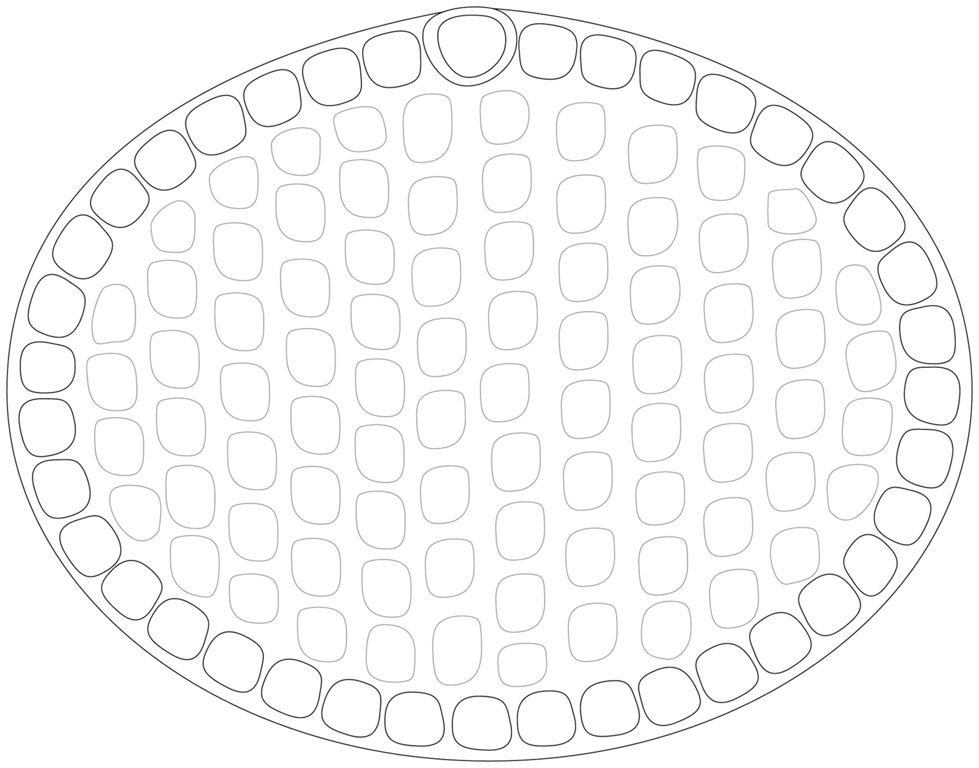
Rhinebothriidea Healy, Caira, Jensen, Webster & Littlewood, 2009; Anthocephaliidae Ruhnke, Caira & Cox, 2015. Worms slightly craspedote, euapolytic. Scolex with four bothridia and cephalic peduncle; apical organ absent. Bothridia stalked, wider than long, with single apical sucker, numerous marginal loculi and fully loculate distal surface; facial loculi arranged in multiple staggered rows (fig. 1); bothridial margins with thin velum. Mature proglottids longer than wide. Testes numerous, arranged in 1–2 columns anterior to cirrus sac. Post-poral field of testes lacking. Vas deferens enters cirrus sac at anterior margin. Genital pores lateral, irregularly alternating; genital atrium present. Cirrus sac thin-walled, recurved posteriorly, containing coiled cirrus; cirrus armed with spinitriches. Vagina sinuous, extending from ootype along midline of proglottid to anterior margin of cirrus sac, then laterally to open into genital atrium anterior to cirrus sac. Ovary H-shaped in frontal view, tetralobed in cross section. Vitellarium follicular; follicles distinctly wider than long, in two lateral bands; each band consisting of one or more columns of follicles, completely interrupted by ovary and cirrus sac. Uterus median, ventral, sacciform, extending from ovarian isthmus to anterior end of proglottid. Parasites of Dasyatidae (Myliobatiformes), Atlantic Ocean and Mediterranean Sea.
Schematic illustration of bothridia characteristic of the genus Alveobothrium.

Remarks
Alveobothrium gen. n. is unique among rhinebothriidean genera in its possession of bothridia bearing simultaneously an apical sucker, marginal loculi and a multiple row of staggered facial loculi. The lack of a myzorhynchus in adult form easily distinguishes Alveobothrium gen. n. from Clydonobothrium Euzet, 1959, Echeneibothrium Van Beneden, 1850, Notomegarhynchus Ivanov & Campbell, 2002, Phormobothrium Alexander, 1963, Pseudanthobothrium Baer, 1956 and Tritaphros Lönnberg, 1889. It can further be distinguished from Zyxibothrium Hayden & Campbell, 1981 and all the eight valid genera of Rhinebothriidae (Healy et al., Reference Healy2009; Caira et al., Reference Caira2017) in its possession, rather than lack, of an apical sucker with rounded protrusions on each bothrium. Beyond the presence of apical sucker, the new genus also differs from Cairaeanthus Kornyushin & Polyakova, 2012 by the bothridia not bifid posteriorly and the presence of some vitelline follicles in the posterior margins of the proglottid. Alveobothrium gen. n. differs from Barbeaucestus Caira, Healy, Marques & Jensen, 2017, Divaricobothrium Caira, Healy, Marques & Jensen, 2017 and Sungaicestus Caira, Healy, Marques & Jensen, 2017 in its possession (rather than lack) of marginal loculi throughout the perimeter of the bothridia. The lack of post-vaginal testes further distinguishes Alveobothrium from Anindobothrium Marques, Brooks & Lasso, 2001.
Alveobothrium most closely resembles Anthocephalum Linton, 1890 and Stillabothrium Healy & Reyda, 2016, but can be distinguished from the former genus by the presence of facial loculi, and from the latter in its possession, rather than lack, of marginal loculi throughout the perimeter of the bothridia. Beyond the differences in the morphology and arrangement of facial loculi described as longer than wide in the posterior region of the bothridia of Stillabothrium, Alveobothrium possesses, rather than lacks, apical suckers. However, the absence of an apical sucker from the bothridia of all species of Stillabothrium remains to be confirmed (e.g. the SEM image of one of the bothridia of S. ashleyae in fig. 3B of Reyda et al. (Reference Reyda2016) certainly seems to suggest the presence of a sucker).
The genus is fully consistent with the diagnosis of the order Rhinebothridea as given by Healy et al. (Reference Healy2009) and the family Anthocephaliidae as presented by Ruhnke et al. (Reference Ruhnke, Caira and Cox2015).
Alveobothrium grabatum sp. n.
Zoobank number for species: urn:lsid:zoobank.org:act:971F4CD5-89A7-474A-AA40-D3DAFF98D624
Taxonomic summary
Type host. Taeniurops grabata (Geoffroy St-Hilaire) (Dasyatidae), round fantail stingray.
Type locality. Zarzis (33°15'N, 11°10'E), Tunisia.
Additional localities. Bizerte (37°30'N, 9°50'E), Tunisia; Sfax (34°47'N, 10°51'E), Tunisia; Gorée Island (14°40'N, 18°25'W), Senegal.
Site of infection. Spiral intestine.
Prevalence in Tunisia. 100% (16 of 16 specimens examined).
Type material. Holotype (MNHN HEL805); 16 paratypes (MNHN HEL805–815).
Etymology. This species is named for its type host, Taeniurops grabata.
Description
Based on 43 whole-mounted specimens and two worms prepared for SEM; measurements taken from 25 specimens.
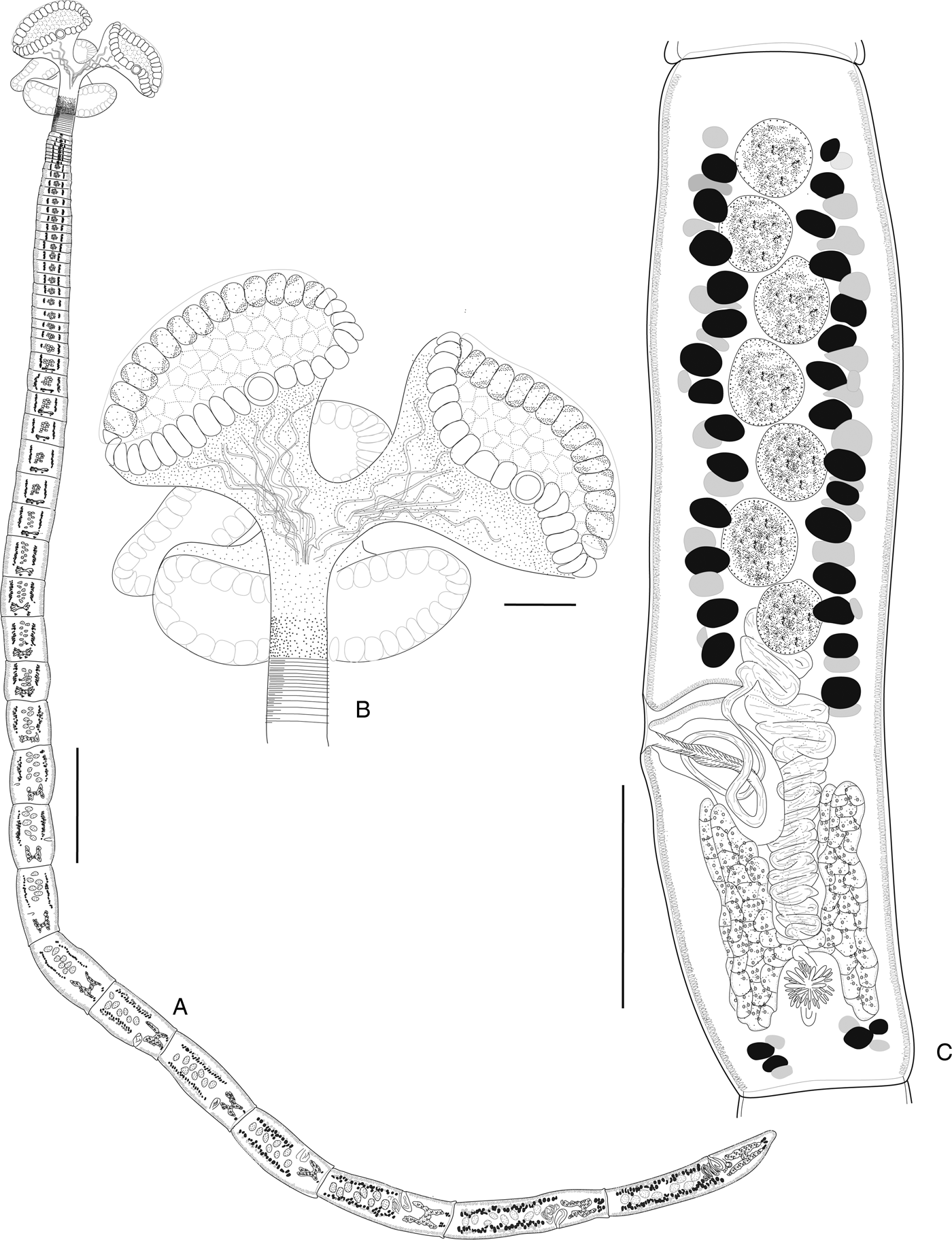
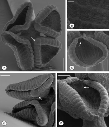
Worms (fig. 2A) slightly craspedote, euapolytic, 6–17 mm (9.6 ± 1.9 mm; n = 25) long, with 62–157 (83 ± 9; n = 25) proglottids; maximum width 350–550 (452 ± 64; n = 25) at level of scolex. Strobila, 5.5–16.8 mm (9.1 ± 2 mm; n = 25), long by 80–150 (124 ± 64; n = 25) wide at level of terminal proglottid. Scolex (figs 2B and 3A) with four bothridia and short cephalic peduncle; myzorhynchus absent. Bothridia (fig. 3C–E) stalked, wider than long, 350–650 (412 ± 26; n = 15) wide, with circular apical sucker, 29–37 (32 ± 2; n = 13) marginal loculi and 13–15 (n = 2) staggered rows of facial loculi (fig. 3E); bothridial margins with thin rim; apical sucker 60–80 (70 ± 4; n = 20) in diameter. Distal bothridial surfaces covered with spinitriches (fig. 3B). Cephalic peduncle 130–190 (155 ± 10; n = 25) long by 70–110 (85 ± 5; n = 25) wide.
Alveobothrium grabatum sp. n. (A) entire worm; (B) scolex, ventral view; (C) mature proglottid, dorsal view. Scale bars: A = 500 μm; B, C = 100 μm.

Scanning electron micrographs of Alveobothrium grabatum sp. n. (A) Scolex, apical view; (B) distal bothridial surface and acicular filitriches; (C, E) bothridium, straggered row of facial loculi; (D) proximal bothridial surface. Arrows indicate the apical sucker. Scale bars: A, C, D, E = 100 μm; B = 1 μm.

Immature proglottids initially wider than long, becoming longer than wide with maturity. Terminal proglottid (fig. 2C) 420–700 (522 ± 54; n = 25) long by 80–150 (124 ± 64; n = 25), wide, length to width ratio 4.5–5.3 (4.7 ± 0.7; n = 25). Gravid proglottids not observed. Testes 6–10 (7 ± 1; n = 25) in number, slightly oblong, 15–30 (24 ± 2; n = 48) long by 14–27 (20 ± 4; n = 48) wide, arranged in single irregular column, extending from anterior margin of proglottid to level of vagina. Post-vaginal testes absent. Vas deferens dorsal, coiled anterior to cirrus sac, expanded and descending to ovarian isthmus. Cirrus sac thin-walled, pyriform, recurved posteriorly, 45–60 (51 ± 2; n = 24) long by 38–55 (43 ± 3; n = 24) wide, containing coiled cirrus; cirrus armed with spinitriches. Genital pores lateral, irregularly alternating, 35–55% (43 ± 3%; n = 25) from posterior end of proglottid; genital atrium not prominent. Vagina slightly sinuous, extending from Mehlis’ gland anteriorly, then ventral and lateral to cirrus sac, opening into common genital atrium. Ovary H-shaped in frontal view, tetralobed in cross-section, slightly asymmetrical, 80–110 (94 ± 5; n = 25) long by 72–96 (82 ± 7; n = 25) wide at level of isthmus; ovarian isthmus in posterior half of ovary; ovicapt not observed. Mehlis’ gland well developed, posterior to ovarian isthmus. Vitelline follicles 20–45 (29 ± 3; n = 30) long by 10–15 (12 ± 2; n = 30) wide, arranged in two lateral bands, each band consisting of one dorsal and one ventral irregular column; not reaching anterior margin of proglottid, interrupted dorsally and ventrally by cirrus sac and ovary. Uterus and gravid proglottids not observed.
Remarks
Alveobothrium grabatum sp. n. is the type species of the genus. In living specimens, the size of the facial loculi varies with degree of bothridial contraction. This probably allows the bothridia to closely fit the shape of intestinal villi and makes anchoring more efficient for bothridia. Reyda et al. (Reference Reyda2016) described a “grid-like” appearance on the bothridia of Stillabothrium hyphantoseptum Hergoz, Bergman & Reyda, 2016 and S. campbelli Delgado, Dedrick & Reyda, 2016, which they interpreted as an extensive overlap of transverse and longitudinal septa. This is not the case for A. grabatum sp. n., in which facial loculi are clearly staggered, so that each loculus meets only two others edge-to-edge (fig. 3A). Furthermore, A. grabatum sp. n. can be distinguished from both species by the presence of a rounded apical sucker and distinctive marginal loculi.
The distribution of A. grabatum sp. n. includes both the Mediterranean Sea and the Atlantic Ocean. Alveobothrium grabatum sp. n. appears to exhibit oioxenic specificity (sensu Euzet and Combes, Reference Euzet, Combes, Bocquet, Genermont and Lmotte1980), in that it is known only from T. grabata.
Alveobothrium zarzisense sp. n.
Zoobank number for species: urn:lsid:zoobank.org:act:04B3C8DA-F26C-4F92-B6BF-E421ED22392C
Taxonomic summary
Type host. Taeniurops grabata (Geoffroy St-Hilaire) (Dasyatidae), round fantail stringray.
Type locality. Zarzis (33°15'N, 11°10'E), Tunisia.
Additional localities. Bizerte (37°30'N, 9°50'E), Tunisia; Sfax (34°47'N, 10°51'E), Tunisia; Gorée Island (14°40'N, 18°25'W), Senegal.
Site of infection. Spiral intestine.
Prevalence in Tunisia: 100% (16 of 16 specimens examined).
Type material. Holotype (MNHN HEL816); 47 paratypes (MNHN HEL816–830).
Etymology. The specific name is after the type locality Zarzis (Tunisia).
Description
Based on 36 whole mounts and two worms prepared for SEM; measurements from 25 specimens.
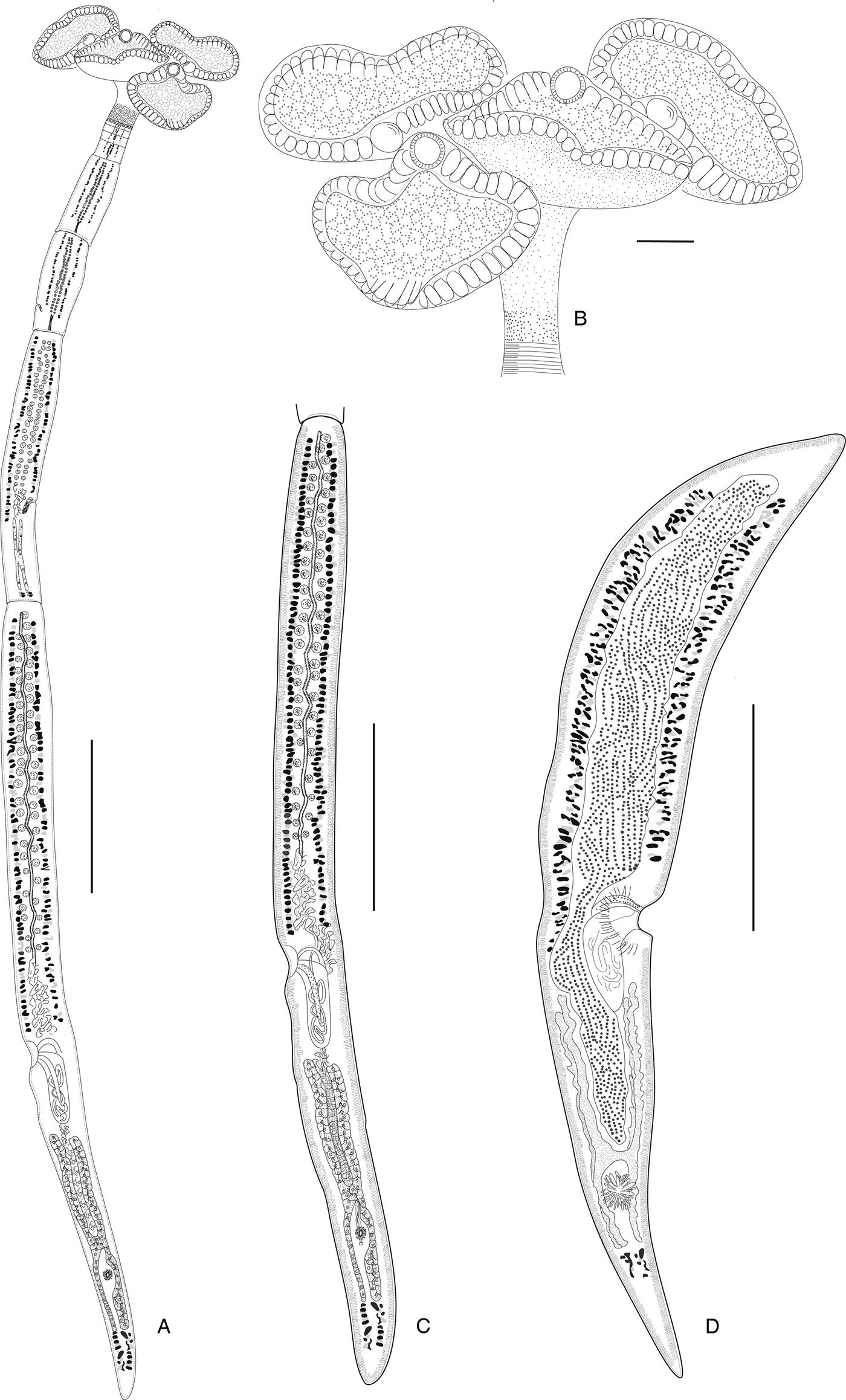
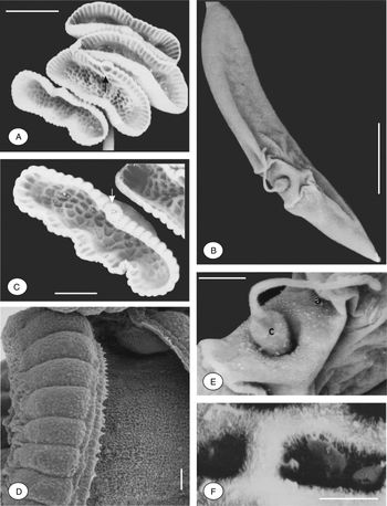
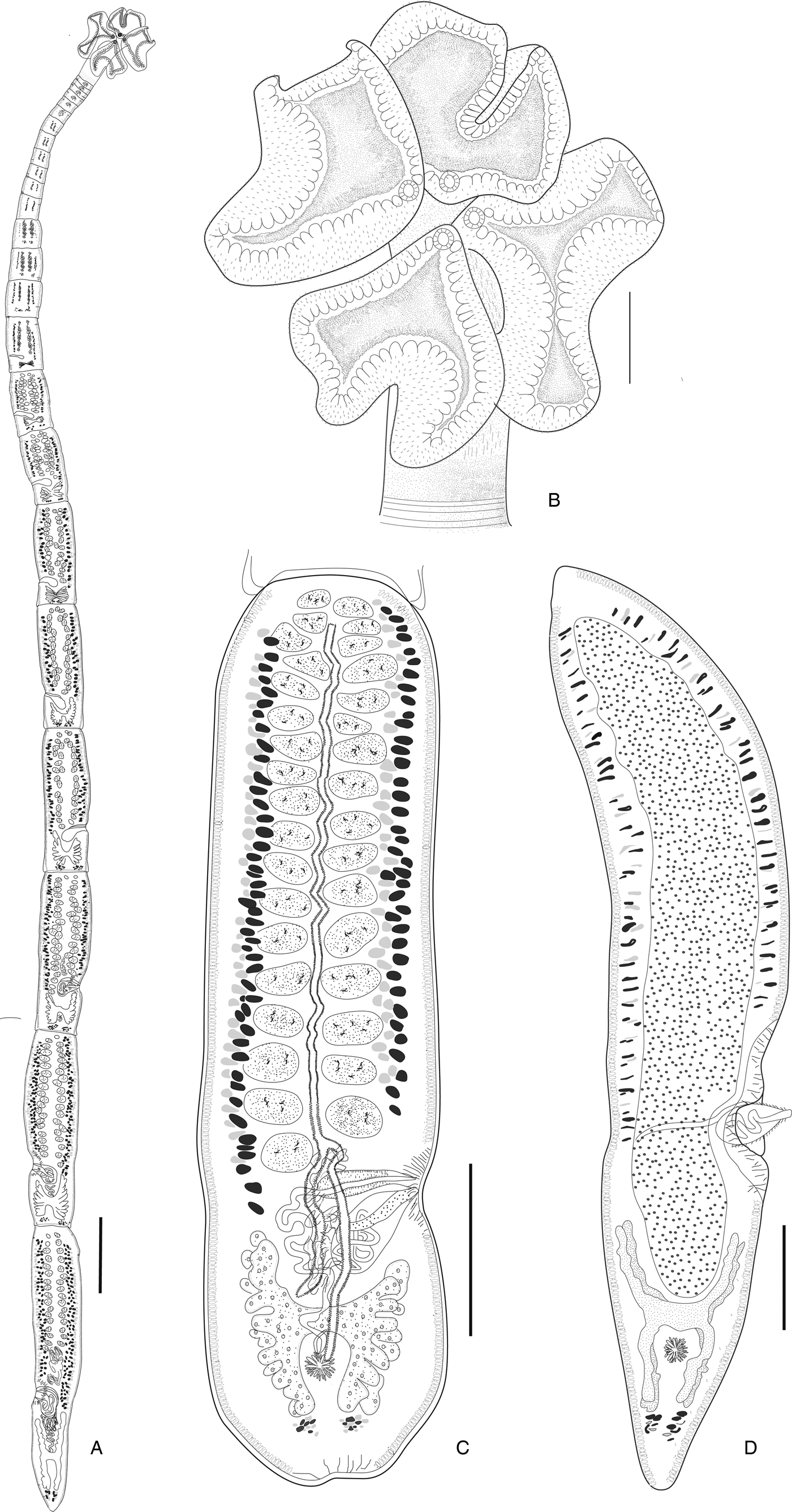
Worms (fig. 4A) slightly craspedote, euapolytic, 3–6 mm (4.1 ± 1.3 mm; n = 25) long, with 6–10 (7 ± 1; n = 25) proglottids; maximum width 320–470 (380 ± 42; n = 25) at level of scolex (fig. 4A). Scolex (figs 4B and 5A) with four bothridia and cephalic peduncle; myzorhynchus absent. Bothridia (fig. 5A, B) stalked, wider than long, 400–620 (524 ± 22; n = 20) wide, with round apical sucker, 47–51 (50 ± 1; n = 20) marginal loculi and 15–17 (n = 8) staggered rows of facial loculi; apical sucker 45–65 (50 ± 4; n = 20) in diameter; bothridial margins with thin rim (fig. 5D). Proximal surface of bothridial rims covered with acicular filitriches (fig. 5D). Distal bothridial surfaces bearing acicular filitriches (fig. 5F). Cephalic peduncle 110–170 (145 ± 10; n = 15) long by 100–130 (117 ± 5; n = 15) wide.
Alveobothrium zarzisense sp. n. (A) Entire worm; (B) scolex, lateral view; (C) mature terminal proglottid; (D) detached gravid proglottid, ventral view, showing chains of eggs. Scale bars: A, C = 500 μm; B = 100 μm; D = 250 μm.

Scanning electron micrographs of Alveobothrium zarzisense sp. n. (A) Scolex in apical view, showing alveolous-like morphology (arrow); (B) detached proglottid with everted cirrus; (C) bothridium, showing facial loculi structure; (D) bothridium, showing marginal loculi and marginal rim; (E) detail of everted cirrus; (F) distal bothridial surface and acicular filitriches. Arrows indicate the apical sucker. a, atrium; c, cirrus. Scale bars: A = 200 μm; B, E = 100 μm; C = 500 μm; D = 20 μm; F = 1 μm.

Immature proglottids initially wider than long, becoming longer than wide with maturity. Terminal proglottid (fig. 4C) 1.6–2.8 mm (2.1 ± 100 mm; n = 25) long by 150–200 (190 ± 12; n = 25) wide, length to width ratio 12.6–14.8 (13.9 ± 1.7; n = 25). Detached gravid proglottids (figs 4D and 5B) 2.5–3 mm (2.5 ± 350 mm; n = 18) long by 240–410 (300 ± 40; n = 18) wide, length to width ratio 7.3–10.4 (8.9 ± 1.7; n = 18). Testes 48–58 (54 ± 2; n = 25) in number, oblong, 25–40 (29 ± 5; n = 25) long by 12–25 (18 ± 2; n = 25) wide, arranged in two columns extending from anterior margin of proglottid to level of vas deferens. Post-vaginal testes absent. Vas deferens dorsal, coiled anterior to cirrus sac, expanded and descending to ovarian isthmus. Cirrus sac thin-walled, conspicuously recurved posteriorly, 180–330 (227 ± 12; n = 25) long, 60–120 (78 ± 7; n = 25) wide, containing coiled cirrus; cirrus armed with inconspicuous spinitriches (fig. 5E). Genital pores lateral, irregularly alternating, 38–48% (42 ± 3%, n = 25) from posterior end of proglottid; genital atrium prominent, becoming more obvious and muscular in terminal and detached proglottids (fig. 5E). Vagina slightly sinuous, extending from Mehlis’ gland to anterior margin of cirrus sac, opening into common genital atrium anterior to cirrus sac. Ovary, follicular, H-shaped in frontal view, tetralobed in cross section, slightly asymmetrical, 500–900 (644 ± 43; n = 25) long by 60–135 (82 ± 12; n = 25) wide at level of isthmus; ovarian isthmus in posterior half of ovary. Ovicapt at posterior margin of ovarian isthmus, 45–55 (48 ± 10; n = 8). Mehlis’ gland well developed, posterior to ovicapt. Vitelline follicles 15–25 (18 ± 13; n = 25) long by 10–15 (11 ± 1; n = 25) wide, arranged in two lateral bands, each band consisting of 1–2 dorsal and 1–2 ventral irregular columns; follicles not reaching anterior margin of proglottid, interrupted dorsally and ventrally by cirrus sac and ovary. Uterus ventral, extending from ovarian isthmus to anterior end of proglottid; uterine duct inconspicuous. Eggs packaged in groups of up to 13–17 eggs within elongate cocoons (fig. 4D); cocoon 340–420 (370 ± 25; n = 30) long.
Remarks
This species is readily distinguished from A. grabatum in its possession of a greater number of marginal loculi (47–51 vs 29–37). It further differs in having a greater number of testes (48–58 vs 6–10) and fewer proglottids per worm (6–10 vs 62–157).
The appearance of the bothridial septa can vary according to the fixative used. In some specimens fixed with 8% formalin and prepared as a whole mount, this morphology was attenuated and sometimes not visible. We emphasize the importance of using SEM to verify this feature.
Anthocephalum jeancadenati sp. n.
Zoobank number for species: urn:lsid:zoobank.org:act:F449FC14-6BE4-418C-9466-4E2267DD4E83
Taxonomic summary
Order Rhinebothriidea Healy, Caira, Jensen, Webster & Littlewood, 2009
Family Anthocephaliidae Runhke, Caira & Cox, 2015
Genus Anthocephalum Linton, 1890
Type host. Taeniurops grabata (Geoffroy St-Hilaire) (Dasyatidae), round fantail stingray.
Type locality. Zarzis (33°15'N, 11°10'E), Tunisia.
Additional localities. Bizerte (37°30'N, 9°50'E), Tunisia; Sfax (34°47'N, 10°51'E), Tunisia; Gorée Island (14°40'N, 18°25'W), Senegal.
Site of infection. Spiral intestine.
Prevalence in Tunisia 100% (16 of 16 specimens examined).
Type material. Holotype (MNHN HEL831); 10 paratypes (MNHN HEL832–839).
Etymology. This species is named after the late Dr Jean Cadenat, a French ichthyologist who created the oceanographic research station at Gorée Island (Senegal) in 1946 and who made available for L. Euzet a rich collection of elasmobranch tapeworms.
Description
Based on 43 whole mounts and two worms prepared for SEM; measurements from 25 specimens.
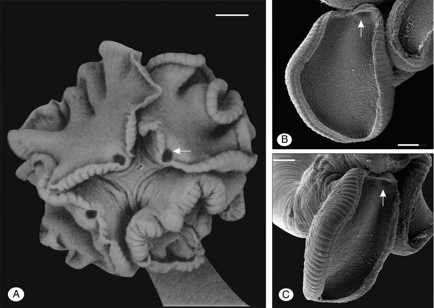
Worms (fig. 6A) slightly craspedote, euapolytic, 6–13 mm (8.5 ± 0.9 mm; n = 25) long, with 16–28 (19 ± 1; n = 25) proglottids; maximum width 450–600 (510 ± 25; n = 25) at level of scolex. Scolex (figs 6B and 7A) with four bothridia and short cephalic peduncle; myzorhynchus absent. Bothridia (fig. 7B, C) stalked, folded when contracted, elongate when relaxed, with 63–79 (72 ± 3; n = 25) marginal loculi and round apical sucker; apical sucker 60–70 (65 ± 3; n = 5) in diameter, (fig. 6B). Microtriches not seen.
Anthocephalum jeancadenati sp. n. (A) Entire worm; (B) scolex, apical view; (C) mature proglottid, ventral view; (D) gravid proglottid, ventral view. Scale bars: A = 500 μm; B, C, D = 250 μm.

Scanning electron micrographs of Anthocephalum jeancadenati sp. n. (A) Scolex in apical view, showing apical sucker; (B) distal bothridial surface; (C) bothridia, showing marginal loculi. Arrows indicate the apical sucker. Scale bars: A = 100 μm; B = 50 μm.

Immature proglottids initially wider than long, rapidly becoming longer than wide. Terminal proglottid (fig. 6C) 1.1–2.7 mm (1.7 ± 160 mm; n = 25) long by 200–450 (278 ± 26; n = 25) wide, length to width ratio 6.2–7.4 (6.9 ± 1.2; n = 25). Detached gravid proglottids (fig. 6D) 1.2–3.2 mm (2.85 ± 107 mm; n = 18) long by 300–610 (472 ± 59; n = 18) wide, length to width ratio 6–6.8 (6.3 ± 1; n = 25) (fig. 6D). Testes 32–46 (36 ± 2; n = 25) in number, oblong, 40–65 (51 ± 2; n = 30) long, by 20–40 (29 ± 2; n = 30) wide, arranged in 2–3 regular columns extending from anterior margin of proglottid to level of genital pore. Post-vaginal testes absent. Vas deferens dorsal, coiled, extending anteriorly from level of ovarian isthmus to enter cirrus sac at the anterior margin. Cirrus sac thin-walled, oval, recurved posteriorly, 120–220 (162 ± 9) long, 70–150 (84 ± 6; n = 25) wide, containing coiled cirrus; basal part of cirrus armed with spinitriches (fig. 6D). Genital pores lateral, muscular, irregularly alternating, 30–47% (35 ± 4%; n = 25) from posterior end of proglottid. Vagina weakly sinuous, extending from ovarian isthmus to anterior margin of cirrus sac. Ovary H-shaped in frontal view, tetralobed in cross section, symmetrically lobed 300–650 (467 ± 43; n = 25) long by 110–190 (137 ± 12; n = 25) wide at the level of isthmus; ovarian margins lobulated. Mehlis’ gland well developed, 50–110 (75 ± 7; n = 20) in diameter, posterior to ovarian isthmus. Vitelline follicles 20–45 (29 ± 3; n = 30) long by 12–30 (16 ± 2; n = 30) wide, arranged in two lateral bands, each band consisting of 2–3 dorsal and 2–3 ventral irregular columns, extending from near anterior to near posterior margin of proglottids, interrupted dorsally and ventrally by cirrus sac and ovary. Uterus ventral, extending from ovarian isthmus to anterior end of proglottid; uterine duct extending from Mehlis’ gland to join uterus anterior to level of vagina.
Remarks
Of the 18 valid species of Anthocephalum described to date (Ruhnke et al., Reference Ruhnke, Caira and Cox2015; Marques and Caira, Reference Marques and Caira2016, Ruhnke et al., Reference Ruhnke, Reyda, Marques, Caira and Jensen2017), Anthocephalum jeancadenati sp. n. bears fewer marginal loculi than A. cairae Ruhnke, 1994, A. currani Ruhnke & Seaman, 2009, A. duszynskii Ruhnke, 1994, A. healyae Ruhnke, Caira & Cox, 2015, A. hobergi (Zamparo, Brooks & Barriga, 1999), A. kingae (Schmidt, 1978), A. lukei Ruhnke & Seaman, 2009, A. mattisi Ruhnke, Caira & Cox, 2015, A. meadowsi Ruhnke, Caira & Cox, 2015, A. odonnellae Ruhnke, Caira & Cox, 2015, A. philruschi Ruhnke, Caira & Cox, 2015 and A. wedli Ruhnke, 2011 (69–79 vs 197–198, 81–110, 95–120, 150–171, > 100, 85, 107–138, 82–95, 98–134, 135–159 and 116–121, respectively) and it bears a greater number of marginal loculi than A. papefayei (69–79 vs 45–60). Anthocephalum jeancadenati sp. n. exhibits a greater number of testes than A. decrisantisorum Ruhnke, Caira & Cox, 2015 and A. jensenae Ruhnke, Caira & Cox, 2015 (32–46 vs 17–24 and 14–20, respectively) and fewer testes than A. gracile (32–46 vs 48–78). This new species most closely resembles A. alicae Ruhnke, 1994 and A. michaeli Ruhnke & Seaman, 2009 collected respectively from Hypanus americanus (Hildebrand & Schroeder) (= Dasyatis americana) and H. longus Garman (= Dasyatis longus). The new species possesses a wider terminal proglottid than A. alicae (200–450 vs 130–170) and fewer proglottids than A. michaeli (14–24 vs 23–41).
Unfortunately, the specimens of A. jeancadenati sp. n. prepared for SEM were not of sufficient quality to assess the microthrix pattern. Nevertheless, given the nature of the microthrix pattern seen across members of this genus, we predict that of this new species will be similar to that of its congeners. However, it would be useful to confirm this hypothesis by examination of material preserved appropriately for examination with SEM.
Ruhnke et al. (Reference Ruhnke, Caira and Cox2015) reported Anthocephalum n. sp. 2 from a specimen of T. grabata collected from Senegal but did not formally describe the species owing to a lack of sufficient material. Given that this specimen was also collected from the coast of Senegal and we found only a single species of Anthocephalum in T. grabata in this region, it seems likely that Anthocephalum n. sp. 2 sensu Ruhnke et al. (Reference Ruhnke, Caira and Cox2015) may be conspecific with this new species.
Discussion
Five valid genera, consisting of Anthocephalum, Barbeaucestus, Sungaicestus, Alveobothrium gen. n. and possibly also Cairaeanthus, are now considered to belong to the Anthocephaliidae (see Ruhnke et al., Reference Ruhnke, Caira and Cox2015, Reference Ruhnke, Reyda, Marques, Caira and Jensen2017; Caira et al., Reference Caira2017). The proglottid anatomy of Alveobothrium gen. n. is relatively consistent with that of the former three genera. The testes are arranged in a few columns anterior to the cirrus sac, the vas deferens extends to the level of ovary, and post-ovarian vitelline follicles are present. As noted by Caira et al. (Reference Caira2017), the proglottid anatomy of Cairaeanthus is distinct, with the presence of multiple columns of small testes, the vitelline follicles do not extend posterior to ovary and the vas deferens apparently does not extend posterior to cirrus sac.
Despite the fact that a molecular analysis was not conducted, owing to the lack of material fixed in ethanol, variation in the morphology and the arrangement of bothridial facial loculi are clearly supported by molecular phylogenetic analysis for anthocephliid genera as revealed by Healy et al. (Reference Healy2009), Rhunke et al. (Reference Ruhnke, Caira and Cox2015) and Marques and Caira (Reference Marques and Caira2016). However, the meaning and the phylogenetic significance of the apical sucker remains problematic. In the genus Divaricobothrium, the anteriormost feature of the bothridia is considered by Caira et al. (Reference Caira2017) as a loculus rather than apical sucker. The apical sucker seems to be absent in the bothridia of Cairaeanthus, but Ruhnke et al. (Reference Ruhnke, Caira and Cox2015) and Caira et al. (Reference Caira2017) stated that the feature may be overlooked in whole-mounted specimens. Hence, examination using scanning electron microscopy is important to verify this. It seems that this character carries no phylogenetic signal and can be regarded as a homoplasy, with independent acquisition of the same trait in unrelated species. Nonetheless, a first SEM examination of the bothridia of Rhinebothrium monodi Euzet, 1954, which also parasitizes the intestine of T. grabata, shows apical sucker and marginal loculi that could lead to a change in the phylogenetic position of this parasite. However, a molecular analysis must be carried out in future studies in order to effectively delineate these species and other rhinebothriidean taxa collected from T. grabata and other Dasyatidae and Rhinobatidae fished in the southern Mediterranean Sea.
According to the recent reclassification of the Dasyatidae by Last et al. (Reference Last, Naylor and Manjaji-Matsumoto2016) and the Torpedinidae by Claeson (Reference Claeson2014), Anthocephalum species are now known to be hosted by nine batoid genera: Bathytoshia Whitley, Fontitrygon Last, Naylor & Manjaji-Matsumoto, Himantura Müller & Henle, Hypanus Rafinesque, Neotrygon Castelnau, Pateobatis Last, Naylor & Manjaji-Matsumoto, Taeniurops Garman, Urobatis Garman, and Torpedo Duméril. The first six genera, Myliobatiformes, are genetically closely related (Last et al., Reference Last, Naylor and Manjaji-Matsumoto2016) whereas the genus Torpedo (Torpediniformes) is more distantly related (Claeson, Reference Claeson2014; Underwood et al., Reference Underwood2015). We hypothesize that the association of A. wedli (as Phyllobothrium gracile Wedl, 1855; see Ruhnke, Reference Ruhnke2011) with Torpedo marmorata Risso is probably ascribed to a lateral transfer (sensu Combes, Reference Combes2005) from a myliobatiform. Indeed, Torpedo marmorata lives in sympatry with other Dasyatidae in the Mediterranean Sea (Bauchot, Reference Bauchot, Fischer, Bauchot and Schneider1987) and thus host switching is possible. The same situation can be observed for the monocolid monogenean genus Empruthotrema (Johnston & Tiegs, 1922), which was found parasitizing the nasal fossae of Myliobatiformes and Torpediformes (e.g. Kearn, Reference Kearn1976; Chisholm and Whittington, Reference Chisholm and Whittington1999). However, cophylogenetic studies must be conducted to formally address this hypothesis.
Taeniurops grabata seems to have a naturally fragmented habitat. It is found in the eastern Atlantic Ocean, southwards from Cape Verde Island to Angola (Capapé and Desoutter, Reference Capapé, Desoutter and Quero1990) and in the central and south-eastern Mediterranean (McEachran and Capapé, Reference McEachran, Capapé and Whitehead1984), but is apparently absent in the western Mediterranean Sea (Postel, Reference Postel1956; Bauchot, Reference Bauchot, Fischer, Bauchot and Schneider1987; Serena, Reference Serena2005) and tends to be a good model in terms of studying the influence of a fragmented host distribution on the diversity of parasites. Fragmentation reduces the local host population, decreases the availability and the probability of parasite transmission and affects the population dynamics of parasites (McCallum and Dobson, Reference McCallum and Dobson2002; Greer and Collins, Reference Greer and Collins2008; Renwick and Lambin, Reference Renwick and Lambin2013), which may lead to the local extinction of parasites (Bush and Kennedy, Reference Bush and Kennedy1994; Strona, Reference Strona2015). For T. grabata, the same anthocephalids are reported from specimens of T. grabata caught from the Mediterranean Sea, in Tunisia, and the Atlantic Ocean, in Senegal. Additionally, the same three species of the monogenean genus Heterocotyle Scott, 1904 were found to be parasitizing the gills of T. grabata from Tunisia and Senegal (Neifar et al., Reference Neifar, Euzet and Ben Hassine1999). However, two assumptions can be made to account for the presence of the same parasite in these two areas: (1) the distribution of T. grabata is not really fragmented and the absence of this batoid from the western Mediterranean Sea may reflect lack of sampling effort. Batoids are generally important mesopredators with high mobility and extensive migrators and can link disparate habitats (Ajemian and Powers, Reference Ajemian and Powers2014). Thus, the absence of T. grabata in the western Mediterranean Sea remains to be verified; (2) the parasites may have been present before the geographical separation of T. grabata in the late-glacial period (c. 11,700 years ago). In this case, we can deduce that the duration of this separation is insufficient to have caused host and parasite speciation. These hypotheses remain to be verified by genetic analysis.
Acknowledgements
We thank Serveis Científics i Tècnics (University of Barcelona, Spain) for their support in the preparation of samples, and Dr Henri Capetta (Université de Montpellier) for valuable discussion on taxonomy of Dasyatidae. We thank the anonymous reviewers for their careful reading of our manuscript and their many insightful comments and suggestions. We are grateful to Wasim Hariz, English professor, for having proofread the manuscript.
Financial support
This work was supported in part by NSF PB&I grants DEB 0818696, and DEB 0818823, and project PCI A/023585/09.
Conflict of interest
None.
Author ORCIDs
L. Boudaya 0000-0002-4562-8358