Introduction
Echinococcosis, a zoonotic disease, has been recognized since time immemorial. The Office International des Epizooties (OIE) has listed the disease for socio-economic impact, public health importance and immense importance in international trade. Human beings acquire infection after ingestion of parasitic ova. Thereafter, metacestodes develop in virtually all organs. In humans surgery is done to cure the disease but chemotherapy has been regarded as a supplementary option in certain cases. Chemotherapy involves benzimidazole compounds (Ammann & Eckert, Reference Ammann, Eckert, Thompson and Lymbery1995).
In general benzimidazole compounds exert anthelmintic activity by binding to β-tubulin. This in turn prevents the polymerization of microtubules (Geerts & Gryseels, Reference Geerts and Gryseels2000). Resistance to benzimidazole compounds in nematodes is accompanied by loss of alleles at the β-tubulin locus and has also been correlated with a conserved point mutation (Driscoll et al., Reference Driscoll, Dean, Reilly, Bergholz and Chalfie1989; Roos et al., Reference Roos, Boersema, Borgsteede, Cornelissen, Taylor and Ruitenberg1990; Kwa et al., Reference Kwa, Veenstra and Roos1993a, Reference Kwa, Veenstra and Roosb, Reference Kwa, Veenstra, Van Dijk and Roos1995). Until now no information has been generated on sequence information and characterization of the β-tubulin gene of Echinococcus granulosus to deduce anthelmintic resistance on the basis of a conserved point mutation. So far the only published report has dealt with sequence information about the β-tubulin gene of E. multilocularis (Brehm et al., Reference Brehm, Kronthaler, Jura and Frosch2000).
Stress response is a phenomenon of adaptation of organisms. Altered patterns of protein synthesis, including expression of heat-shock proteins (HSP), might occur on exposure to natural and anthropogenic stressors. Stress proteins are synthesized in cells in response to diverse circumstances such as physiological conditions and the presence of environmental stressors like ultraviolet light, heat and chemicals (Del Razo et al., Reference Del Razo, Quintanilla-Vega, Brambila-Colombres, Calderón-Aranda, Manno and Albores2001). Due to its responsiveness to diverse forms of stress, heat-shock protein expression has been widely used in biomonitoring and environmental toxicology (Feder & Hofmann, 1999).
In vitro evaluation of benzimidazole resistance in nematodes is achieved by egg hatch assay. Heat-shock protein (HSP) has been suggested as a drug read-out system for proper anthelmintic selection against cestode (Martínez et al., Reference Martínez, Pérez-Serrano, Bernadina and Rodriguez-Caabeiro1999a, Reference Martínez, Pérez-Serrano, Bodega, Casado and Rodríguez-Caabeirob). Previously, researchers have proved this hypothesis on the basis of biochemical mining, and the viability of organisms has been assessed by phenotypic characterization.
The present study was conducted on β-tubulin gene isoforms to determine the candidate gene for benzimidazole compound resistance, based on point mutation. Relative expression of molecular chaperones has been deduced both by molecular and biochemical mining, and viability of protoscolices of E. granulosus has been correlated with the 14-3-3 protein signature.
Materials and methods
Extraction of genomic DNA from protoscolices and host cells
Cyst samples were collected from cattle, buffalo, sheep and goats from Tangra slaughter house, Kolkata, West Bengal, India. A total of 46 and ten cysts were collected from buffalo and cattle, respectively. Twenty and 34 samples were collected from goat and sheep, respectively. Isolated protoscolices, after washing with phosphate buffered saline (PBS) (pH 7.2), were preserved in 70% ethanol (v/v) at − 20°C. Genomic DNA from the organism and host species were isolated using a Genome® DNA kit (Q-BIO gene, California, USA). DNA samples were dissolved in nuclease-free water (NFW) and kept at − 70°C.
Design of primers
The following primers were designed from available sequence information (accession numbers AJ249548, AJ249549, AJ249550) to amplify tubulin gene 1 (Tub1), tubulin gene 2 (Tub2) and tubulin gene 3 (Tub3).
Tub1: Forward: 5′GGATTGCTCTCAGCTTTGAAAACTA3′
Reverse: 5′CACGGTACTGTTGACTGCCACGACT3′
Tub2: Forward: 5′CCTCCAGGGCTTCCAGCTCACCCAC3′
Reverse: 5′GAAGTGCAGGCGCGGGAATGGAACC3′
Tub3: Forward: 5′TTTAGCAGGTGACCAGCCCTTCTAA3′
Reverse: 5′GAGGTTCGACTGGCGAGGGGCGCAA3′
Amplification was carried out with an initial denaturation step at 95°C for 5 min followed by 35 cycles. Annealing was done at 56–60°C followed by elongation at 72°C.
Single-stranded conformation polymorphism (SSCP)
Seven microlitres of each polymerase chain reaction (PCR) product was used for the test, as described by Xie et al. (Reference Xie, Ho and Ma1997). Denatured products were separated on 10% acrylamide:bisacrylamide (30:1) non-denaturing Tris–borate–EDTA (TBE) gels. Patterns of relative migration (Mr) were visualized by ethidium bromide staining.
Cloning, sequencing and alignment of amino acid sequences
All SSCP conformers were cloned in TA cloning vector (Fermentas, Glen Burnie, Maryland, USA) and sequence information was generated by the University of Delhi, South Campus, New Delhi, India. Alignment of three isoforms of β-tubulin genes were made using online software (MEGA 4, Tamura et al., Reference Tamura, Dudley, Nei and Kumar2007) and data were compared with respective sequence information for E. multilocularis, Fasciola hepatica, Homo sapiens and Haemonchus contortus, to deduce conserved point mutations.
In vitro culture of protoscolices and drug induction
Isolated protoscolices were washed three times in Hank's balanced salt solution. Subsequently, viability of organisms was tested as described earlier (Casado et al., Reference Casado, Rodriguez-Caabeiro and Fernandez1986). The protoscolices were further harvested in M199 medium (Hi-Media, India) supplemented with 100 U penicillin/ml, 100 μg streptomycin and 10% fetal calf serum (Hi-Media). The organisms were incubated at 37°C with 5% CO2 tension. For in vitro evaluation of albendazole, the pure compound (99.62% pure) (batch no. TAFAAAP70025) provided by M/S Alembic Pharmaceuticals, India, was used at a concentration of 20 μg/ml in 48-well tissue culture plates (Axygen, India). Control cultures were maintained with and without dimethyl sulphoxide. Supernatants were collected at every 24 h interval up to 72 h and stored at − 20°C until use.
RNA extraction and cDNA preparation
Total RNA was extracted from protoscolices at 24 h intervals using TRI Reagent (Sigma, St. Louis, Missouri, USA). After extraction and quantification of RNA, cDNA was prepared using High-capacity cDNA Reverse Transcription Kit (Applied Biosystems, Foster City, California, USA).
Design of primer and semi-quantitative RT-PCR
The following primers were designed to amplify actin II, 14-3-3 protein, HSP 60 and HSP 70 from available sequence information of actin (accession number L07774) and molecular chaperones (accession numbers AY942149, EF201887, U26448).
Actin II: Forward: 5′GGAGTGATGGTTGGTATGGGACA3′
Reverse: 5′TGCCTGGATGGCTACATACATCG3′
14-3-3 protein: Forward: 5′ATGTCTTCTCTCAGTAAGCGCG3′
Reverse: 5′CTTTTCAAGTTGCGCTCAGTTC3′
HSP 60: Forward: 5′ATGTCAAGGCCCGTTACTACA3′
Reverse: 5′GCAACGAGCACCCTTCTCTGTG3′
HSP 70: Forward: 5′CGGCTGTAGGCATTGATTT3′
Reverse: 5′ACGCTCCGTATCCGTGAACG3′
The amplification reaction was carried out using each dNTP (200 μm), 10 × PCR buffer, 2.5 mm MgCl2, 10 pmol of each gene specific primer and an equal volume of cDNA as template. Actin II was used as an internal control to normalize expression of PCR templates of molecular chaperones. Relative absorbance of molecular chaperones was corrected against absorbance obtained for actin II.
Data analysis
Data were expressed as mean ± standard error (SE) and examined for their statistically significant differences with analysis of variance (ANOVA). P values less than 0.05 were considered to be statistically significant.
ELISA, SDS-PAGE and Western blot analysis
Enzyme-linked immunosorbent assay (ELISA) plates were coated with 50 μl supernatant (collected at 24 h intervals) diluted with coating buffer (10 mm sodium phosphate, pH 7.0). The plates were incubated overnight at 4°C. After incubation, the plates were washed with PBS and blocking was done using 3% bovine serum albumin (w/v). Subsequently, to determine the presence of HSPs, monoclonal anti-heat-shock protein 70 (clone BRM-22, Sigma) and monoclonal anti-heat-shock protein 60 (clone BRM-LK2, Sigma) were probed at 1:4000 and 1:200 dilutions, respectively. After incubation for 1 h, rabbit anti-mouse IgG–horseradish peroxidase (HRP) conjugate (Genei, Bangalore, India) was added after washing with PBS, and was incubated for 1 h. Optical density was measured after addition of substrate–chromogen complex.
Subsequently culture supernatants were separated on 12.5% discontinuous gel and transferred to nitrocellulose membrane (0.45 μm) (Schleicher and Schuell, Germany) (Towbin et al., Reference Towbin, Staehelin and Gordon1979). After washing with PBS, blocking was done using 5% skimmed milk powder. The nitrocellulose membrane was further incubated with monoclonal anti-heat-shock protein 70 (clone BRM-22, Sigma) and monoclonal anti-heat-shock protein 60 (clone BRM-LK2, Sigma). After washing with PBS, rabbit anti-mouse–HRP conjugate (Genei) was added. After an incubation period, colour was developed using a substrate–chromogen complex.
Results and discussion
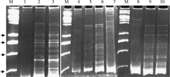
On the basis of SSCP analysis, two SSCP conformers could be detected within Tub1 and three conformers within Tub2 and Tub3 isoforms (fig. 1) of buffalo and sheep isolates. Based on the banding pattern, SSCP conformers were not detected in cattle and goat isolates. Amplicon sizes of conformers of Tub1 were 440 bp (FJ997219-220). Size of the PCR product of Tub2 (FJ997221-222, FJ997225) and Tub 3 (FJ997226, FJ997228, FJ997331) were 439–440 and 439, respectively.
SSCP banding patterns of tubulin isoforms of Echinococcus granulosus of buffalo and sheep isolates Lanes. M, 1 kb DNA ladder, arrows from the bottom indicate 250, 500, 750, 1000 bp fragments; 1, 3, SSCP conformers of buffalo isolates of tubulin; 2, SSCP conformer of sheep isolate of tubulin 1; 4, 6, 7, SSCP conformers of buffalo isolate of tubulin 2; 5, SSCP conformers of sheep isolate of tubulin 2; 8, 10, SSCP conformers of buffalo isolate of tubulin 3; 9, SSCP conformer of sheep isolate of tubulin 3.

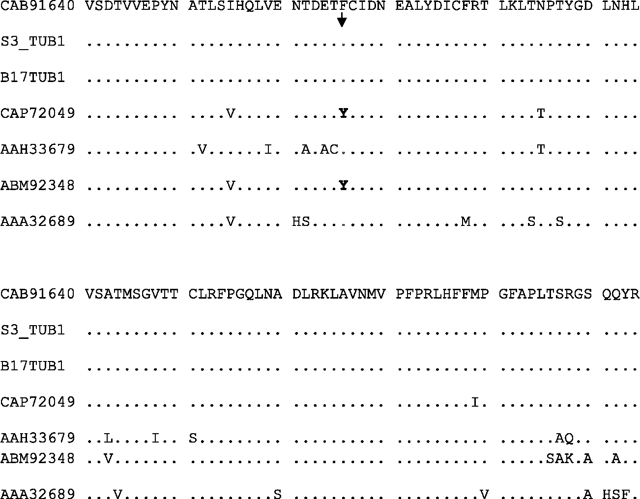
A perusal of available literature suggests that there was a tyrosine (Y) residue at position 200 of the Tub2 isoform in a vertebrate host (Sullivan, Reference Sullivan1988). Presence of this amino acid residue at position 200 after point mutation was indicative of benzimidazole resistance in Haemonchus contortus and Caenorhabditis elegans (Kwa et al., Reference Kwa, Veenstra, Van Dijk and Roos1995; Roos et al., Reference Roos, Kwa and Grant1995). In contrast, a phenylalanine (F) residue at position 200 of Tub1 and Tub3 indicated benzimidazole sensitivity (Kwa et al., Reference Kwa, Veenstra, Van Dijk and Roos1995; Thomas et al., Reference Thomas, Neff and Botstein1995; Elard et al., Reference Elard, Comes and Humbert1996). This has been further elaborated in the literature: the presence of a tyrosine (Y) residue at position 167 of Tub2 indicated benomyl resistance of Saccharomyces cerevisiae (Kwa et al., Reference Kwa, Veenstra, Van Dijk and Roos1995); and thiabendazole resistance has been indicated in Aspergillus nidulans when valine (V) has been detected in position 167 within the Tub2 isoform ( Jung & Oakley, Reference Jung and Oakley1990).
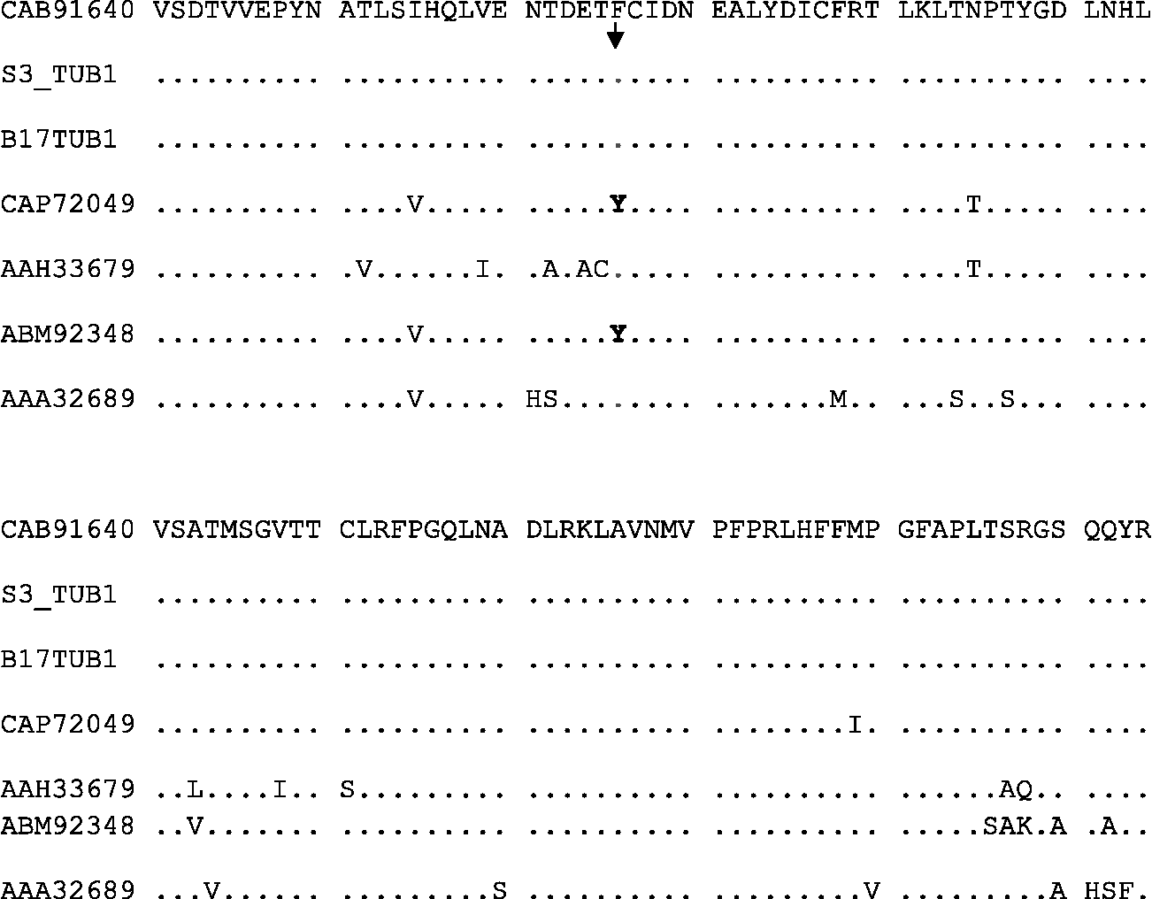
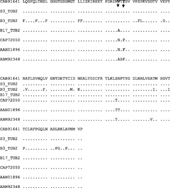
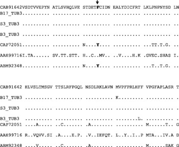
During the present study (see figs 2–4) a conserved point mutation was not detected at position 200 of Tub2 and positions 165 and 167 of Tub1 and Tub3, respectively. However, a phenylalanine (F) residue at position 200 of Tub1 and Tub3 sequences indicated sensitivity to benzimidazole compound (figs 2 and 4). On the contrary, valine (V) and tyrosine (Y) residues were present in positions 165 and 167 of Tub2 isoform (fig. 3). These results suggested that Tub2 isoform can be used as a candidate gene to deduce anthelmintic resistance. Jung & Oakley (Reference Jung and Oakley1990) proposed a similar result in a study conducted with yeast and fungus.
Predicted amino acid sequence of two conformers of Tubulin 1. Dots denote homology with CAB91640 (Tubulin 1 of E. multilocularis). CAP72049 (Fasciola hepatica tubulin 1), AAH33679 (Homo sapiens tubulin 1), ABM92348 (Haemonchus contortus tubulin 1), AAA32689 (Aspergillus flavus tubulin 1) sequences were used as outgroups. Arrow indicates presence of phenylalanine (F) residue at position 200. S3_TUB1 (FJ997219) and B17 TUB1 (FJ997220) have GenBank accession numbers.
Predicted amino acid sequence of three conformers of Tubulin 2. Dots denote homology with CAB91641 (Tubulin 2 of E. multilocularis). CAP72050 (Fasciola hepatica tubulin 2), AAH01896 (Homo sapiens tubulin), ABM92348 (Haemonchus contortus tubulin) sequences were used as outgroups. Arrows indicate valine (v) and tyrosine residues (Y) at positions 165 and 167, respectively. S3_TUB2 (FJ997225), B3_TUB2 (FJ997221) and B17_TUB2 (FJ997222) have GenBank accession numbers.

Predicted amino acid sequence of three conformers of Tubulin 3. Dots denote homology with CAB91642 (Tubulin 3 of E. multilocularis). CAP72051 (Fasciola hepatica tubulin 3), AAH99716 (Homo sapiens tubulin 3 like), ABM92348 (Haemonchus contortus tubulin isotype 1) sequences were used as outgroups. Arrow indicates presence of phenylalanine (F) residue at position 200. B17_TUB3 (FJ997231), S3_TUB3 (FJ997228) and B3_TUB3 (FJ997226) have GenBank accession numbers.

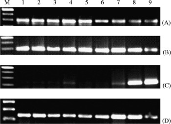
Product sizes of the housekeeping gene actin II (FJ897573), 14-3-3 protein (FJ897574), HSP 70 (FJ897572) and HSP 60 (FJ897575) were 291, 295, 134 and 240 bp, respectively. When HSP and 14-3-3 gene transcription were deduced by semi-quantitative RT-PCR, it was found that transcription of 14-3-3 protein, HSP 70 and HSP 60 were increased after albendazole treatment compared to the control and the DMSO-treated groups, and the result was statistically significant. Further it was found that transcription of HSP 60 was highest after 72 h of treatment (5.38 ± 0.147) in albendazole-treated protoscolices, followed by transcription of 14-3-3 protein (2.04 ± 0.046) and HSP 70 (1.62 ± 0.059) (table 1, fig. 5).
Relative expression of E. granulosus (Eg)14-3-3, HSP 70, HSP 60 in control, DMSO and albendazole (ABZ) treated groups at 24 h intervals.

Significant at ***, P < 0.001; **, P < 0.01; *, P < 0.05.
mRNA expression levels of actin II (A), Eg14-3-3 (B), HSP 60 (C) and HSP 70 (D) of protoscolices of Echinococcus granulosus with or without albendazole treatment (mRNA levels were detected by reverse transcriptase (RT)-PCR using specific primers). Lanes: M, 100 bp DNA ladder; 1–3, controls after 24 h, 48 h, 72 h incubation; 4–6, controls with DMSO after 24 h, 48 h, 72 h incubation; 7–9, albendazole treated after 24 h, 48 h, 72 h intervals. The amplified cDNA was resolved on 1.5% (w/v) agarose gel and visualized by ethidium bromide.

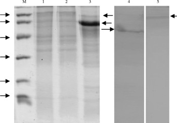
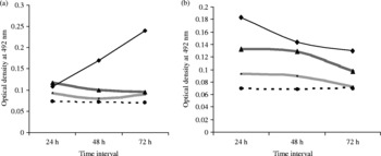
The protein profile of the culture supernatant harvested in the presence of albendazole displayed an intense band with a relative molecular weight (Mr) of 60 kDa. A faint band was observed with a Mr of 70 kDa. Both peptides reacted with anti-HSP 60 and anti-HSP 70 monoclonal antibodies (fig. 6). Protoscolices treated with albendazole exhibited a gradual increase in OD value at 24 h intervals (0.109–0.240) in comparison to control and DMSO-treated groups when HSP 60 monoclonal antibody was used in indirect ELISA (fig. 7a). OD value decreased gradually at 24 h intervals (0.183–0.130) when anti-HSP 70 was used (fig. 7b). The result of semi-quantitative RT-PCR and primary binding assay confirmed HSP 60 as a marker protein after albendazole treatment in vitro. The role of HSPs in host–parasite interaction in eliciting an immune response has received some attention (Polla, Reference Polla1991; Kaufmann, Reference Kaufmann1992; Maresca & Kobayashi, Reference Maresca and Kobayashi1994). Previous studies have indicated that expression of HSP 60 increased in protoscolices after treatment with anthelmintics (Martínez et al., Reference Martínez, Pérez-Serrano, Bernadina and Rodriguez-Caabeiro1999a, Reference Martínez, Pérez-Serrano, Bodega, Casado and Rodríguez-Caabeirob). However, the previous results were interpreted on the basis of biochemical mining and the present investigation has identified HSP 60 as a biomarker based both on biochemical and molecular mining.
Protein profiles of concentrated cell culture supernatant of control (lane 1), control with DMSO (lane 2), and albendazole-treated groups (arrows from bottom indicate 60 and 70 kDa peptides) (lane 3). M, medium-range protein molecular weight marker (arrows indicate, from bottom, 14.3, 20.1, 29, 43, 66 and 97.4 kDa). Arrows indicate 60 kDa (lane 4) and 70 kDa (lane 5) immunoreactive peptides confirmed by monoclonal anti-HSP 60 and anti-HSP 70 antibodies.

Optical density (OD) values of the cell culture supernatant of control ( ), negative control (
 ), DMSO (
 ) and albendazole treated (ABZ) (
 ) groups at 24 h intervals with (a) HSP 60 and (b) HSP 70 used as secondary antibodies.

14-3-3 protein is a highly conserved acidic protein (Siles-Lucas et al., Reference Siles-Lucas, Merli and Gottstein2008). In mammals seven isoforms have been identified (van Heusden & Steensma, Reference van Heusden and Steensma2006). Two different isoforms of 14-3-3 protein have been identified in E. granulosus (Siles-Lucas et al., Reference Siles-Lucas, Merli and Gottstein2008). During the present study, 14-3-3 protein (FJ897574) zeta isoform was identified. In general this intracellular protein plays an active role during signal transduction, cell cycle control, cell differentiation and cell survival (Fu et al., Reference Fu, Subramanian and Masters2000; van Hemert et al., Reference van Hemert, Steensma and van Heusden2001). During the present investigation, more expression of this biomolecule after albendazole treatment in comparison to control and DMSO-treated groups indicated that albendazole might have played a role as a sensory signal molecule. Survivability of protoscolices remained unquestioned due to expression of actin II throughout the experimental study. Actin II is constitutively expressed in normal living cells and its transcription level is thought to represent the normal viability of the cells (Matsumoto et al., Reference Matsumoto, Müller, Hemphill, Oku, Kamiya and Gottstein2006).
Therefore, in the present investigation HSP 60 has been identified as a biomarker of the drug read-out system, β-tubulin isoform 2 has been distinguished as a candidate gene for anthelmintic resistance and viability of the organism has been confirmed on the basis of expression of actin II and 14-3-3 protein.
Acknowledgements
The authors are grateful to the Director, Indian Veterinary Research Institute, for provision of necessary facilities.